This website uses cookies to ensure you get the best experience on our website.


- Table of Contents
Plan chromogenic IHC on paraffin sections using cytoplasmic staining in lymphatic organs as a reference (HPA tissue IHC). Start the IHC-validated antibody A03282-1 at 1:100–1:300 (datasheet), and compare staining with the listed positive and negative cell populations (HPA tissue IHC).

Expected localisation, validated protocols, controls and antibodies — the at-a-glance facts below, then the full design guide.
| Expected localisation | Cytoplasmic staining in lymphatic organs (HPA tissue IHC) | |
| Staining pattern | Abundant cytoplasmic staining in lymphatic organs (HPA tissue IHC) | |
| Antigen retrieval | Citrate pH 6.0 HIER, 95–98 °C, 20 min (rule: cytoplasmic / membrane antigen) | |
| Positive control | Testis+4 more · see all | |
| Negative control | Adipose tissue+4 more · see all |
| Fixation | Keep fixation consistent across sections. (standard IHC practice; not target-specific) | |
| Caveat | EGF can shift Girdin from cytosol to membrane (UniProt) | |
| Regulation | Expression regulation is unspecified (UniProt) | |
| Isoform / epitope | Five isoforms; check antibody epitope coverage (UniProt) |
The catalog antibody’s IHC-P protocol is accompanied by published CCDC88A protocols for pancreatic ductal adenocarcinoma (PMC5139074) and hepatocellular carcinoma (PMC9043556).
| Sample | Paraffin-embedded human brain tissue; fixative not specified (datasheet A03282-1) |
| Fixation | Image fixative and duration unreported (datasheet A03282-1); verify before use. |
| Sectioning | 4–5 µm sections on charged slides (standard) |
| Deparaffinisation | Xylene, graded ethanol series to water (standard) |
| Antigen retrieval | Heat-induced epitope retrieval in citrate buffer, pH 6.0, 20 min at 95–98 °C (standard rule: cytoplasmic / membrane antigen) |
| Peroxidase block | 3% H2O2, 10 min, room temperature (standard) |
| Blocking | 10% normal serum of the secondary host, 30 min, room temperature (standard) |
| Primary antibody | Rabbit anti-CCDC88A, 1:100 - 1:300 (datasheet A03282-1) |
| Primary incubation | Overnight at 4 °C (standard) |
| Detection | HRP-polymer secondary, DAB chromogen 5–10 min (standard) |
| Counterstain | Hematoxylin, blue, dehydrate and mount (standard) |
| Expected result | CCDC88A-positive staining in pachytene spermatocytes of testis (HPA tissue IHC: High). HPA tissue profile: Abundant cytoplasmic expression in lymphatic organs. No signal in the no-primary control. |
CCDC88A is a soluble protein found in the cytosol and at membranes and cytoskeletal structures; it has no transmembrane segment (UniProt Q3V6T2). In paraffin section IHC, expect chiefly cytoplasmic staining in pachytene spermatocytes and selected lymphoid cells or lung macrophages (HPA: tissue IHC). HPA rates its tissue IHC profile Enhanced, with medium consistency between antibody staining and RNA expression (HPA: tissue IHC).
| Distinct cytoplasmic staining in pachytene spermatocytes; weaker staining in germinal center cells or lung macrophages. | This matches the reported cell distribution: High in testis pachytene spermatocytes and Medium in lymph node and tonsil germinal center cells and lung macrophages (HPA: tissue IHC). Compare cells within their tissue context; these levels do not specify a universal staining intensity across laboratories. |
| Predominantly nuclear staining in an IHC section, with little cytoplasmic signal in expected positive cells. | Review the result as a possible compartment mismatch: UniProt places CCDC88A in cytosol and associated structures (UniProt Q3V6T2). HPA reports nucleoplasm in ICC-IF, but marks that location uncertain (HPA: subcellular ICC-IF). That IF observation alone does not establish a predominantly nuclear IHC pattern. |
| Strong staining in cells reported as undetected, such as adipocytes or bronchial respiratory epithelium. | HPA reports no detection in those specified cell types (HPA: tissue IHC). Check cell identification and controls before calling the result CCDC88A; antibody cross-reactivity or endogenous chromogenic detection activity are possible general IHC explanations. An undetected cell type does not make every cell in that tissue a negative control. |
| A broad, even deposit across tissue, including cells expected to be negative. | This obscures the cell-specific pattern needed for interpretation (HPA: tissue IHC). In general IHC practice, excess primary or detection reagent, incomplete blocking, or inadequate washing can produce diffuse background. Examine the no-primary control and compare signal with the expected positive cell population before scoring. |
| No convincing signal in pachytene spermatocytes on a testis section. | That conflicts with the High signal reported for these cells (HPA: tissue IHC). Confirm that the relevant cells are present and that the positive control and detection run worked; then review the antibody's documented IHC use and general retrieval and dilution conditions. HPA supplies no CCDC88A-specific fixation sensitivity here. |
| Topology and compartment | CCDC88A has no transmembrane segment and is reported in cytosol, at the cell membrane, and at cytoskeletal structures (UniProt Q3V6T2). Interpret a cytoplasmic pattern as compatible with the target; membrane association may occur without a membrane-spanning protein. |
| Cell state | UniProt reports cytosolic localization in unstimulated cells and increased membrane localization after EGF stimulation (UniProt Q3V6T2). A membrane-associated component can therefore fit the record, but this does not predict the stimulus state or intensity of a fixed tissue section. |
| Tissue and antibody evidence | HPA calls the tissue IHC profile Enhanced but notes medium consistency with RNA data (HPA: tissue IHC). Its antibody records rate HPA038102 Enhanced and HPA038101 Supported for IHC (HPA: antibodies). Use those grades to frame confidence, not as proof that every stained cell is specific. |
| Isoforms and epitope coverage | UniProt lists 5 CCDC88A isoforms (UniProt Q3V6T2). The supplied sources give no epitope map for the antibody used here, so isoform-specific detection or absence cannot be inferred from staining; check the antibody's documented epitope if an isoform claim matters. |
| IF/ICC Q: Is nucleoplasmic or centrosomal staining expected? | A: HPA reports centrosome and basal body as supported ICC-IF locations, plasma membrane as an additional supported location, and nucleoplasm as uncertain (HPA: subcellular ICC-IF). These cell-image observations belong to IF/ICC interpretation and do not replace the tissue IHC pattern. |
| Situation | Likely cause | Next action |
|---|---|---|
| The testis positive control is blank. | Pachytene spermatocytes may be absent from the examined area, or a general IHC staining step may have failed (HPA: High in pachytene spermatocytes; general IHC practice). | Locate the relevant cells on the counterstain, verify the detection control, then review primary antibody dilution and antigen retrieval against the antibody's documented IHC conditions. Do not assign a CCDC88A-specific fixation effect without evidence. |
| Germinal center cells stain faintly while testis stains clearly. | The difference may reflect the reported tissue pattern: germinal center cells are Medium and pachytene spermatocytes High (HPA: tissue IHC). | Score identified cell populations separately and compare with a run-matched positive control. Avoid increasing detection intensity solely to force equal staining across these tissues (HPA: tissue IHC; general IHC practice). |
| Bronchial respiratory epithelium or adipocytes stain strongly. | Those specific cell types are reported as not detected (HPA: tissue IHC); cross-reactivity or endogenous detection activity is a possible general IHC cause. | Confirm cell identity, inspect the no-primary control for endogenous chromogenic activity, and reassess antibody specificity before assigning the signal to CCDC88A (general IHC practice). |
| The whole section has diffuse brown background. | Nonspecific reagent binding, insufficient blocking or washing, or overdevelopment can obscure cell-level contrast (general IHC practice). | Compare no-primary and positive controls, then adjust blocking, washing, antibody concentration, or chromogen development within the documented IHC workflow. Reassess the reported positive cell types after background is reduced (HPA: tissue IHC; general IHC practice). |
| Signal appears mainly nuclear in paraffin section IHC. | The dominant compartment differs from UniProt's cytosolic localization; HPA's nucleoplasm assignment comes from ICC-IF and is uncertain (UniProt Q3V6T2; HPA: subcellular ICC-IF). | Check the counterstain and no-primary control, then review staining specificity and section morphology before scoring nuclear signal as CCDC88A (general IHC practice). |
| A brain section is weak despite tissue-enhanced brain RNA. | RNA and cell-level protein staining need not match: HPA reports brain-enhanced RNA but no detection in caudate glial cells or cerebral cortex endothelial cells, and Low staining in cerebellar Purkinje cells (HPA: tissue IHC). | Interpret the named cells separately and use the listed testis positive population to check the IHC run; do not infer a uniformly strong brain protein signal from the RNA category (HPA: tissue IHC). |


Comprehensive Human Protein Atlas IHC scoring per tissue (reliability: Enhanced — Medium consistency between antibody staining and RNA expression data.). Rows are taken directly from the HPA tissue chart — click any row's HPA link to view the source.
| Tissue | Cell type | Level | Evidence | Source |
|---|---|---|---|---|
| Testis | Pachytene spermatocytes | High | Protein (IHC) | HPA → |
| Appendix | Lymphoid tissue | Medium | Protein (IHC) | HPA → |
| Lung | Macrophages | Medium | Protein (IHC) | HPA → |
| Lymph node | Germinal center cells | Medium | Protein (IHC) | HPA → |
| Tonsil | Germinal center cells | Medium | Protein (IHC) | HPA → |
| Tissue | Cell type | Level | Evidence | Source |
|---|---|---|---|---|
| Adipose tissue | Adipocytes | Not detected | Protein (IHC) | HPA → |
| Adrenal gland | Glandular cells | Not detected | Protein (IHC) | HPA → |
| Breast | Adipocytes | Not detected | Protein (IHC) | HPA → |
| Bronchus | Respiratory epithelial cells | Not detected | Protein (IHC) | HPA → |
| Caudate | Glial cells | Not detected | Protein (IHC) | HPA → |
Troubleshoot CCDC88A (Girdin) staining in paraffin sections by checking retrieval, compartment, cell type and controls before comparing scores.
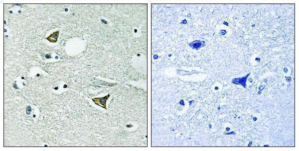
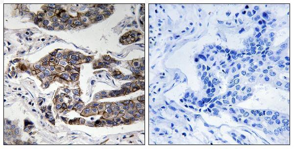
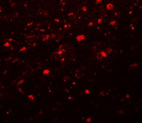
Anti-CCDC88A antibodies have IHC images from paraffin-embedded human brain and breast carcinoma, and an IF image from human brain (catalog figure captions). Listed reactivity covers human, mouse, and rat (catalog: reactivity).
A03282-1 shows IHC in paraffin-embedded human brain, with synthesized-peptide blocking (A03282-1 figure caption); P03282 shows phospho-Ser1417 IHC in paraffin-embedded human breast carcinoma, with phospho-peptide blocking (P03282 figure caption). A03282-2 shows IF in human brain at 20 μg/mL (A03282-2 figure caption).
Which to pick: For tissue IHC, choose A03282-1 for total Girdin or P03282 for phospho-Ser1417; both are rabbit polyclonals listed for human and mouse IHC (catalog: titles, dilution data, applications, reactivity). For IF/ICC, A03282-1 and P03282 list both applications, while A03282-2 has a human-brain IF image and lists IF but not ICC (catalog: applications; A03282-2 figure caption). A03282-2 also lists rat reactivity, though its pictured IF sample is human (catalog: reactivity; A03282-2 figure caption); the IHC captions specify paraffin sections but report no fixative (A03282-1 and P03282 figure captions).