This website uses cookies to ensure you get the best experience on our website.


- Table of Contents
Plan CCNA2 chromogenic IHC around nuclear staining in a varying fraction of cells (HPA tissue IHC). This guide covers consistent fixation (standard IHC practice), cell-cycle localisation (UniProt), and the catalog antibody’s 1:50 IHC dilution (datasheet M00700-1).

Expected localisation, validated protocols, controls and antibodies — the at-a-glance facts below, then the full design guide.
| Expected localisation | Nuclear in a varying fraction of tissue cells (HPA tissue IHC) | |
| Staining pattern | Nuclear staining in a varying fraction of cells in most tissues (HPA tissue IHC) | |
| Antigen retrieval | EDTA pH 8.0 HIER, heat-mediated (datasheet M00700-1) | |
| Positive control | Appendix+4 more · see all | |
| Negative control | Adipose tissue+4 more · see all |
| Fixation | Matched tissue-IHC evidence does not establish the fixation claim. Validate the specimen-specific method before use. (selected-SKU IHC image M00700-1) | |
| Caveat | Prophase can shift signal from nucleus to cytoplasm (UniProt) | |
| Regulation | Staining varies with the cycling-cell fraction (UniProt) | |
| Isoform / epitope | No annotated isoforms; one full-length chain (UniProt) |
The catalog antibody’s IHC-P protocol (datasheet: M00700-1) is followed by published CCNA2 protocols for prostate tissue chips (PMC11211272) and follicular-fluid cell smears (PMC12563169).
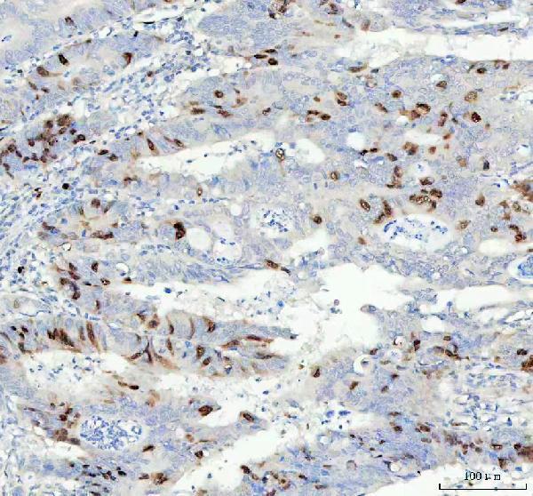
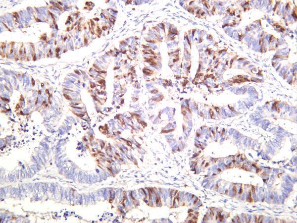
| Sample | Paraffin-embedded human colorectal adenocarcinoma tissue; fixative not specified (datasheet M00700-1) |
| Fixation | Image fixative and duration unreported (datasheet M00700-1); verify before use. |
| Sectioning | 4–5 µm sections on charged slides (standard) |
| Deparaffinisation | Xylene, graded ethanol series to water (standard) |
| Antigen retrieval | Heat retrieval: EDTA pH 8.0 (datasheet M00700-1); 20 min, 95–100 °C (standard) |
| Peroxidase block | 3% H2O2, 10 min, room temperature (standard) |
| Blocking | 10% goat serum (datasheet M00700-1) |
| Primary antibody | Rabbit monoclonal (clone HOC-3) anti-CCNA2, 1:50 (datasheet M00700-1) |
| Primary incubation | Overnight at 4 °C (datasheet M00700-1) |
| Detection | HRP-conjugated secondary, DAB chromogen (datasheet M00700-1) |
| Counterstain | Hematoxylin, blue, dehydrate and mount (standard) |
| Expected result | CCNA2-positive staining in endocrine cells of appendix (HPA tissue IHC: High). HPA tissue profile: Nuclear expression in a varying fraction of cells in most tissues. No signal in the no-primary control. |
CCNA2 staining should be predominantly nuclear in a varying fraction of cells across most tissues (HPA tissue IHC; UniProt P20248: interphase localisation). High staining is reported in gastrointestinal endocrine cells, while several other listed cell populations are not detected (HPA tissue IHC). The tissue IHC profile has Enhanced reliability, with medium consistency between antibody staining and RNA expression (HPA tissue IHC). CCNA2 has no transmembrane segment (UniProt P20248 topology).
| Distinct nuclear staining appears in only some cells; appendix, colon, duodenum, rectum or small-intestine endocrine cells stain strongly. | This fits the variable nuclear fraction reported across tissues and the High level reported in those endocrine populations (HPA tissue IHC). Score the stained cell population and nuclear compartment, rather than treating unstained neighbours as a failed run (HPA tissue IHC; UniProt P20248: interphase localisation). |
| Broad, strong cytoplasmic staining dominates cells whose nuclei show little or no signal. | That pattern warrants an artefact check because CCNA2 is exclusively nuclear during interphase (UniProt P20248). Cytoplasmic signal is possible at prophase or with SCAPER, and ICC-IF also reports cytosol; judge it with cell context and controls (UniProt P20248; HPA subcellular ICC-IF). |
| Adipocytes or liver cholangiocytes stain strongly when the intended positive cells do not. | HPA reports CCNA2 as Not detected in those cell populations (HPA tissue IHC). Consider antibody cross-reactivity or endogenous chromogenic activity, then compare a known-positive section and detection controls (HPA tissue IHC; general IHC practice). |
| Colour spreads across stroma, lumina or many unrelated cells without clear nuclear boundaries. | This is difficult to score as CCNA2 because the tissue profile describes nuclear staining in a varying fraction of cells (HPA tissue IHC). Diffuse deposit can arise from nonspecific binding or detection background; inspect the no-primary control (general IHC practice). |
| No nuclear signal appears in appendix endocrine cells, despite an otherwise readable section. | Those cells are reported High and offer a useful positive comparison, though HPA levels are observations rather than a guarantee for every section (HPA tissue IHC). Check that the population is present, then review assay controls and detection steps (general IHC practice). |
| Cell-cycle localisation | Interphase CCNA2 is exclusively nuclear; nucleus and cytoplasm are reported at prophase, and association with SCAPER can retain CCNA2 in cytoplasm (UniProt P20248). Interpret occasional cytoplasmic cells in that context rather than requiring every stained cell to look identical. |
| Tissue and cell population | HPA reports High staining in endocrine cells of appendix, colon, duodenum, rectum and small intestine; breast glandular cells and endometrial stromal cells are Medium (HPA tissue IHC). The observed fraction and compartment matter more than a whole-section average. |
| Antibody validation | CAB000114 has Enhanced IHC validation; HPA020626 has no IHC status in the supplied record (HPA antibodies). Enhanced reflects reproduction by independent antibodies or orthogonal data, while the tissue profile still reports medium RNA–staining consistency (HPA antibodies; HPA tissue IHC). |
| Processing and topology | UniProt lists one CCNA2 chain spanning residues 1–432, with no signal peptide, propeptide, transmembrane segment or annotated isoforms (UniProt P20248). These annotations give no basis to expect a secreted or membrane staining pattern; epitope-specific retrieval behaviour is unreported. |
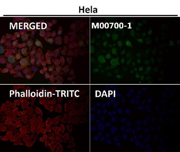
| IF/ICC: What pattern should I expect? | HPA reports enhanced nucleoplasm and cytosol localisation in ICC-IF (HPA subcellular ICC-IF). This supports an IF localisation check, while paraffin-section interpretation should follow the tissue IHC pattern and the cell-cycle context (HPA tissue IHC; UniProt P20248). |
| Situation | Likely cause | Next action |
|---|---|---|
| No nuclear staining in a section containing appendix endocrine cells. | A missed assay step or insufficient staining is possible; the cell population is reported High, but its presence alone does not validate a run (HPA tissue IHC; general IHC practice). | Verify the cells are present, compare a concurrent positive section, and check antigen retrieval, primary incubation and detection against the validated IHC workflow (general IHC practice). |
| Strong cytoplasmic colour appears in nearly every cell, with little nuclear definition. | The distribution conflicts with interphase nuclear localisation; prophase or SCAPER-associated cytoplasm may explain individual cells, not an automatic whole-section call (UniProt P20248). | Compare nuclear counterstain and cell context, then inspect no-primary and detection controls for diffuse background (general IHC practice). |
| Adipocytes stain while the intended nuclear positive population does not. | HPA reports adipocytes as Not detected; nonspecific antibody staining or endogenous detection activity is possible (HPA tissue IHC; general IHC practice). | Run a no-primary control and a known-positive tissue, then review blocking and endogenous-peroxidase control for the chromogenic method (general IHC practice). |
| Colour covers tissue broadly and obscures individual nuclei. | Diffuse deposit is inconsistent with a clearly scoreable, variable nuclear pattern (HPA tissue IHC); excess background can result from nonspecific binding or detection conditions (general IHC practice). | Check the no-primary control, review blocking and washes, and adjust staining conditions within the validated assay (general IHC practice). |
| A few cells show both nuclear and cytoplasmic staining. | Nuclear and cytoplasmic CCNA2 is reported at prophase, and cytoplasmic localisation can occur with SCAPER (UniProt P20248). HPA ICC-IF also reports nucleoplasm and cytosol (HPA subcellular ICC-IF). | Record the compartments and frequency, compare adjacent interphase-appearing cells, and avoid calling isolated cytoplasmic signal artefact without controls (UniProt P20248; general IHC practice). |
| Different antibody preparations give different IHC patterns. | The supplied validation is antibody-specific: CAB000114 is IHC Enhanced, while HPA020626 has no supplied IHC status (HPA antibodies). | Anchor interpretation to an IHC-validated antibody, its controls and the reported nuclear tissue pattern; investigate discordant staining before scoring it as CCNA2 (HPA antibodies; HPA tissue IHC; general IHC practice). |
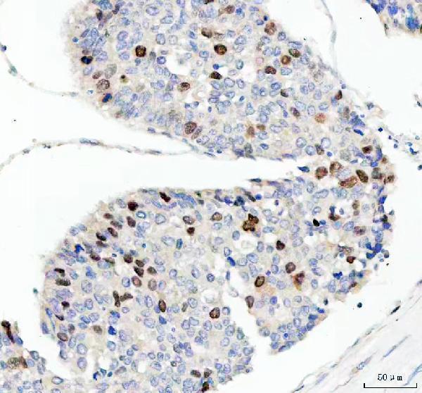


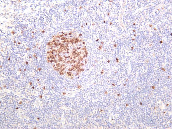
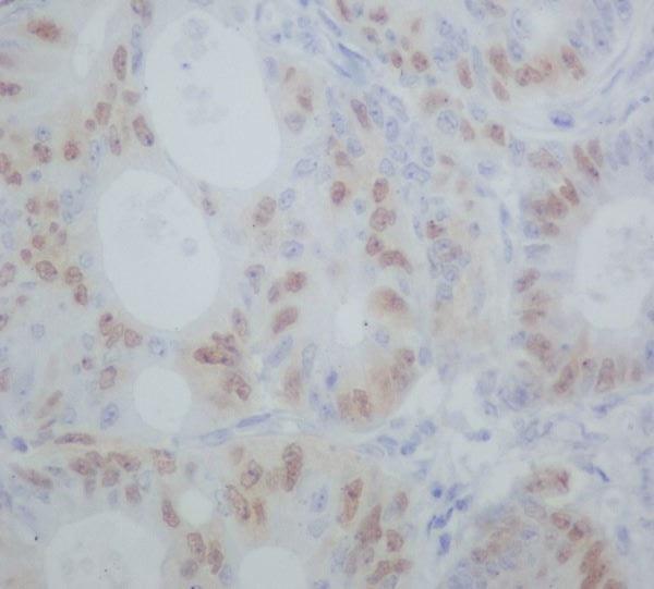
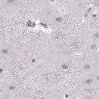


Comprehensive Human Protein Atlas IHC scoring per tissue (reliability: Enhanced — Medium consistency between antibody staining and RNA expression data.). Rows are taken directly from the HPA tissue chart — click any row's HPA link to view the source.
| Tissue | Cell type | Level | Evidence | Source |
|---|---|---|---|---|
| Appendix | Endocrine cells | High | Protein (IHC) | HPA → |
| Colon | Endocrine cells | High | Protein (IHC) | HPA → |
| Duodenum | Endocrine cells | High | Protein (IHC) | HPA → |
| Rectum | Endocrine cells | High | Protein (IHC) | HPA → |
| Small intestine | Endocrine cells | High | Protein (IHC) | HPA → |
| Tissue | Cell type | Level | Evidence | Source |
|---|---|---|---|---|
| Adipose tissue | Adipocytes | Not detected | Protein (IHC) | HPA → |
| Caudate | Glial cells | Not detected | Protein (IHC) | HPA → |
| Cerebellum | Cells in granular layer | Not detected | Protein (IHC) | HPA → |
| Cerebral cortex | Endothelial cells | Not detected | Protein (IHC) | HPA → |
| Epididymis | Glandular cells | Not detected | Protein (IHC) | HPA → |
Troubleshoot CCNA2 staining by checking retrieval, cellular compartment, cell cycle context, and the source of background before interpreting positive cells.
Catalog antibodies cover human paraffin-section IHC, including colorectal adenocarcinoma, and IF/ICC in A431 cells (catalog image captions); several list human, mouse and rat reactivity (catalog reactivity).
M00700-1 and PB9424 each show IHC in human paraffin-embedded colorectal adenocarcinoma; PB9424 also shows IF/ICC in A431 cells (respective image captions). M00700-3 shows IHC in human colon carcinoma and tonsil, while M00700 shows IHC in paraffin-embedded human colon cancer and recognizes Cyclin A1/A2 (respective image captions; M00700 catalog title).
Which to pick: For CCNA2 tissue IHC, start with M00700-1: it is a rabbit monoclonal with human, mouse and rat reactivity, and its own human paraffin-section image specifies EDTA retrieval at pH 8.0 and 1:50 primary dilution (M00700-1 catalog; M00700-1 IHC caption). For IF/ICC, PB9424 has an A431 cell image at 5 μg/mL; M00700-1 also lists IF/ICC, and M00700-3 lists IF with human, mouse and rat reactivity (PB9424 IF caption; M00700-1 catalog; M00700-3 catalog). For cross-species IHC, M00700-1 and M00700-3 list human, mouse and rat reactivity, but their supplied IHC images show human samples; fixation is unreported in those captions, so establish retrieval and dilution for each specimen (respective catalog reactivity and IHC captions).