Click image to see more details
-

-
-
-
-
+6
Product Info Summary
| SKU: | PB9424 |
|---|---|
| Size: | 100 μg/vial |
| Reactive Species: | Human, Mouse, Rat |
| Host: | Rabbit |
| Application: | Flow Cytometry, IF, IHC, ICC, WB |
Customers Who Bought This Also Bought

Product info
Product Name
Anti-Cyclin A2/CCNA2 Antibody Picoband®
SKU/Catalog Number
PB9424
Size
100 μg/vial
Form
Lyophilized
Description
Boster Bio Anti-Cyclin A2/CCNA2 Antibody Picoband® catalog # PB9424. Tested in Flow Cytometry, IF, IHC, ICC, WB applications. This antibody reacts with Human, Mouse, Rat. The brand Picoband indicates this is a premium antibody that guarantees superior quality, high affinity, and strong signals with minimal background in Western blot applications. Only our best-performing antibodies are designated as Picoband, ensuring unmatched performance.
Storage & Handling
Store at -20˚C for one year from date of receipt. After reconstitution, at 4˚C for one month. It can also be aliquotted and stored frozen at -20˚C for six months. Avoid repeated freeze-thaw cycles.
Cite This Product
Anti-Cyclin A2/CCNA2 Antibody Picoband® (Boster Biological Technology, Pleasanton CA, USA, Catalog # PB9424)
Host
Rabbit
Contents
Each vial contains 4 mg Trehalose, 0.9 mg NaCl and 0.2 mg Na2HPO4.
Clonality
Polyclonal
Isotype
Rabbit IgG
Immunogen
E.coli-derived human Cyclin A2 recombinant protein (Position: A10-K168). Human Cyclin A2 shares 74.5% amino acid (aa) sequence identity with mouse Cyclin A2.
Cross-reactivity
No cross-reactivity with other proteins
Reactive Species
PB9424 is reactive to CCNA2 in Human, Mouse, Rat
Observed Molecular Weight
55 kDa
Calculated molecular weight
48.6 kDa
Background of CCNA2
Cyclin A2, known as CCNA2, is mapped to 4q27. The protein encoded by this gene belongs to the highly conserved cyclin family, whose members are characterized by a dramatic periodicity in protein abundance through the cell cycle. Cyclins function as regulators of CDK kinases. Different cyclins exhibit distinct expression and degradation patterns which contribute to the temporal coordination of each mitotic event. In contrast to cyclin A1, which is present only in germ cells, this cyclin is expressed in all tissues tested. This cyclin binds and activates CDC2 or CDK2 kinases, and thus promotes both cell cycle G1/S and G2/M transitions. And Cyclin A2 is synthesized at the onset of S phase and localizes to the nucleus, where the cyclin A2-CDK2 complex is implicated in the initiation and progression of DNA synthesis. Phosphorylation of CDC6 and MCM4 by the cyclin A2-CDK2 complex prevents re-replication of DNA during the cell cycle.
Antibody Validation
Boster validates all antibodies on WB, IHC, ICC, Immunofluorescence, and ELISA with known positive control and negative samples to ensure specificity and high affinity, including thorough antibody incubations.
Application & Images
Applications
PB9424 is guaranteed for Flow Cytometry, IF, IHC, ICC, WB Boster Guarantee
Recommend Dilution
| Application | Dilution | Species |
|---|---|---|
| Western blot | 0.1-0.5μg/ml | Human, Mouse, Rat |
| Immunohistochemistry (Paraffin-embedded Section) | 2-5μg/ml | Human |
| Immunocytochemistry/Immunofluorescence | 5μg/ml | Human |
| Flow Cytometry (Fixed) | 1-3μg/1x106 cells | Human |
Tested application
Suggested blocking solution with 5% non-fat milk or BSA; (*)Recommended protein loading: 20-40 µg per lane
Use TE buffer pH 9.0 for antigen retrieval; (*) citrate buffer pH 6.0 is an alternative.
Validation Images & Assay Conditions
Click image to see more details
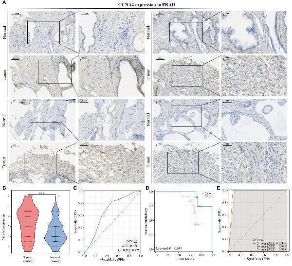
CCNA2 is highly expressed in PRAD and is associated with poor patient prognosis. (A, B) Differential expression of CCNA2 in PRAD. (C) Diagnostic predictive value of CCNA2 in PRAD. (D) KM curve of overall survival of CCNA2 in PRAD. (E) Prognostic predictive value of CCNA2 in PRAD.
Index in PubMed under a CC BY license. PMID: 38947325

Click image to see more details
CCNA2 has a high binding capacity to PRAD-targeted drugs. (A) Analysis of the binding capacity of CCNA2 to PD1 inhibitors. (B) Analysis of the binding capacity of CCNA2 to bicalutamide. (C) Analysis of the binding capacity of CCNA2 to enzalutamide. (D) Analysis of the binding capacity of CCNA2 to abiraterone.
Index in PubMed under a CC BY license. PMID: 38947325

Click image to see more details
CCNA2 positively correlates with monocyte infiltration levels. (A) Correlation analysis of CCNA2 and monocyte infiltration levels. (B, C) Analysis of CCNA2 correlation with monocyte markers. (D) Mendelian randomization analysis of high HLA-DR expressing monocytes in relation to prostate cancer. (E–I) Single-cell analysis of the correlation between CCNA2 and immune cell infiltration.
Index in PubMed under a CC BY license. PMID: 38947325

Click image to see more details
CCNA2 identified as the best prognostic gene among monocyte-associated genes. (A, B) Functional analysis of monocyte-related prognostic genes. (C, D) GBM and RandomForest algorithms to screen key prognostic genes in the TCGA-PRAD dataset. (E, F) GBM and RandomForest algorithms to screen key prognostic genes in the GSE16560 dataset. (G, H) KM curves of CCNA2 and ACSM3 in the TCGA-PRAD dataset. (I, J) KM curves of CCNA2 and ACSM3 in the GSE16560 dataset.
Index in PubMed under a CC BY license. PMID: 38947325

Click image to see more details
Functional analysis of CCNA2 in PRAD. (A) KEGG analysis of CCNA2 in PRAD. (B–J) GSEA analysis of CCNA2 in PRAD.
Index in PubMed under a CC BY license. PMID: 38947325

Click image to see more details
Western blot analysis of Cyclin A2 using anti-Cyclin A2 antibody (PB9424).
Electrophoresis was performed on a 5-20% SDS-PAGE gel at 70V (Stacking gel) / 90V (Resolving gel) for 2-3 hours. The sample well of each lane was loaded with 30 ug of sample under reducing conditions.
Lane 1: human K562 whole cell lysates,
Lane 2: human HL-60 whole cell lysates,
Lane 3: human MCF-7 whole cell lysates,
Lane 4: rat stomach tissue lysates,
Lane 5: rat testis tissue lysates,
Lane 6: mouse stomach tissue lysates.
After electrophoresis, proteins were transferred to a nitrocellulose membrane at 150 mA for 50-90 minutes. Blocked the membrane with 5% non-fat milk/TBS for 1.5 hour at RT. The membrane was incubated with rabbit anti-Cyclin A2 antigen affinity purified polyclonal antibody (Catalog # PB9424) at 0.5 μg/mL overnight at 4°C, then washed with TBS-0.1%Tween 3 times with 5 minutes each and probed with a goat anti-rabbit IgG-HRP secondary antibody at a dilution of 1:5000 for 1.5 hour at RT. The signal is developed using an Enhanced Chemiluminescent detection (ECL) kit (Catalog # EK1002) with Tanon 5200 system. A specific band was detected for Cyclin A2 at approximately 55 kDa. The expected band size for Cyclin A2 is at 49 kDa.

Click image to see more details
Flow Cytometry analysis of U20S cells using anti-Cyclin A2 antibody (PB9424).
Overlay histogram showing U20S cells stained with PB9424 (Blue line). To facilitate intracellular staining, cells were fixed with 4% paraformaldehyde and permeabilized with permeabilization buffer. The cells were blocked with 10% normal goat serum. And then incubated with rabbit anti-Cyclin A2 Antibody (PB9424, 1 μg/1x106 cells) for 30 min at 20°C. DyLight®488 conjugated goat anti-rabbit IgG (BA1127, 5-10 μg/1x106 cells) was used as secondary antibody for 30 minutes at 20°C. Isotype control antibody (Green line) was rabbit IgG (1 μg/1x106) used under the same conditions. Unlabelled sample without incubation with primary antibody and secondary antibody (Red line) was used as a blank control.

Click image to see more details
IHC analysis of Cyclin A2 using anti-Cyclin A2 antibody (PB9424).
Cyclin A2 was detected in a paraffin-embedded section of human colorectal adenocarcinoma tissue. Heat mediated antigen retrieval was performed in EDTA buffer (pH 8.0, epitope retrieval solution). The tissue section was blocked with 10% goat serum. The tissue section was then incubated with 2 μg/ml rabbit anti-Cyclin A2 Antibody (PB9424) overnight at 4°C. Peroxidase Conjugated Goat Anti-rabbit IgG was used as secondary antibody and incubated for 30 minutes at 37°C. The tissue section was developed using HRP Conjugated Rabbit IgG Super Vision Assay Kit (Catalog # SV0002) with DAB as the chromogen.

Click image to see more details
IF analysis of Cyclin A2 using anti-Cyclin A2 antibody (PB9424).
Cyclin A2 was detected in an immunocytochemical section of A431 cells. Enzyme antigen retrieval was performed using IHC enzyme antigen retrieval reagent (AR0022) for 15 mins. The cells were blocked with 10% goat serum. And then incubated with 5 μg/mL rabbit anti-Cyclin A2 Antibody (PB9424) overnight at 4°C. DyLight®488 Conjugated Goat Anti-Rabbit IgG (BA1127) was used as secondary antibody at 1:100 dilution and incubated for 30 minutes at 37°C. The section was counterstained with DAPI. Visualize using a fluorescence microscope and filter sets appropriate for the label used.

Click image to see more details
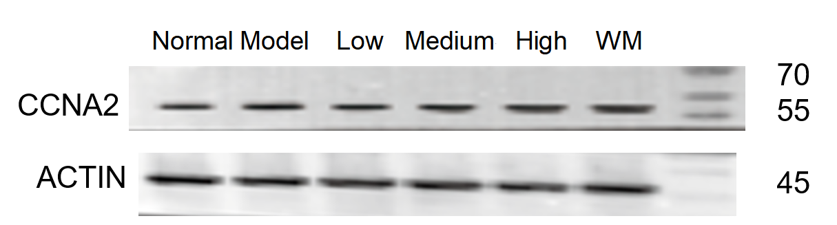
Western blot analysis of Cyclin A2 using anti-Cyclin A2 antibody (PB9424).
Electrophoresis was performed on a 5-20% SDS-PAGE gel at 70V (Stacking gel) / 90V (Resolving gel) for 2-3 hours. The sample well of each lane was loaded with 30 ug of sample under reducing conditions.
Lane 1: Normal group-rat colon tissue lysates,
Lane 2: Model group-rat colon tissue lysates,
Lane 3: Triditional Chinese medicine treatment (low dose)-rat colon tissue lysates,
Lane 4: Triditional Chinese medicine treatment (medium dose)-rat colon tissue lysates,
Lane 5: Triditional Chinese medicine treatment(high dose)-rat colon tissue lysates,
Lane 6: Western medicine-rat colon tissue lysates.
After electrophoresis, proteins were transferred to a nitrocellulose membrane at 150 mA for 50-90 minutes. Blocked the membrane with 5% non-fat milk/TBS for 1.5 hour at RT. The membrane was incubated with rabbit anti-Cyclin A2 antigen affinity purified polyclonal antibody (Catalog # PB9424) at 1:1000 overnight at 4°C, then washed with TBS-0.1%Tween 3 times with 5 minutes each and probed with a goat anti-rabbit IgG-HRP secondary antibody at a dilution of 1:5000 for 1 hour at RT. The signal is developed using an Enhanced Chemiluminescent detection (ECL) kit (Catalog # EK1002) with ChemiDoc MP system. A specific band was detected for Cyclin A2 at approximately 55 kDa. The expected band size for Cyclin A2 is at 49 kDa.
Specific Publications For Anti-Cyclin A2/CCNA2 Antibody Picoband® (PB9424)
Loading publications
Recommended Resources
Here are featured tools and databases that you might find useful.
- Boster's Pathways Library
- Protein Databases
- Bioscience Research Protocol Resources
- Data Processing & Analysis Software
- Photo Editing Software
- Scientific Literature Resources
- Research Paper Management Tools
- Molecular Biology Software
- Primer Design Tools
- Bioinformatics Tools
- Phylogenetic Tree Analysis
Customer Reviews
Have you used Anti-Cyclin A2/CCNA2 Antibody Picoband®?
Share your experimental results or join a short interview to earn up to $1,000 in product credits or other rewards.
1 Reviews For Anti-Cyclin A2/CCNA2 Antibody Picoband®
This antibody is highly efficient and specific, suitable for detecting CCNA2 protein in rat colon by Western blot, with only minimal nonspecific bands.
Excellent
| SKU | PB9424 |
|---|---|
| Application | Western Blot |
| Sample | rat colon tissue |
| Sample Processing Description | RIPA lysis buffer with protease inhibitor PMSF (100:1) was used to lyse the sample for 10 minutes, followed by centrifugation at 12,000 rpm for 15 minutes. The supernatant was mixed with 5× loading buffer, denatured at 100°C for 10 minutes, and then loaded onto SDS-PAGE. |
| Other Reagents | Blocking buffer |
| Primary Antibody | Cyclin A2/CCNA2 Antibody Picoband® |
| Primary Incubation | 1:1000, overnight at 4 ℃ |
| Secondary Antibody | HRP Conjugated AffiniPure Goat Anti-Rabbit IgG (H+L) |
| Secondary Incubation | 1:5000, 1 hour in room temperature |
| Detection | Substrate: ECL, Imaging system:ChemiDoc MP |
| Results Summary | The figure shows a schematic of Western blot results for the target protein CCNA2 and the internal control Actin in rat colon across different groups. Expression was increased in the model group. Among the low, medium, and high doses of the herbal treatment, the low-dose group showed the best effect. The target bands are clear, and the experimental results are satisfactory. |
Shiyu Zhang, LUTCM
Verified customer
Submitted 2026-01-05
Customer Q&As
Have a question?
Find answers in Q&As, reviews.
Can't find your answer?
Submit your question
1 Customer Q&As for Anti-Cyclin A2/CCNA2 Antibody Picoband®
Question
We are currently using anti-Cyclin A2/CCNA2 antibody PB9424 for human tissue, and we are well pleased with the IF results. The species of reactivity given in the datasheet says human, rat. Is it likely that the antibody can work on bovine tissues as well?
Verified Customer
Verified customer
Asked: 2020-04-03
Answer
The anti-Cyclin A2/CCNA2 antibody (PB9424) has not been validated for cross reactivity specifically with bovine tissues, but there is a good chance of cross reactivity. We have an innovator award program that if you test this antibody and show it works in bovine you can get your next antibody for free. Please contact me if I can help you with anything.
Boster Scientific Support
Answered: 2020-04-03
