CCND1 / G1/S-specific cyclin-D1 · IHC design guide

Design Immunohistochemistry for CCND1

Plan CCND1 chromogenic IHC around the general nuclear tissue pattern (HPA tissue IHC). Use the catalog antibody’s paraffin-section protocol and compare staining across defined cell populations, including lymphoid cells that can stain strongly (datasheet PB9370; HPA tissue IHC).

Evidence assembled Oct 2026 · For research use; verify linked source records and product datasheet before use
Immunohistochemistry protocol sheet for CCND1 (IHC for CCND1): expected localisation General nuclear staining in tissue (HPA tissue IHC), antibody PB9370, validated IHC image, and IHC protocol steps
Printable CCND1 IHC protocol sheet — expected localisation General nuclear staining in tissue (HPA tissue IHC), antibody PB9370, controls and protocol steps. Open the full CCND1 IHC guide →

CCND1 Immunohistochemistry Experimental Design Guide

Expected localisation, validated protocols, controls and antibodies — the at-a-glance facts below, then the full design guide.

Must know before staining
Expected localisation General nuclear staining in tissue (HPA tissue IHC)
Staining pattern Nuclear staining across multiple tissue cell types (HPA tissue IHC)
Antigen retrieval EDTA pH 8.0 HIER, heat-mediated (datasheet PB9370)
Positive control ⓘ Appendix+4 more · see all
Negative control ⓘ Prostate
Important caveats
Reasons your staining may differ from the expected pattern.
Fixation Keep fixation consistent across sections (standard IHC practice; not target-specific)
Caveat High lymphoid-cell staining may complicate scoring (HPA tissue IHC)
Regulation Mitogenic state may affect CCND1 signal (UniProt)
Isoform / epitope No annotated isoforms; one full-length chain (UniProt)
Section 1

Recommended CCND1 IHC & IF Protocols

The catalog antibody’s IHC-P protocol is accompanied by published CCND1 IHC methods for chondroid lipoma (PMC3087950), endometrial cancer (PMC11816803), and plasma cell myeloma (PMC6676137).

Recommended immunohistochemistry (IHC-P) protocol parameters
SampleParaffin-embedded human colon cancer tissue; fixative not specified (datasheet PB9370)
FixationImage fixative and duration unreported (datasheet PB9370); verify before use.
Sectioning4–5 µm sections on charged slides (standard)
DeparaffinisationXylene, graded ethanol series to water (standard)
Antigen retrievalHeat retrieval: EDTA pH 8.0 (datasheet PB9370); 20 min, 95–100 °C (standard)
Peroxidase block3% H2O2, 10 min, room temperature (standard)
Blocking10% goat serum (datasheet PB9370)
Primary antibodyRabbit anti-CCND1, 2-5μg/ml (datasheet PB9370)
Primary incubationOvernight at 4 °C (datasheet PB9370)
DetectionHRP-conjugated secondary, DAB chromogen (datasheet PB9370)
CounterstainHematoxylin, blue, dehydrate and mount (standard)
Expected resultCCND1-positive staining in lymphoid tissue of appendix (HPA tissue IHC: High). HPA tissue profile: General nuclear expression. No signal in the no-primary control.
💡Decision noteStart with heat-mediated EDTA retrieval at pH 8.0 for the catalog antibody (datasheet: PB9370); use each published retrieval condition with its corresponding protocol.
Section 2

What Is the Expected CCND1 Staining Pattern?

CCND1 staining should be predominantly nuclear in the cell populations that express it; HPA describes general nuclear expression in tissue IHC, with Supported reliability and medium agreement with RNA data (HPA tissue IHC). Nucleoplasm is the supported ICC-IF location, while UniProt also lists cytoplasm and the nuclear membrane (HPA subcellular; UniProt P24385). CCND1 has no transmembrane segment, so a cell-surface pattern is unexpected (UniProt P24385 topology).

What am I looking at on my slide?
Distinct nuclear staining in breast myoepithelial cells or appendix lymphoid tissue.This matches cell populations scored High by HPA; interpret the stained cells rather than treating every cell in the section as a positive control (HPA tissue IHC).
Predominant cell-surface or extracellular staining, with little nuclear signal.The compartment conflicts with HPA’s general nuclear IHC pattern and CCND1’s lack of a transmembrane segment (HPA tissue IHC; UniProt P24385 topology). Review morphology, detection background and antibody specificity (standard IHC practice).
Strong staining in a cell population outside the expected pattern for the chosen tissue.Consider cross-reactivity or endogenous detection activity, particularly if the signal lacks convincing nuclear localisation (standard IHC practice; HPA tissue IHC). HPA’s examples do not establish that every unlisted cell type must be negative (HPA tissue IHC).
Diffuse colour across nuclei, cytoplasm and surrounding tissue.Poor compartment contrast makes the result difficult to interpret; check blocking, washes and detection controls before scoring cells as positive (standard IHC practice). Cytoplasmic CCND1 is listed by UniProt, so cytoplasmic signal alone is not proof of artefact (UniProt P24385).
No nuclear signal in a chosen HPA High cell population.A failed positive control is possible; confirm that the relevant cells are present, then assess antibody dilution, retrieval and detection with appropriate controls (HPA tissue IHC; standard IHC practice). HPA’s Supported tissue profile does not guarantee staining in every preparation (HPA tissue IHC).
💡Expected CCND1 appearanceCall a result positive when identifiable HPA High cells show clear, predominantly nuclear staining—for example, breast myoepithelial or appendix lymphoid cells; widespread extracellular colour or cell-surface outlines are suspect (HPA tissue IHC; UniProt P24385 topology; standard IHC practice).
How each factor affects the staining
Tissue and cell selectionHPA scores breast myoepithelial, appendix lymphoid and several other populations High, but prostate glandular cells are Not detected (HPA tissue IHC). Use the annotated cell population when judging a control; a whole-section average can hide the expected pattern (standard IHC practice).
Antibody validationCAB080462 has Supported IHC status; HPA027802 has Supported ICC status, with no IHC status supplied for it (HPA antibodies). These application labels should be kept distinct when interpreting a paraffin-section result (HPA antibodies).
CCND1 location and topologyUniProt lists nucleus, cytoplasm and nuclear membrane, and describes nuclear translocation of cyclin D–CDK4 complexes (UniProt P24385). HPA’s tissue pattern remains generally nuclear; a purely cytoplasmic result needs control-based assessment rather than an automatic positive call (HPA tissue IHC; standard IHC practice).
Protein featuresUniProt records one 295 aa chain, no signal peptide or propeptide, no annotated isoforms and phosphorylation at Thr286 (UniProt P24385). The supplied sources do not show how these features affect IHC retrieval, antibody binding or staining intensity (UniProt P24385; HPA tissue IHC).
IF/ICC Q&A: what pattern is expected?A: HPA supports nucleoplasmic localisation and lists BJ and U2OS ICC-IF images (HPA subcellular). That observation can guide compartment interpretation, but it does not supply an IF/ICC protocol or replace paraffin-section IHC validation (HPA subcellular; HPA antibodies).
Why is my staining missing, weak or wrong?
SituationLikely causeNext action
No signal in breast myoepithelial cells or appendix lymphoid tissue.The chosen positive cells may be absent from the field, or an IHC workflow step may have failed (HPA tissue IHC; standard IHC practice).Locate the annotated cells on the counterstained section; then check the antibody dilution, retrieval, detection reagents and a concurrently processed positive control (standard IHC practice).
Prostate glandular cells stain strongly.HPA reports this population as Not detected; cross-reactivity or endogenous detection activity is possible (HPA tissue IHC; standard IHC practice).Confirm the stained cell identity and nuclear location, then compare an antibody-omission control and the positive control before assigning specificity (standard IHC practice).
Colour outlines cells or fills extracellular spaces.That distribution conflicts with general nuclear tissue staining and the absence of a transmembrane segment (HPA tissue IHC; UniProt P24385 topology).Review tissue morphology, blocking and detection controls; score only convincing cell-associated signal with appropriate localisation (standard IHC practice; HPA tissue IHC).
Cytoplasmic colour dominates while nuclei are faint.UniProt permits cytoplasmic localisation, but HPA describes general nuclear tissue IHC; the discrepancy is unresolved by the supplied sources (UniProt P24385; HPA tissue IHC).Compare nuclear and cytoplasmic signal in the HPA High control cells and check background controls before interpreting the cytoplasmic component (HPA tissue IHC; standard IHC practice).
Diffuse background obscures individual nuclei.Insufficient blocking, washing or control of endogenous chromogenic detection activity can reduce contrast (standard IHC practice).Inspect antibody-omission and detection controls, adjust blocking and washes, and reassess whether discrete nuclear staining emerges (standard IHC practice; HPA tissue IHC).
A low-staining cell population gives an ambiguous weak result.HPA lists several populations as low and rates the overall tissue pattern Supported with medium RNA agreement; weak colour may be hard to distinguish from background (HPA tissue IHC).Judge it alongside a HPA High population and a background control; report an unresolved weak result rather than assigning a categorical positive or negative call (HPA tissue IHC; standard IHC practice).

Sample controls for CCND1 IHC & IF

🧪Run breast first and confirm staining in myoepithelial cells (HPA: High in breast myoepithelial cells). Use prostate glandular cells as the negative tissue (HPA: Not detected in prostate glandular cells); on the breast slide, treat any adjacent cells without nuclear signal as an internal background comparison only after confirming their identity (UniProt P24385: nucleus; standard IHC control practice).
Positive control tissue: Appendix (Lymphoid tissue, HPA High)
Negative control tissue: Prostate (HPA Not detected)
ICC-IF cell lines (HPA subcellular resource): HPA ICC-IF images show CCND1 in BJ [Human fibroblast], U2OS, with annotated localisation: Nucleoplasm (supported) (HPA subcellular).
Technical controls: Include no-primary (secondary-only) and concentration-matched rabbit isotype controls, plus CCND1 knockout material or peptide blocking if a validated blocking peptide is available (PB9370 caption: rabbit primary; standard IHC control practice). Quench endogenous peroxidase and check background before interpreting HRP/DAB staining in breast tissue (PB9370 caption: HRP/DAB; standard IHC practice).
⚠️Feasibility: The selected PB9370 paraffin-section caption does not report a fixative; no target-specific fixation window or fixation effect is reported in the supplied evidence (PB9370 tissue-IHC caption). Its reported IHC conditions use heat retrieval in EDTA at pH 8.0, but they do not establish that retrieval is required under other conditions (PB9370 tissue-IHC caption). The evidence does not establish that frozen sections or tissue IF are easier; ICC-IF images support nucleoplasmic localisation in BJ and U2OS, while breast sections require care when assigning nuclear staining to myoepithelial rather than adjacent cells (HPA: nucleoplasm supported; HPA: High in breast myoepithelial cells).

HPA tissue IHC evidence for CCND1

Comprehensive Human Protein Atlas IHC scoring per tissue (reliability: Supported — Medium consistency between antibody staining and RNA expression data.). Rows are taken directly from the HPA tissue chart — click any row's HPA link to view the source.

Positive expression · recommended positive controls

TissueCell typeLevelEvidenceSource
Appendix Lymphoid tissue High Protein (IHC) HPA →
Bone marrow Hematopoietic cells High Protein (IHC) HPA →
Breast Myoepithelial cells High Protein (IHC) HPA →
Caudate Neuronal cells High Protein (IHC) HPA →
Cerebellum Cells in granular layer High Protein (IHC) HPA →

Undetected expression · recommended negative controls

TissueCell typeLevelEvidenceSource
Prostate Glandular cells Not detected Protein (IHC) HPA →
Section 3

Advanced CCND1 IHC Tips

Troubleshoot CCND1 staining in paraffin sections by checking retrieval, nuclear localisation, controls and scoring; IF/ICC considerations are included separately.

How should I troubleshoot weak CCND1 staining after antigen retrieval?
Start with heat-mediated retrieval in EDTA at pH 8.0 for paraffin sections (datasheet PB9370). The selected image used that condition before overnight incubation at 4°C with 2 μg/ml primary antibody, so reproduce the sequence when investigating weak nuclear staining (caption PB9370). Compare adjacent sections with and without retrieval, judging nuclear signal in the same cell population alongside tissue morphology (standard IHC practice; HPA tissue IHC). If signal remains weak, adjust heating duration in small increments while keeping EDTA and pH 8.0 constant, and reject conditions that damage morphology (standard IHC practice).
Could fixation explain inconsistent nuclear CCND1 staining between blocks?
The selected tissue caption identifies a paraffin section but does not report its fixative, so target-specific fixation sensitivity is unknown (caption PB9370). Nuclear expression and phosphorylation at residue 286 do not establish an optimal fixation schedule (HPA tissue IHC; UniProt P24385). For new blocks, record fixative, delay before fixation, fixation duration and processing, then compare matched sections using the same EDTA pH 8.0 retrieval (standard IHC practice; datasheet PB9370). Include a known positive control and assess nuclear signal together with morphology, because fixation-related signal loss and inadequate retrieval can appear similar (standard IHC practice).
Should cytoplasmic or nuclear-rim staining count as positive CCND1?
Score nuclear staining as the primary finding: tissue IHC shows general nuclear expression, and the supported subcellular location is the nucleoplasm (HPA tissue IHC; HPA subcellular). Cytoplasm and nuclear membrane are also annotated locations, while cyclin D–CDK4 complexes can accumulate at the nuclear membrane before nuclear translocation (UniProt P24385). Record those patterns separately from nuclear positivity rather than combining compartments in one score (standard IHC practice). Compare their distribution with nuclear staining on the same section and with a no-primary control; diffuse staining across unrelated cells warrants investigation before a compartment-specific interpretation (standard IHC practice).
How can I assess whether the antibody recognises the intended CCND1 epitope?
The supplied record lists 0 isoforms, a cyclin N-terminal domain at residues 28–152, and phosphothreonine at residue 286 (UniProt P24385). These annotations do not locate the catalog antibody’s epitope or show whether phosphorylation changes its binding (UniProt P24385; caption PB9370). If staining is unexpected, check the antibody’s documented immunogen or epitope before attributing signal differences to a domain or modification (standard IHC practice). Compare matched sections under identical EDTA pH 8.0 retrieval and, where available, use an independently validated antibody recognising a documented different epitope as an orthogonal check (datasheet PB9370; standard IHC practice).
How should I adapt CCND1 localisation checks for multiplex IF?
For secondary IF/ICC work, expect predominantly nucleoplasmic signal; HPA reports supported nucleoplasmic localisation and ICC/IF images in BJ and U2OS cells (HPA subcellular). In a breast tissue multiplex, pair CCND1 with a separately validated myoepithelial marker to identify the expected cell population (HPA tissue IHC; standard IF practice). Select a far-red fluorophore when shorter wavelengths show tissue autofluorescence, and inspect unstained and no-primary controls in each channel (standard IF practice). CCND1 has no transmembrane segment, so permeabilise the plasma and nuclear membranes to access intracellular epitopes; optimise that step for the IF preparation rather than assuming the IHC retrieval conditions transfer (UniProt P24385; standard IF practice).
What should I check when DAB staining obscures CCND1 nuclei?
The selected paraffin-section image used 10% goat serum blocking, a peroxidase-conjugated secondary and DAB development (caption PB9370). Check a no-primary section for secondary-related signal and apply an endogenous peroxidase block before chromogenic detection, particularly if pigment appears outside nuclei (standard IHC practice). Compare background after adjusting blocking, washes and development time one variable at a time, while retaining EDTA pH 8.0 retrieval for the initial comparison (standard IHC practice; datasheet PB9370). Judge improvement by clearer nuclear boundaries and preserved tissue morphology, because general nuclear expression is the reported tissue pattern (HPA tissue IHC; standard IHC practice).
How should I quantify heterogeneous CCND1 staining across a section? ⚠ ANSWER MARKED FOR VERIFICATION
Define the cell population and anatomical region before scoring, because HPA reports general nuclear expression with different staining levels across cell types (HPA tissue IHC). Report the percentage of positive nuclei and, when intensity matters, a nuclear H-score from 0–300 using consistent intensity thresholds (standard IHC practice). Normalise each measure to all evaluable nuclei of the specified cell type, excluding necrotic areas and damaged section edges (standard IHC practice). If spatial abundance matters, report positive nuclei per mm² of evaluable tissue alongside the percentage, and keep staining and image-analysis settings consistent across compared sections (standard IHC practice).
How can I distinguish genuine CCND1 positivity from section artefacts?
True signal should have a plausible nuclear distribution in the relevant cells: HPA reports general nuclear expression, including high staining in appendix lymphoid tissue and no detection in prostate glandular cells (HPA tissue IHC). Evaluate those controls by cell type rather than assuming every cell in either tissue must share the same result (HPA tissue IHC; standard IHC practice). Treat staining confined to section edges or necrotic regions as suspect, and compare it with intact central tissue (standard IHC practice). Investigate diffuse cytoplasmic DAB or signal on a no-primary section with an endogenous peroxidase block, while recording any reproducible nuclear-rim pattern separately from nuclear positivity (standard IHC practice; UniProt P24385).
Boster reagents

Best CCND1 / G1/S-specific cyclin-D1 IHC Antibodies

Anti-CCND1 antibodies cover human, mouse and rat IHC and IF applications (catalog: application and reactivity lists). All three have tissue IHC figures; PB9370 and M00149-1 also have cell IF figures (catalog: image captions).

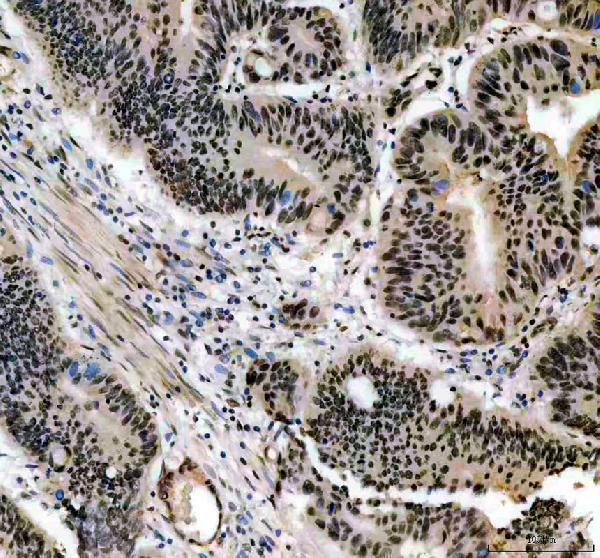
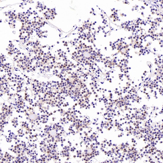
Real IHC data IHC analysis of CCND1 using anti-CCND1 antibody (PB9370). CCND1 was detected in a paraffin-embedded section of human colon cancer tissue. Heat mediated antigen retrieval was performed in EDTA buffer (pH 8.0, epitope retrieval solution). The tissue section was blocked with 10% goat serum. The tissue section was then incubated with 2 μg/ml rabbit anti-CCND1 Antibody (PB9370) overnight at 4°C. Peroxidase Conjugated Goat Anti-rabbit IgG was used as secondary antibody and incubated for 30 minutes at 37°C. The tissue section was developed using HRP Conjugated Rabbit IgG Super Vision Assay Kit (Catalog # SV0002) with DAB as the chromogen.
Anti-Cyclin D1/CCND1 Antibody ®
Cat # PB9370
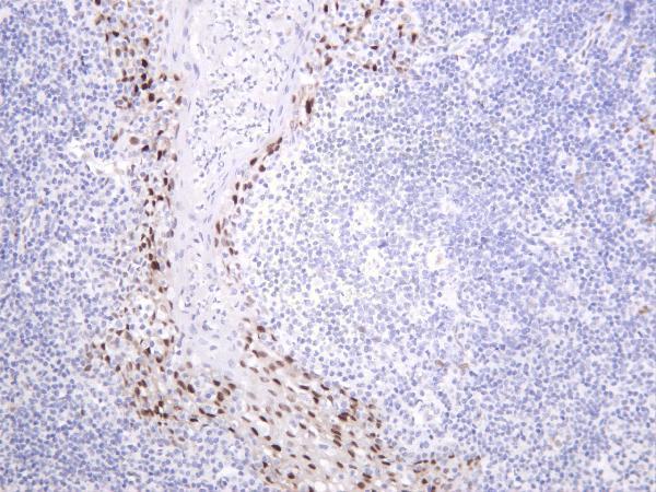
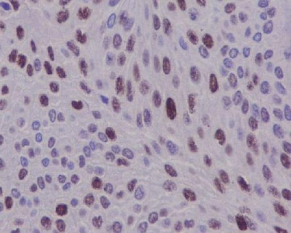
Real IHC data Human esophagus was stained with anti-Cyclin D1 rabbit antibody
Anti-Cyclin D1 Rabbit Monoclonal Antibody
Cat # M00149-5
Real IHC data Immunohistochemical analysis of paraffin-embedded human bladder, using Cyclin D1 Antibody.
Anti-Cyclin D1 CCND1 Rabbit Monoclonal Antibody
Cat # M00149-1

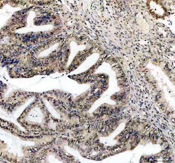
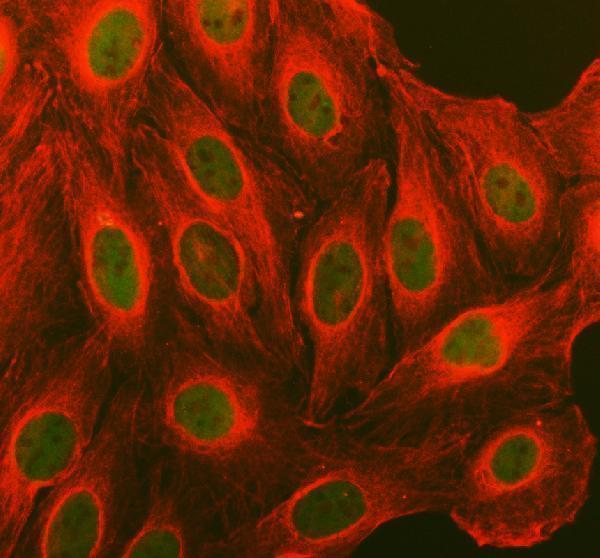
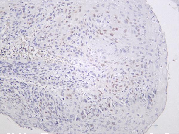
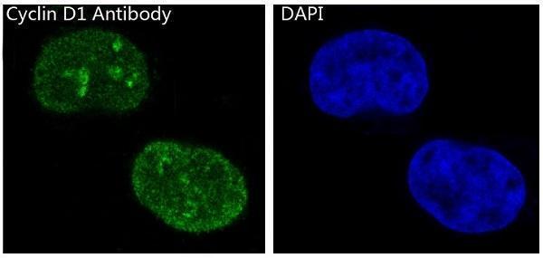
PB9370 has paraffin-section IHC figures from human colon and endometrial cancers and rat lung, plus IF in U2OS cells (PB9370: image captions); M00149-5 has IHC figures from human esophagus and tonsil, rat spleen and mouse colon (M00149-5: image captions). M00149-1 has paraffin-section IHC figures from human bladder, pituitary tumor, squamous cell carcinoma and ovarian cancer, plus IF in MCF-7 cells (M00149-1: image captions).

Which to pick: For tissue IHC, choose PB9370 when a documented paraffin-section workflow is useful: its human colon cancer caption specifies EDTA retrieval at pH 8.0, 2 μg/ml primary antibody and DAB detection (PB9370: IHC image caption). For IF/ICC, M00149-1 has both applications listed and an MCF-7 IF figure (M00149-1: application list and IF image caption). For IHC across species, M00149-5 has tissue figures from human, mouse and rat samples (M00149-5: IHC image captions); the fixative is unreported in these IHC captions (catalog: IHC image captions).

Each figure is that product's own IHC / IF validation image from its datasheet.

References

  1. UniProt Consortium. UniProt entry P24385 (CCND1_HUMAN, G1/S-specific cyclin-D1).
  2. Human Protein Atlas. CCND1 tissue IHC expression (reliability: Supported).
  3. Human Protein Atlas. CCND1 subcellular location (ICC-IF): Localized to the nucleoplasm..
  4. Human Protein Atlas. CCND1 antibody validation summary (2 antibodies).
  5. Delineation of chondroid lipoma: an immunohistochemical and molecular biological analysis. Sarcoma 2011 — PMC3087950.
  6. Germline mutations in MSR1, ASCC1, and CTHRC1 in patients with Barrett esophagus and esophageal adenocarcinoma. JAMA 2011 — PMC3574553.
  7. Assessment of Cyclin D1 Expression: Prognostic Value and Functional Insights in Endometrial Cancer: In Silico Study. International journal of molecular sciences 2025 — PMC11816803.
  8. Immunohistochemistry for identification of CCND1, NSD2, and MAF gene rearrangements in plasma cell myeloma. Cancer science 2019 — PMC6676137.
  9. PubMed PMID:1826542 — UniProt-cited evidence.
  10. PubMed PMID:1833066 — UniProt-cited evidence.
  11. PubMed PMID:1827756 — UniProt-cited evidence.