Click image to see more details
-

-
-
-
-
+10
Product Info Summary
| SKU: | M00149-1 |
|---|---|
| Size: | 100 μl |
| Reactive Species: | Human, Mouse, Rat |
| Host: | Rabbit |
| Application: | IP, IF, IHC, ICC, WB |
Customers Who Bought This Also Bought

Product info
Product Name
Anti-Cyclin D1 CCND1 Rabbit Monoclonal Antibody
SKU/Catalog Number
M00149-1
BM4272 is an alternative SKU for this antibody, used in previous lots.
Size
100 μl
Form
Liquid
Description
Boster Bio Anti-Cyclin D1 CCND1 Rabbit Monoclonal Antibody catalog # M00149-1. Tested in WB, IHC, ICC/IF, IP applications. This antibody reacts with Human, Mouse, Rat.
Storage & Handling
Store at -20°C for one year. For short term storage and frequent use, store at 4°C for up to one month. Avoid repeated freeze-thaw cycles.
Cite This Product
Anti-Cyclin D1 CCND1 Rabbit Monoclonal Antibody (Boster Biological Technology, Pleasanton CA, USA, Catalog # M00149-1)
Host
Rabbit
Contents
Rabbit IgG in stabilizing components, phosphate buffered saline, pH 7.4, 150mM NaCl, 0.02% sodium azide and 50% glycerol.
*This antibody is supplied in a stabilized formulation.
Compatibility with conjugation reactions depends on the chemistry of the conjugation method used.
For conjugation methods that are not compatible with the stabilizing components present in this formulation, a carrier-free antibody format is required.
Clonality
Monoclonal
Clone Number
DAD-3
Isotype
Rabbit IgG
Immunogen
A synthesized peptide derived from human Cyclin D1
Reactive Species
M00149-1 is reactive to CCND1 in Human, Mouse, Rat
Observed Molecular Weight
34 kDa
Calculated molecular weight
33.7 kDa
Antibody Validation
Boster validates all antibodies on WB, IHC, ICC, Immunofluorescence, and ELISA with known positive control and negative samples to ensure specificity and high affinity, including thorough antibody incubations.
Application & Images
Applications
M00149-1 is guaranteed for IP, IF, IHC, ICC, WB Boster Guarantee
Recommend Dilution
WB 1:500-2000
IHC 1:50-200
ICC/IF 1:50-200
IP 1:20
Tested application
Suggested blocking solution with 5% non-fat milk or BSA; (*)Recommended protein loading: 20-40 µg per lane
Use TE buffer pH 9.0 for antigen retrieval; (*) citrate buffer pH 6.0 is an alternative.
Validation Images & Assay Conditions
Click image to see more details
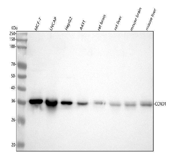
Western blot analysis of Cyclin D1 using anti-Cyclin D1 antibody (M00149-1).
Electrophoresis was performed on a 5-20% SDS-PAGE gel at 70V (Stacking gel) / 90V (Resolving gel) for 2-3 hours. The sample well of each lane was loaded with 30 ug of sample under reducing conditions.
Lane 1: human MCF-7 whole cell lysates,
Lane 2: human LNCAP whole cell lysates,
Lane 3: human HepG2 whole cell lysates,
Lane 4: human A431 whole cell lysates,
Lane 5: rat brain tissue lysates,
Lane 6: rat liver tissue lysates,
Lane 7: mouse brain tissue lysates,
Lane 8: mouse liver tissue lysates.
After electrophoresis, proteins were transferred to a nitrocellulose membrane at 150 mA for 50-90 minutes. Blocked the membrane with 5% non-fat milk/TBS for 1.5 hour at RT. The membrane was incubated with rabbit anti-Cyclin D1 antigen affinity purified monoclonal antibody (Catalog # M00149-1) at 1:1000 overnight at 4°C, then washed with TBS-0.1%Tween 3 times with 5 minutes each and probed with a goat anti-rabbit IgG-HRP secondary antibody at a dilution of 1:1000 for 1.5 hour at RT. The signal is developed using an Enhanced Chemiluminescent detection (ECL) kit (Catalog # EK1002) with Tanon 5200 system. A specific band was detected for Cyclin D1 at approximately 34 kDa. The expected band size for Cyclin D1 is at 34 kDa.

Click image to see more details
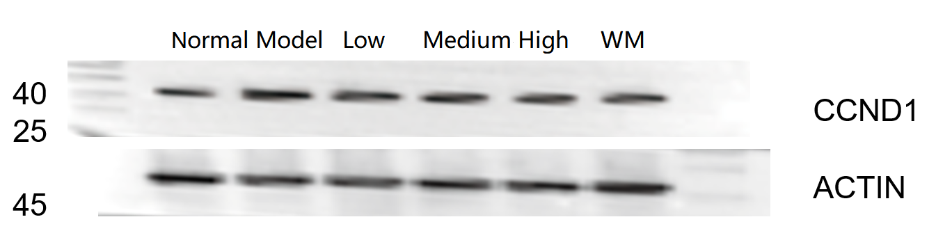
Western blot analysis of Cyclin D1 using anti-Cyclin D1 antibody (M00149-1).
Electrophoresis was performed on a 5-20% SDS-PAGE gel at 70V (Stacking gel) / 90V (Resolving gel) for 2-3 hours. The sample well of each lane was loaded with 30 ug of sample under reducing conditions.
Lane 1: Normal group-rat colon tissue lysates,
Lane 2: Model group-rat colon tissue lysates,
Lane 3: Triditional Chinese medicine treatment (low dose)-rat colon tissue lysates,
Lane 4: Triditional Chinese medicine treatment (medium dose)-rat colon tissue lysates,
Lane 5: Triditional Chinese medicine treatment(high dose)-rat colon tissue lysates,
Lane 6: Western medicine-rat colon tissue lysates.
After electrophoresis, proteins were transferred to a nitrocellulose membrane at 150 mA for 50-90 minutes. Blocked the membrane with 5% non-fat milk/TBS for 1.5 hour at RT. The membrane was incubated with rabbit anti-Cyclin D1 antigen affinity purified monoclonal antibody (Catalog # M00149-1) at 1:1000 overnight at 4°C, then washed with TBS-0.1%Tween 3 times with 5 minutes each and probed with a goat anti-rabbit IgG-HRP secondary antibody for 1 hour at RT. The signal is developed using an Enhanced Chemiluminescent detection (ECL) kit (Catalog # EK1002) with ChemiDoc MP system. A specific band was detected for Cyclin D1 at approximately 36 kDa. The expected band size for Cyclin D1 is at 34 kDa.

Click image to see more details
Immunohistochemical analysis of paraffin-embedded human bladder, using Cyclin D1 Antibody.

Click image to see more details
Immunohistochemical analysis of paraffin-embedded Human pituitary tumor, using the Antibody at 1:100 dilution.

Click image to see more details
Immunohistochemical analysis of paraffin-embedded Human squamous cell carcinoma , using the Antibody at 1:100 dilution.

Click image to see more details
Immunohistochemical analysis of paraffin-embedded Human ovarian cancer, using the Antibody at 1:500 dilution.

Click image to see more details
Immunofluorescent analysis of MCF-7 cells, using Cyclin D1 Antibody.

Click image to see more details
Immunofluorescent analysis using the Antibody at 1:50 dilution.

Click image to see more details
Combined effects of FSH and SCF on the expressions of cell cycling proteins CCND1 and CDK2. (A–D) 4-day-old chickens were treated by FSH in vivo . Brown staining represents the immunohistochemical antigen. (E,F) 4-day-old chicken ovaries were treated by FSH or SCF for 2 days in vitro . (A) Arrowheads represent oocytes stained with CDK2 antibody. (B) Arrowheads and arrows represent oocytes and somatic cells stained with CCND1 antibody. (C,D) Western blot and gray analysis indicate that FSH promoted the CDK2 and CCND1 protein expression in vivo . (E,F) Western blot and gray analysis indicate that SCF enhanced the effect of FSH on promoting the CDK2 and CCND1 protein expression in vitro . T -tests were used to determine statistically significant differences. The values are the mean ± SEM of six experiments. Asterisks indicate significant differences (* P < 0.05, ** P < 0.01). Scale bars: 50 μm.
Index in PubMed under a CC BY license. PMID: 30837955

Click image to see more details
AIF blocks hypoxia-induced progression of PH in vitro and in vivo. A Hypoxia increased the viability of PASMCs after growth arrest for 24 h, and this effect was decreased by AIF (n = 4). B Pretreatment with an AIF overexpression plasmid blocked the effects of hypoxia on EdU incorporation in cells (n = 6). Scale bars: 50 μm. C Cell cycle analysis by flow cytometry indicated that hypoxia stimulated cell progression into G 2 /M + S phase, and this effect was inhibited by AIF overexpression (n = 3). D Effects of AIF on the expression of PCNA, Cyclin A and Cyclin D under hypoxia (n = 4–5). E Represents weight ratio of the right ventricular (RV)/left ventricular (LV) + Septum (n = 6); F Represents the right ventricular systolic pressure (RVSP) from mice (n = 5); G pulmonary artery velocity time integral (PAVTI), pulmonary artery acceleration time (PAAT) and left ventricular ejection fraction (LVEF) of the hypoxic mouse model infected with AAV5-NC and AAV5-AIF (n = 6). All data are presented as the means ± standard deviation. *p < 0.05; **p < 0.01; ***p < 0.001; Nor normoxia, Hyp hypoxia, NC negative control
Index in PubMed under a CC BY license. PMID: 35090552

Click image to see more details
The MAT1A/CCND1 signaling axis promotes glycolysis and tumorigenesis in NSCLC. The xenograft tumor model in nude mice established by A549 cells that interfered with MAT1A and CCND1 expression. After 39 days of monitoring, A tumor volume and B tumor weight was measured. C HE, IHC staining and D WB analysis was performed to determine the expression of MAT1A, CCND1, proliferation marker Ki67 and glycolytic enzymes (ALDOC, HK2) after tumor tissue sections or protein extraction.
Index in PubMed under a CC BY license. PMID: 39438468

Click image to see more details
MAT1A enhances the glycolysis of NSCLC cells through CCND1. The metabolic profile of glycolysis in MAT1A knockdown A549 cells was analyzed, including ( A ) glucose utilization, lactic acid production, PA, PGK, GAPDH activity and ( B ) expression of pivotal glycolytic enzymes (PKM2, ALDOC, ADH6). C , D Measurement of glucose utilization, lactic acid production and ( D ) expression of pivotal glycolytic enzymes (PKM2, ALDOC, ADH6) in A549 cells (NC, as negative control; MAT1A-overexpressing; CCND1 knockdown; MAT1A-overexpressing and CCND1 knockdown). E , F MAT1A-overexpresed A549 cells treated with AKT inhibitor (perifosine) and measurement of ( E ) glucose utilization, lactic acid production and ( F ) expression of pivotal glycolytic enzymes (PKM2, ALDOC, ADH6). G , H MAT1A-overexpresed A549 cells treated with glycolysis inhibitor 2-deoxy-d-glucose (2-DG) and measurement of ( G ) glucose utilization, lactic acid production and ( H ) expression of pivotal glycolytic enzymes (PKM2, ALDOC, ADH6). Results were shown as mean ± SD. * p < 0.05, ** p < 0.05, *** p < 0.001.
Index in PubMed under a CC BY license. PMID: 39438468

Click image to see more details
MAT1A inhibited the ubiquitination of CCND1 by blocking the binding of SKP2 to CCND1. In co-immunoprecipitation experiments, the total lysates of A549 and NCI-H1299 cells were immunized with ( A ) anti-CCND1 or ( B ) anti-SKP2 and WB, respectively. In the co-immunoprecipitation experiments, total lysates of A549 and NCI-H1299 cells were immunized with ( C ) anti-MAT1A or ( D ) anti-SKP2 and WB. E MAT1A knockdown and control A549 and NCI-H1299 cells were treated with protein synthesis inhibitor (CHX, cycloheximide from microbial, 10 μmol/L) and then subjected to cycloheximide chase assays. F SKP2 overexpressed and control A549 and NCI-H1299 cells were treated with CHX (10 μmol/L) to detect the relative protein levels of CCND1 at indicated time points. CCND1 protein level was determined in ( G ) MAT1A-depleted or ( H ) SKP2-overexpresed A549 and NCI-H1299 cells treated with or without proteasome inhibitor (MG132, 10 μmol/L). The ubiquitin of CCND1 immunoprecipitated in ( I ) MAT1A-depleted or ( J ) SKP2-overexpresed A549 and NCI-H1299 cells was detected. K Total lysates of A549 and NCI-H1299 cells with or without MAT1A knockdown were immunized with anti-CCND1 and WB.
Index in PubMed under a CC BY license. PMID: 39438468

Click image to see more details
MAT1A facilitated NSCLC cell proliferation and migration through CCND1. A Relative mRNA levels of top 4 DEGs were detected by qPCR analysis in A549 and NCI-H1299 cell after MAT1A depletion. B Relative protein levels of CCND1, BIRC5, E2F1 and MCM2 in A549 and NCI-H1299 cell after MAT1A depletion were determined by WB assays. C Cell viabilities of A549 cell after depletion of CCND1, BIRC5, E2F1 and MCM2 were assessed by Celigo cell count assays at indicated times. D CCND1 expression levels in human NSCLC cell lines (A549, NCI-H1299 and NCI-H1944) and normal human lung epithelial cell line (BEAS-2B) were determined by qPCR. A549 and NCI-H1299 cells were grouped as NC (empty vector, as negative control), MAT1A (MAT1A-overexpressing), shCCND1 (CCND1 knockdown) and MAT1A+shCCND1 (MAT1A-overexpressing and CCND1-knockdown) groups, performed ( E ) Celigo cell count assays, ( F ) flow cytometry, ( G ) wound-healing assays. Results were shown as mean ± SD. * p < 0.05, ** p < 0.05, *** p < 0.001.
Index in PubMed under a CC BY license. PMID: 39438468
Specific Publications For Anti-Cyclin D1 CCND1 Rabbit Monoclonal Antibody (M00149-1)
Loading publications
Recommended Resources
Here are featured tools and databases that you might find useful.
- Boster's Pathways Library
- Protein Databases
- Bioscience Research Protocol Resources
- Data Processing & Analysis Software
- Photo Editing Software
- Scientific Literature Resources
- Research Paper Management Tools
- Molecular Biology Software
- Primer Design Tools
- Bioinformatics Tools
- Phylogenetic Tree Analysis
Customer Reviews
Have you used Anti-Cyclin D1 CCND1 Rabbit Monoclonal Antibody?
Share your experimental results or join a short interview to earn up to $1,000 in product credits or other rewards.
1 Reviews For Anti-Cyclin D1 CCND1 Rabbit Monoclonal Antibody
This antibody exhibits high efficiency and specificity and is suitable for Western blot detection of CCND1 protein in rat colon tissue, with only minor nonspecific bands observed.
Excellent
| SKU | M00149-1 |
|---|---|
| Application | Western Blot |
| Sample | rat colon tissue |
| Sample Processing Description | RIPA lysis buffer with protease inhibitor PMSF (100:1) was used to lyse the sample for 10 minutes, followed by centrifugation at 12,000 rpm for 15 minutes. The supernatant was mixed with 5× loading buffer, denatured at 100°C for 10 minutes, and then loaded onto SDS-PAGE. |
| Other Reagents | Blocking buffer |
| Primary Antibody | Cyclin D1 CCND1 Rabbit Monoclonal Antibody |
| Primary Incubation | 1:1000, overnight at 4 ℃ |
| Secondary Antibody | HRP Conjugated AffiniPure Goat Anti-Rabbit IgG (H+L) |
| Secondary Incubation | 1 hour in room temperature |
| Detection | Substrate: ECL, Imaging system:ChemiDoc MP |
| Results Summary | The figure shows the Western blot results of the target protein CCND1 and the internal control Actin in rat colon tissue across the following groups: normal, disease model, low/middle/high dose traditional Chinese medicine treatment, and Western medicine treatment. The target bands are clear and distinct, and the experimental results are satisfactory. |
Shiyu Zhang, LUTCM
Verified customer
Submitted 2026-01-06
Customer Q&As
Have a question?
Find answers in Q&As, reviews.
Can't find your answer?
Submit your question
1 Customer Q&As for Anti-Cyclin D1 CCND1 Rabbit Monoclonal Antibody
Question
Could you ask Abways for the immunogen sequence of M00149-1 (CY5404) Cyclin D1?
Verified Customer
Verified customer
Asked: 2020-04-30
Answer
The immunogen sequence of M00149-1 is VDLACTPTDVRDVDI.
Boster Scientific Support
Answered: 2020-04-30
