This website uses cookies to ensure you get the best experience on our website.
- Table of Contents
16 Q&As
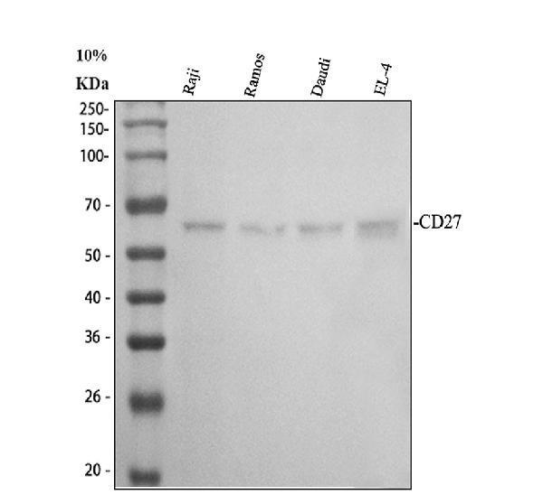
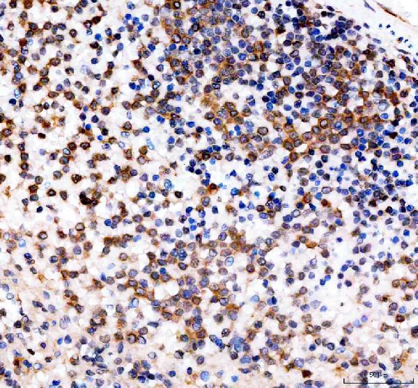
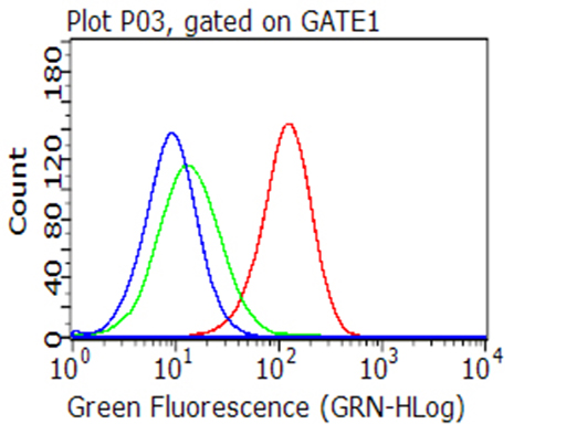
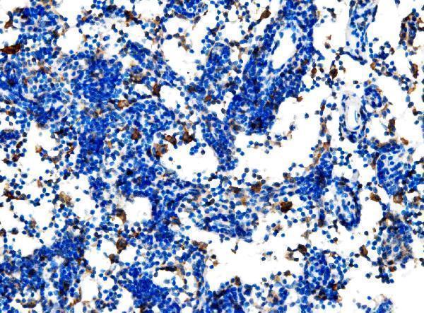
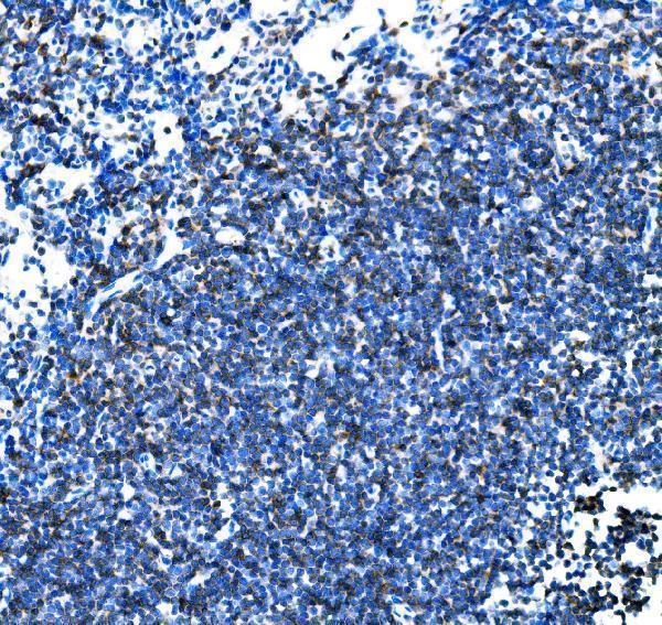
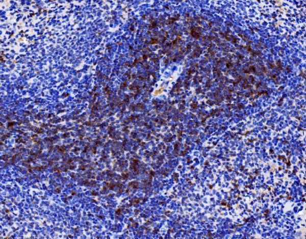
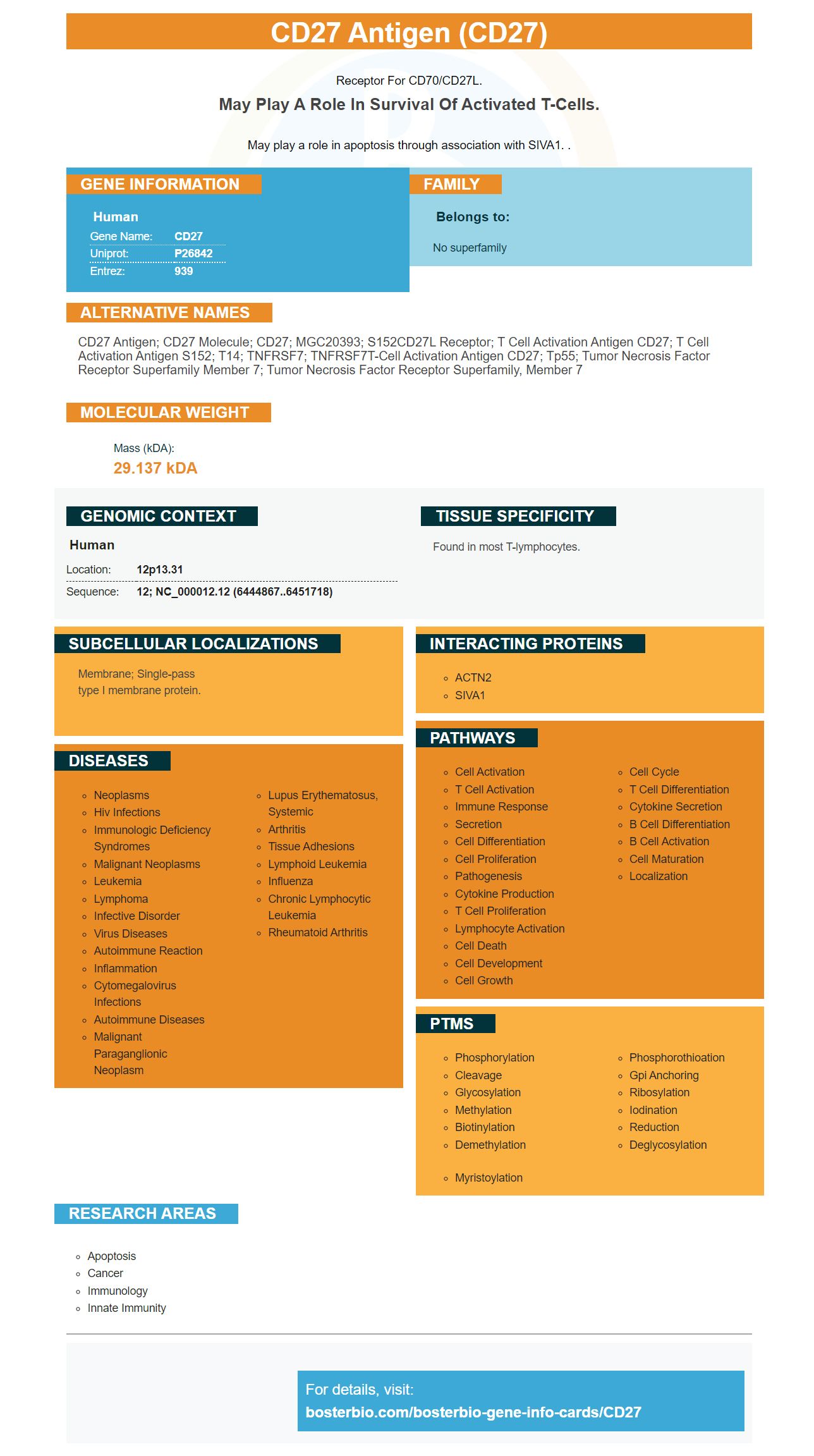
Facts about CD27 antigen.
Receptor for CD70/CD27L.
May play a role in survival of activated T-cells.May play a role in apoptosis through association with SIVA1. .
Human | |
---|---|
Gene Name: | CD27 |
Uniprot: | P26842 |
Entrez: | 939 |
Belongs to: |
---|
No superfamily |
CD27 antigen; CD27 molecule; CD27; MGC20393; S152CD27L receptor; T cell activation antigen CD27; T cell activation antigen S152; T14; TNFRSF7; TNFRSF7T-cell activation antigen CD27; Tp55; Tumor necrosis factor receptor superfamily member 7; tumor necrosis factor receptor superfamily, member 7
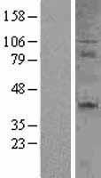
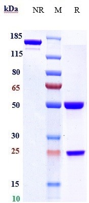
Mass (kDA):
29.137 kDA
Human | |
---|---|
Location: | 12p13.31 |
Sequence: | 12; NC_000012.12 (6444867..6451718) |
Found in most T-lymphocytes.
Membrane; Single-pass type I membrane protein.
This article will cover the IHC protocol, biomarkers relevant for Varlilumab as well as the CD27 marker which is part of the TNFR superfamily. You'll also learn how CD27 can aid in the research of varlilumb. You'll also learn which CD27 biomarkers are the best for your research. We hope these posts can be of help to you and your patients!
The CD27 marker is among the most important markers of T-cell biology. It interacts with CD70 on antigen-presenting cell lines to stimulate T cells ' activation, and affect the differentiation and survival of CD4+ T cell lines, and boost their longevity once activated. Furthermore, genetic mutations in CD27 or CD70 could result in chronic symptomatic infections with EBV, or in lymphoproliferative diseases. Moreover, the expression level of CD27 in T cells correlates strongly with their suppressive power.
In vitro studies, CD27+CD70+ Tregs were co-cultured with the total DC population as well as an anti-CD27 mAb. Both systems were mAb-positive against CD70 that hindered the growth and proliferation of T cells in response. It was interesting to note that the suppression of proliferation was the same regardless of whether DCs were cocultured with CD27+CD70+ Tregs.
In vitro experiments evaluated CD27 and CD70 expression on Tregs that were freshly isolated ex vivo , or Tregs that had been expanded for 2 to 4 weeks. After each stimulation, CD27 and CD70 expression was determined using IHC. Multiple comparisons were analysed by using a Friedman test and Dunn's post-test. Representative plots were constructed to show the intensity of the fluorescence and percentages of CD27+ cells from one donor.
The IHC protocol for the CD27 marker utilizes an mAb that targets human CD27. This procedure makes use of a mixture of mAbs that block anti-CD27 mAbs as well as non-mutated anti–human Ig. In addition, they are cross-linked using isotype-matched antibody. The CD27+CD70+ Tregs were extremely efficient in reducing CD4+, CD8+ T-cell proliferation.
Depending on the dose and duration of the treatment, a patient may require multiple infusions. This process is called dose escalation. In dose escalation, patients were given an initial dose of 10 percent of the total dose infusion and then waited for an hour of observation. The remaining dose was given. The duration of observation was four to six hours for the first two infusions, and two hours for subsequent doses.
In addition to blood, tumor samples can also function as biomarkers. These biomarkers can aid doctors in determining the most effective treatment. For instance, the CRISU2C Dream Team looked into a combination of two biomarkers relating to killer T cells and the PD-1/PD-L1 biomarker. Despite the fact that the detection of killer T cells is traditionally a invasive biopsy, this combination offered greater predictive power. A CRI-funded postdoctoral fellow developed a new technique for getting information without a biopsy.
However, PD1 expression is not the only marker that can be used to evaluate patient response to anti-PD1 antibody. Other biomarkers such as PD-L1 levels and PD1 expression might be of interest as well. The research suggests that biomarkers correlated with the PD-1 level could enhance the accuracy of immunotherapy predictions.
One patient who had had three previous treatment options for stage IV renal cell carcinoma received one dose of varlilumab. At three months she experienced complete tumor regression. At the age of four she continued to receive the biologic, despite the absence of additional disease-related toxicity. The patient also showed complete recovery of her paraortic lymph lesions. After five years, she was found to have no more cancer, which was a positive outcome for the treatment.
Other studies suggest that a patient's tumor's mutational burden could be a relevant biomarker of response. Patients with tumors with high levels of mutations react better to nivolumab that patients with other types. This is also applicable to patients with mutational burdens within the first two months of treatment. Researchers have found that patients with tumors with mutations within the first five to eight weeks have a higher response rate than patients who have no tumor mutations.
The KI67 index, and the BCL1 protein are key biomarkers for varlilumab. Ki67 index is the proportion of MCL cells that exhibit an elevated level of Ki67, a proliferative marker. A high Ki67 index can be a sign of poor prognosis or treatment response. It is used in many workup protocols and has significant correlation with survival time. This biomarker does not have any connection with LDH levels B symptoms, levels, or performance status.
The superfamily of tumor necrosis factor receptors (TNFRSF) comprises several genes that are part of it, including CD27. The members of the TNF superfamily are signaling-competent and play a role in distinct signaling pathways. In turn, TNFRs are activated by the soluble TNFLs, which occur as trimeric transmembrane type II proteins or as soluble ligand trimers.
A number of members of the TNFR superfamily that include CD27, play key roles in cell differentiation, growth, and Apoptosis. Additionally, ligation of these proteins leads to the death of cells in a programmed manner. This process requires that the proteins possess an 80-aa continuous stretch, called the death domain. Additionally, CD27 is known to induce apoptosis within certain cells, including tumor cells. However, co-crosslinking of surface Ig receptors could enhance CD27's apoptosis-inducing activity.
Although CD27 is found in subpopulations (T and B cells) but it isn't yet understood how it contributes to the immune system. Studies have demonstrated that it plays an essential role in regulating T - and B-cell proliferation , by providing costimulatory signals. A new human protein, Siva (605567), has been discovered. It interacts with the receptor CD27 to trigger apoptosis on various cell types. While more research is required to understand the basic principles of the role played by CD27 within the immune system is well established.
Flow cytometric analysis can be used to study the expression of CD27 receptors in living cells. It has been established that CD27 can co-express with GFP for up to two days. Similar to flow cytometry. This method permits the detection of CD27 receptors on a variety of cells. Interestingly, CD27 is also part of the TNF superfamily and the expression of the protein is comparable to flow cytometric analyses.
A recent study has discovered that ligation of TNFR60 activates the SAPK/JNK pathway. This activation is dependent on TRAF2 and TRAF3 and CD27 interacts with these signaling molecules. TRAF2 and TRAF5 inhibitors inhibit the co-stimulatory signal through CD27. In addition, the kinase activities of these molecules was determined through immunoprecipitation with GST-c-Jun-(1-79) as a substrate.
Co-stimulation is also a method to enhance the effects of other members of TNFR superfamily. Co-stimulation of CD27 and GITR with 4-1BB increased the response to both ligands. Although it is not known whether CD27 co-stimulation with T cells can cause synergy or hinder the antiviral response, this research suggests that CD27 and GITR can both stimulate the effects of IL-2.
Co-stimulation using CD27 has been shown to increase the first phase of cytokines. Surprisingly, CD27 co-stimulation of 4-1BB also increases the production of cytokines in the same timescale as T-cell adaption to co-stimulation, but without co-stimulation.
PMID: 1655907 by Camerini D., et al. The T cell activation antigen CD27 is a member of the nerve growth factor/tumor necrosis factor receptor gene family.
PMID: 1334106 by Loenen W.A., et al. Genomic organization and chromosomal localization of the human CD27 gene.