Click image to see more details
Product Info Summary
| SKU: | A01148-2 |
|---|---|
| Size: | 100 μg/vial |
| Reactive Species: | Mouse, Rat |
| Host: | Rabbit |
| Application: | ELISA, Flow Cytometry, IHC |
Customers Who Bought This Also Bought

Product info
Product Name
Anti-Cd27 Antibody
SKU/Catalog Number
A01148-2
Size
100 μg/vial
Form
Lyophilized
Description
Boster Bio Anti-Cd27 Antibody catalog # A01148-2. Tested in ELISA, Flow Cytometry, IHC applications. This antibody reacts with Mouse, Rat.
Storage & Handling
At -20°C for one year from date of receipt. After reconstitution, at 4°C for one month. It can also be aliquotted and stored frozen at -20°C for six months. Avoid repeated freezing and thawing.
Cite This Product
Anti-Cd27 Antibody (Boster Biological Technology, Pleasanton CA, USA, Catalog # A01148-2)
Host
Rabbit
Contents
Each vial contains 4 mg Trehalose, 0.9 mg NaCl, 0.2 mg Na2HPO4.
Clonality
Polyclonal
Isotype
Rabbit IgG
Immunogen
E.coli-derived mouse Cd27 recombinant protein (Position: P24-F187).
Cross-reactivity
No cross-reactivity with other proteins.
Reactive Species
A01148-2 is reactive to Cd27 in Mouse, Rat
Calculated molecular weight
28.2 kDa
Background of Cd27
CD27 is a member of the tumor necrosis factor receptor superfamily. This receptor is required for generation and long-term maintenance of T cell immunity. It binds to ligand CD70, and plays a key role in regulating B-cell activation and immunoglobulin synthesis. And this receptor transduces signals that lead to the activation of NF-kappaB and MAPK8/JNK. Adaptor proteins TRAF2 and TRAF5 have been shown to mediate the signaling process of this receptor. CD27-binding protein (SIVA), a proapoptotic protein, can bind to this receptor and is thought to play an important role in the apoptosis induced by this receptor.
Antibody Validation
Boster validates all antibodies on WB, IHC, ICC, Immunofluorescence, and ELISA with known positive control and negative samples to ensure specificity and high affinity, including thorough antibody incubations.
Application & Images
Applications
A01148-2 is guaranteed for ELISA, Flow Cytometry, IHC Boster Guarantee
Recommend Dilution
| Application | Dilution | Species |
|---|---|---|
| Immunohistochemistry(Paraffin-embedded Section) | 2-5 μg/ml | Mouse, Rat |
| Flow Cytometry (Fixed) | 1-3 μg/1x106 cells | Mouse |
| ELISA | 0.1-0.5 μg/ml | - |
Tested application
Use TE buffer pH 9.0 for antigen retrieval; (*) citrate buffer pH 6.0 is an alternative.
Validation Images & Assay Conditions
Click image to see more details
IHC analysis of Cd27 using anti-Cd27 antibody (A01148-2).
Cd27 was detected in a paraffin-embedded section of mouse lymphaden tissue. Heat mediated antigen retrieval was performed in EDTA buffer (pH 8.0, epitope retrieval solution). The tissue section was blocked with 10% goat serum. The tissue section was then incubated with 2 μg/ml rabbit anti-Cd27 Antibody (A01148-2) overnight at 4°C. Peroxidase Conjugated Goat Anti-rabbit IgG was used as secondary antibody and incubated for 30 minutes at 37°C. The tissue section was developed using HRP Conjugated Rabbit IgG Super Vision Assay Kit (Catalog # SV0002) with DAB as the chromogen.

Click image to see more details
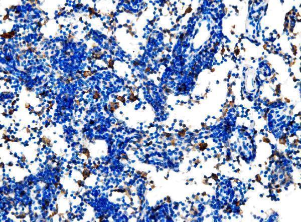
IHC analysis of Cd27 using anti-Cd27 antibody (A01148-2).
Cd27 was detected in a paraffin-embedded section of rat lymphaden tissue. Heat mediated antigen retrieval was performed in EDTA buffer (pH 8.0, epitope retrieval solution). The tissue section was blocked with 10% goat serum. The tissue section was then incubated with 2 μg/ml rabbit anti-Cd27 Antibody (A01148-2) overnight at 4°C. Peroxidase Conjugated Goat Anti-rabbit IgG was used as secondary antibody and incubated for 30 minutes at 37°C. The tissue section was developed using HRP Conjugated Rabbit IgG Super Vision Assay Kit (Catalog # SV0002) with DAB as the chromogen.

Click image to see more details
IHC analysis of Cd27 using anti-Cd27 antibody (A01148-2).
Cd27 was detected in a paraffin-embedded section of rat spleen tissue. Heat mediated antigen retrieval was performed in EDTA buffer (pH 8.0, epitope retrieval solution). The tissue section was blocked with 10% goat serum. The tissue section was then incubated with 2 μg/ml rabbit anti-Cd27 Antibody (A01148-2) overnight at 4°C. Peroxidase Conjugated Goat Anti-rabbit IgG was used as secondary antibody and incubated for 30 minutes at 37°C. The tissue section was developed using HRP Conjugated Rabbit IgG Super Vision Assay Kit (Catalog # SV0002) with DAB as the chromogen.

Click image to see more details
Flow Cytometry analysis of mouse PBMC cells using anti-Cd27 antibody (A01148-2).
Overlay histogram showing mouse PBMC cells stained with A01148-2 (Blue line). The cells were fixed with 4% paraformaldehyde and blocked with 10% normal goat serum. And then incubated with rabbit anti-Cd27 Antibody (A01148-2, 1 μg/1x106 cells) for 30 min at 20°C. DyLight®488 conjugated goat anti-rabbit IgG (BA1127, 5-10 μg/1x106 cells) was used as secondary antibody for 30 minutes at 20°C. Isotype control antibody (Green line) was rabbit IgG (1 μg/1x106) used under the same conditions. Unlabelled sample without incubation with primary antibody and secondary antibody (Red line) was used as a blank control.
Specific Publications For Anti-Cd27 Antibody (A01148-2)
Loading publications
Recommended Resources
Here are featured tools and databases that you might find useful.
- Boster's Pathways Library
- Protein Databases
- Bioscience Research Protocol Resources
- Data Processing & Analysis Software
- Photo Editing Software
- Scientific Literature Resources
- Research Paper Management Tools
- Molecular Biology Software
- Primer Design Tools
- Bioinformatics Tools
- Phylogenetic Tree Analysis
Customer Reviews
Have you used Anti-Cd27 Antibody?
Share your experimental results or join a short interview to earn up to $1,000 in product credits or other rewards.
0 Reviews For Anti-Cd27 Antibody
Customer Q&As
Have a question?
Find answers in Q&As, reviews.
Can't find your answer?
Submit your question
