CD3E / T-cell surface glycoprotein CD3 epsilon chain · IHC design guide

Design Immunohistochemistry for CD3E

Plan chromogenic CD3E IHC on paraffin sections using tonsil as a positive control (HPA tissue IHC; datasheet PB9093). Score selective cytoplasmic staining in T cells while considering CD3E's membrane location and activation linked internalization (HPA tissue IHC; UniProt).

Evidence assembled Oct 2026 · For research use; verify linked source records and product datasheet before use
Immunohistochemistry protocol sheet for CD3E (IHC for CD3E): expected localisation T-cell cytoplasm observed; membrane expected (HPA tissue IHC; UniProt), antibody PB9093, validated IHC image, and IHC protocol steps
Printable CD3E IHC protocol sheet — expected localisation T-cell cytoplasm observed; membrane expected (HPA tissue IHC; UniProt), antibody PB9093, controls and protocol steps. Open the full CD3E IHC guide →

CD3E Immunohistochemistry Experimental Design Guide

Expected localisation, validated protocols, controls and antibodies — the at-a-glance facts below, then the full design guide.

Must know before staining
Expected localisation T-cell cytoplasm observed; membrane expected (HPA tissue IHC; UniProt)
Staining pattern Selective cytoplasmic staining in T cells (HPA tissue IHC)
Antigen retrieval Citrate pH 6 HIER, heat-mediated (datasheet PB9093)
Positive control ⓘ Appendix+4 more · see all
Negative control ⓘ Adipose tissue+4 more · see all
Important caveats
Reasons your staining may differ from the expected pattern.
Fixation Matched tissue-IHC evidence does not establish the fixation claim. Validate the specimen-specific method before use. (selected-SKU IHC image PB9093)
Caveat Cytoplasmic IHC may differ from membrane expectations (HPA tissue IHC; UniProt)
Regulation Activation promotes CD3E internalization (UniProt)
Isoform / epitope 0 isoforms listed; distinguish extracellular from cytoplasmic epitopes (UniProt)
Section 1

Recommended CD3E IHC & IF Protocols

The catalog antibody’s paraffin IHC protocol uses citrate pH 6 retrieval (datasheet PB9093). Three published CD3E IHC protocols provide additional conditions (PMC4013007; PMC5351587; PMC12529491).

Recommended immunohistochemistry (IHC-P) protocol parameters
SampleParaffin-embedded human tonsil tissues; fixative not specified (datasheet PB9093)
FixationImage fixative and duration unreported (datasheet PB9093); verify before use.
Sectioning4–5 µm sections on charged slides (standard)
DeparaffinisationXylene, graded ethanol series to water (standard)
Antigen retrievalHeat retrieval: Citrate pH 6, 20 min (datasheet PB9093)
Peroxidase block3% H2O2, 10 min, room temperature (standard)
Blocking10% goat serum (datasheet PB9093)
Primary antibodyRabbit anti-CD3E, 0.5-1μg/ml (datasheet PB9093)
Primary incubationOvernight at 4 °C (datasheet PB9093)
DetectionStreptavidin-biotin complex (SABC), DAB chromogen (datasheet PB9093)
CounterstainHematoxylin, blue, dehydrate and mount (standard)
Expected resultCD3E-positive staining in lymphoid tissue of appendix (HPA tissue IHC: High). HPA tissue profile: Selective cytoplasmic expression in T-cells. No signal in the no-primary control.
💡Decision noteStart with citrate pH 6 for the catalog antibody (datasheet PB9093). Tris-EDTA pH 9.0 is a published alternative for paraffin liver sections (PMC12529491).
Section 2

What Is the Expected CD3E Staining Pattern?

CD3E is a cell-membrane protein with an extracellular segment at residues 23–126 and a cytoplasmic segment at 153–207 (UniProt P07766 topology). In paraffin-section IHC, expect staining in T-cells; HPA describes selective cytoplasmic expression and rates its tissue pattern Enhanced, reflecting high consistency with RNA data (HPA tissue IHC). Interpret that observed cytoplasmic pattern alongside CD3E’s membrane topology rather than requiring a crisp membrane rim in every cell.

What am I looking at on my slide?
Discrete stained lymphocytes in appendix lymphoid tissue or the non-germinal-center regions of lymph node or tonsil, with little staining in adjacent non-lymphoid cells (HPA tissue IHC).This fits the reported high staining in those compartments and selective T-cell expression (HPA tissue IHC). Assess the distribution across cells as well as intensity: a convincing result identifies a lymphocyte population rather than coloring an entire tissue compartment (HPA tissue IHC; general IHC interpretation).
Predominantly nuclear staining, with no convincing lymphocyte-associated cytoplasmic or membrane signal.A nuclear-only pattern does not fit the reported IHC profile or membrane topology (HPA tissue IHC; UniProt P07766 topology). Treat it as suspect and compare it with a known-positive section and detection controls before interpreting it as CD3E (general IHC practice).
Broad staining of adipocytes or respiratory epithelial cells instead of a selective lymphocyte population.HPA reports CD3E as not detected in those cell types (HPA tissue IHC: adipocytes; respiratory epithelial cells). Consider antibody cross-reactivity or endogenous detection activity, and check controls; an occasional stained lymphocyte within an otherwise negative tissue has a different interpretation (general IHC practice; HPA tissue IHC).
A diffuse wash of chromogen obscures cell boundaries across positive and negative areas.The distribution cannot establish selective T-cell staining (HPA tissue IHC; general IHC interpretation). Background can arise from detection activity, inadequate blocking, or excessive antibody concentration; inspect a no-primary control and adjust the general IHC workflow before scoring cells (general IHC practice).
No stained lymphocytes in appendix lymphoid tissue, lymph node, or tonsil, while the section and counterstain remain visible.These are reported high-staining sites, so a blank result warrants a technical check (HPA tissue IHC: High). Confirm that lymphocyte-rich areas are present, then review antibody identity, controls, retrieval and detection conditions; their optimal settings are not established by the supplied target evidence (general IHC practice).
💡Expected CD3E appearanceA positive IHC result shows selective, readily discernible staining of T-cell-rich lymphocyte populations, often appearing cytoplasmic in paraffin sections; broad staining of HPA-negative non-lymphoid cells or isolated nuclear staining is suspect (HPA tissue IHC; UniProt P07766 topology).
How each factor affects the staining
Which compartment should guide IHC scoring?UniProt places CD3E at the cell membrane, with a transmembrane segment at 127–152, while HPA describes selective cytoplasmic T-cell staining in tissue IHC (UniProt P07766 topology; HPA tissue IHC). Use the tissue pattern and cell identity together; a perfectly outlined membrane is not required to match the reported IHC appearance.
Which tissue areas provide a useful comparison?HPA reports High staining in appendix lymphoid tissue and non-germinal-center cells of lymph node and tonsil, and Medium staining in bone-marrow hematopoietic cells and splenic red-pulp cells (HPA tissue IHC). Compare like compartments; these levels do not predict uniform intensity across every cell.
How strong is the tissue-pattern evidence?HPA rates the tissue IHC pattern Enhanced and lists five antibodies with Enhanced IHC validation: HPA040957, HPA043955, CAB000010, CAB072863 and CAB072864 (HPA tissue IHC; HPA antibodies). Validation supports the reported pattern but does not replace controls for the antibody and run in use (general IHC practice).
Does the record establish an antigen-retrieval condition?Target-specific fixation effects are not established by the supplied assay evidence. Verify with a matched IHC source before attributing a result to fixation.
Can intracellular signal be biologically plausible?CD3E participates in TCR-CD3 internalization through sequences in its cytosolic region (UniProt P07766 function). That fact and HPA’s cytoplasmic IHC profile support considering intracellular staining in the right cells; they do not make diffuse staining of unrelated cells specific (UniProt P07766; HPA tissue IHC).
For IF/ICC, where has CD3E been seen?HPA reports plasma-membrane and endoplasmic-reticulum localization, with additional Golgi localization, in its ICC-IF subcellular record (HPA subcellular). Those observations answer the localization question for IF/ICC; they are a separate assay record and do not set a paraffin-section IHC protocol or require every tissue cell to show all three compartments.
Why is my staining missing, weak or wrong?
SituationLikely causeNext action
The expected lymphocyte population is weak or absent in a positive section.The run may have insufficient usable signal, or the examined area may lack the reported positive compartment (HPA tissue IHC; general IHC practice).First locate appendix lymphoid tissue or an appropriate lymph-node or tonsil region, then check the IHC-validated antibody’s instructions, retrieval, dilution and detection controls (HPA tissue IHC; general IHC practice). Do not infer a CD3E-specific fixation failure from this result.
Signal appears only in nuclei.Nuclear-only localization conflicts with the membrane topology and selective cytoplasmic tissue profile (UniProt P07766 topology; HPA tissue IHC).Compare a known-positive lymphocyte area with a no-primary control and review detection background before calling the nuclear signal specific (general IHC practice).
Adipocytes or respiratory epithelium stain broadly.Those cell types are listed as not detected by HPA; cross-reactivity or endogenous detection activity is possible (HPA tissue IHC; general IHC practice).Check a no-primary control and the distribution in a positive lymphoid compartment. If staining persists without primary antibody, review blocking and detection steps; otherwise reassess antibody specificity (general IHC practice).
Diffuse chromogen makes individual lymphocytes hard to identify.Background obscures the selective cell pattern reported for CD3E (HPA tissue IHC). Excess primary antibody or detection background are general possibilities, not established CD3E-specific effects (general IHC practice).Use a no-primary control, then review blocking, washing, antibody concentration and development time according to the IHC workflow (general IHC practice). Score only cells whose signal remains distinguishable from background.
A membrane rim is faint, but discrete lymphocytes show cytoplasmic staining.HPA’s paraffin-section tissue profile describes selective cytoplasmic T-cell expression despite CD3E’s membrane topology (HPA tissue IHC; UniProt P07766 topology).Judge cell identity, tissue distribution and control results together; do not reject an otherwise matching IHC pattern solely because the membrane rim is faint (HPA tissue IHC; general IHC interpretation).
The negative comparison tissue contains a few stained small cells.HPA’s not-detected entries refer to specified cell types, such as adipocytes and respiratory epithelial cells, rather than every cell present in the tissue (HPA tissue IHC).Identify which cells stain and compare their morphology with lymphocytes in a positive section. Use the negative compartment and a no-primary control to assess nonspecific signal (HPA tissue IHC; general IHC practice).

Sample controls for CD3E IHC & IF

🧪Run tonsil first: non-germinal center cells should stain strongly (HPA: High in tonsil non-germinal center cells), and the catalog antibody has a paraffin-section tonsil example (PB9093 tissue-IHC caption). Use adipose tissue as a negative, scoring adipocytes rather than any resident lymphocytes (HPA: Not detected in adipocytes); non-T-cell profiles on the tonsil slide should lack specific CD3E staining (UniProt P07766: CD3E is part of the T-lymphocyte surface TCR-CD3 complex).
Positive control tissue: Appendix (Lymphoid tissue, HPA High)
Negative control tissue: Adipose tissue (HPA Not detected)
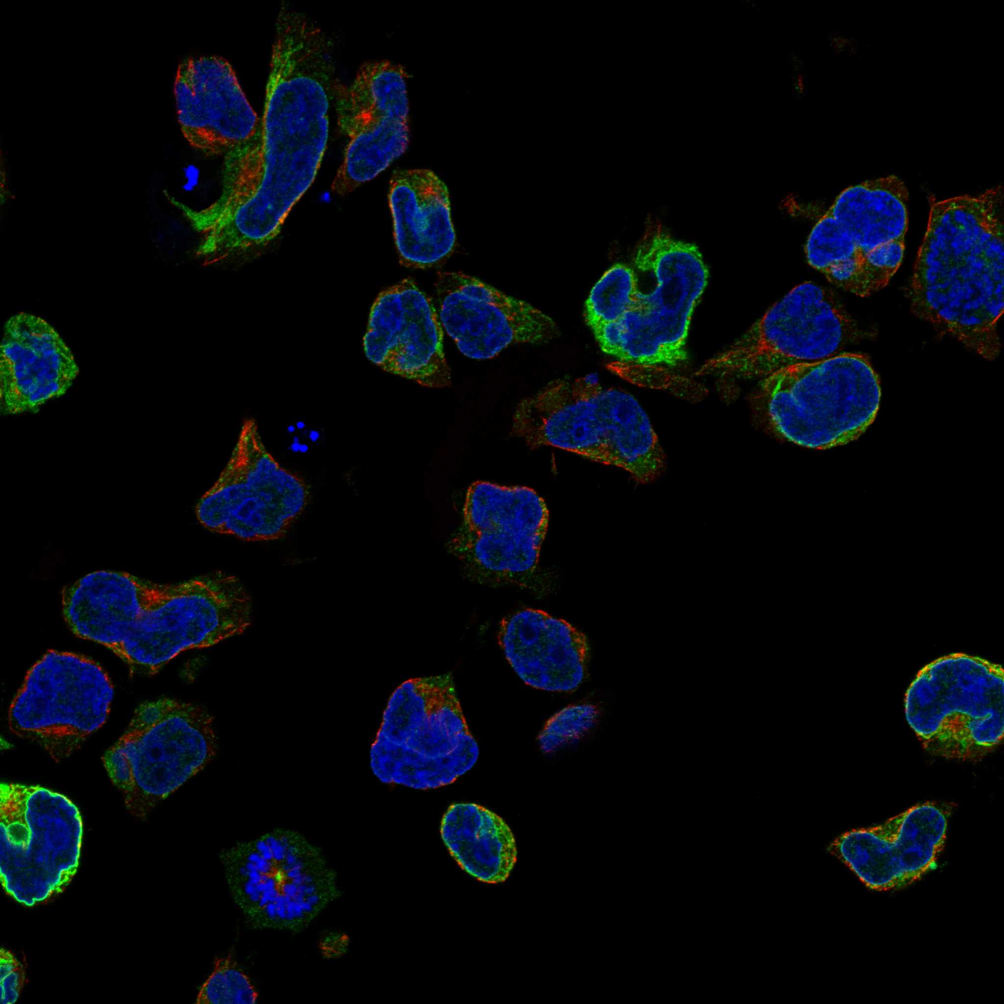
ICC-IF cell lines (HPA subcellular resource): HPA ICC-IF images show CD3E in JURKAT, U2OS, with annotated localisation: Endoplasmic reticulum (approved), Plasma membrane (supported) (HPA subcellular).
Technical controls: Include a no-primary, secondary-only control; a host- and clonality-matched isotype control; and, if available, CD3E-knockout tissue as a biological negative (standard IHC practice). For chromogenic tonsil IHC, quench endogenous peroxidase and check background from the biotin-based detection system used in the example (standard IHC practice; PB9093 tissue-IHC caption: biotin-based detection with DAB).
⚠️Feasibility: A target-specific fixation window or fixation effect is unreported, and the selected PB9093 paraffin-section caption does not state the fixative (PB9093 tissue-IHC caption). That caption reports heat retrieval in citrate buffer at pH 6 for 20 minutes; it supports trying that retrieval condition but does not establish that retrieval is required under every processing condition (PB9093 tissue-IHC caption). The supplied evidence does not establish whether frozen sections or IF are easier for this target; assess tonsil background alongside specific staining because the example uses biotin-based DAB detection (PB9093 tissue-IHC caption; standard IHC practice).

HPA tissue IHC evidence for CD3E

Comprehensive Human Protein Atlas IHC scoring per tissue (reliability: Enhanced — High consistency between antibody staining and RNA expression data.). Rows are taken directly from the HPA tissue chart — click any row's HPA link to view the source.

Positive expression · recommended positive controls

TissueCell typeLevelEvidenceSource
Appendix Lymphoid tissue High Protein (IHC) HPA →
Lymph node Non-germinal center cells High Protein (IHC) HPA →
Tonsil Non-germinal center cells High Protein (IHC) HPA →
Bone marrow Hematopoietic cells Medium Protein (IHC) HPA →
Spleen Cells in red pulp Medium Protein (IHC) HPA →

Undetected expression · recommended negative controls

TissueCell typeLevelEvidenceSource
Adipose tissue Adipocytes Not detected Protein (IHC) HPA →
Adrenal gland Glandular cells Not detected Protein (IHC) HPA →
Breast Adipocytes Not detected Protein (IHC) HPA →
Bronchus Respiratory epithelial cells Not detected Protein (IHC) HPA →
Caudate Glial cells Not detected Protein (IHC) HPA →
Section 3

Advanced CD3E IHC Tips

Troubleshoot CD3E staining in paraffin sections by checking retrieval, cell distribution, compartment, controls and scoring before interpreting chromogenic signal.

How should I adjust retrieval when CD3E staining is weak in paraffin sections?
Start with heat-mediated retrieval in citrate buffer at pH 6 for 20 minutes (PB9093 tissue-IHC caption). The PB9093 tonsil example used that retrieval before overnight incubation with 1 µg/mL primary antibody at 4°C, so keep those conditions together for the first comparison (PB9093 tissue-IHC caption). If staining remains weak, compare a slightly longer heating interval or an EDTA-based retrieval condition on adjacent sections, while watching for tissue damage (standard IHC practice). Include tonsil as a positive control and compare T-cell staining with background under identical chromogenic development (PB9093 tissue-IHC caption; HPA: High in tonsil non-germinal center cells; standard IHC practice).
Could fixation explain weak or uneven CD3E staining?
The PB9093 image describes a paraffin-embedded tonsil section but does not report its fixative or fixation duration (PB9093 tissue-IHC caption). Target-specific CD3E sensitivity to fixation is therefore unknown from the supplied IHC evidence; do not assign weak staining to a particular fixative without a controlled comparison (PB9093 tissue-IHC caption). Record the fixative, duration and processing history for each specimen, then compare similarly processed sections with the same pH 6 citrate retrieval and chromogenic development (standard IHC practice; PB9093 tissue-IHC caption). If a positive-control section stains while a test section does not, inspect processing and tissue preservation before changing the antibody concentration (standard IHC practice).
Should CD3E appear on membranes or in cytoplasm by chromogenic IHC?
CD3E is a cell-membrane component of the TCR-CD3 complex, with an extracellular region at residues 23–126 and a cytoplasmic region at 153–207 (UniProt P07766 topology and function). Tissue IHC reports selective cytoplasmic expression in T cells, so cytoplasmic chromogen alone does not rule out CD3E staining in a section (HPA: tissue-IHC profile). Plasma membrane and endoplasmic reticulum are reported subcellular locations, with Golgi localisation also reported (HPA: subcellular). Judge signal by its distribution among lymphoid cells and the quality of the accompanying control, while treating broad epithelial or stromal staining as a reason to revisit specificity and background controls (HPA: tissue-IHC profile; standard IHC practice).
How can epitope position change my CD3E IHC result?
The supplied CD3E record lists 0 isoforms, so it offers no annotated splice variant to explain a staining difference (UniProt P07766 record). CD3E has an extracellular segment at residues 23–126, a transmembrane segment at 127–152 and a cytoplasmic segment at 153–207 (UniProt P07766 topology). Its cytoplasmic ITAM spans residues 178–205 and includes phosphotyrosines at 188 and 199, but the supplied PB9093 caption does not identify the recognized epitope (UniProt P07766 domains and modified residues; PB9093 tissue-IHC caption). Check epitope documentation before attributing variable IHC staining to phosphorylation or membrane sidedness, then compare matched positive controls after the same retrieval (standard IHC practice).
How should I assess CD3E in a multiplex IF experiment?
Plan CD3E IF as a separate validation from the chromogenic paraffin protocol, and include a T-cell marker in another channel to assess expected cell-type overlap (UniProt P07766 function; standard IF practice). Choose fluorophores after measuring tissue autofluorescence in an unstained section, favoring a well-separated, brighter channel for the weaker signal (standard IF practice). CD3E spans the membrane, with extracellular residues 23–126 and cytoplasmic residues 153–207; permeabilisation needs depend on which side contains the antibody epitope, which the supplied caption does not specify (UniProt P07766 topology; PB9093 tissue-IHC caption). Run single-stain controls to check bleed-through and compare membrane-associated with intracellular signal (HPA: subcellular; standard IF practice).
What should I check when CD3E DAB staining is widespread?
First compare the stained section with a primary-antibody omission control to separate detection-system background from primary-antibody-associated signal (standard IHC practice). The PB9093 example used 10% goat serum blocking, a biotinylated secondary, streptavidin-biotin detection and DAB, so inspect background at each relevant detection step (PB9093 tissue-IHC caption). For a peroxidase-based DAB workflow, use an appropriate endogenous-peroxidase block and check whether endogenous biotin contributes signal when using avidin-biotin chemistry (standard IHC practice). Reassess primary concentration, washes and chromogen development if staining spreads beyond lymphoid cells; the caption used 1 µg/mL primary antibody (PB9093 tissue-IHC caption; HPA: tissue-IHC profile; standard IHC practice).
How should I quantify CD3E-positive cells across tissue sections? ⚠ ANSWER MARKED FOR VERIFICATION
Define the tissue compartment before counting, because CD3E is associated with T lymphocytes and HPA reports high staining in tonsil non-germinal center cells (UniProt P07766 function; HPA: tissue-IHC profile). Report the percentage of positive nucleated cells or positive-cell density per mm² within a consistent region of interest (standard IHC practice). If intensity matters, use an H-score with fixed intensity thresholds, and report it alongside cell counts so stronger DAB does not masquerade as more T cells (standard IHC practice). Normalize density to evaluated viable tissue area, exclude necrosis and folds consistently, and hold retrieval and development conditions constant across sections (standard IHC practice).
When is a CD3E-positive IHC signal credible?
A credible result shows staining in morphologically appropriate lymphoid cells, supported by a positive-control section and a clean primary-antibody omission control (UniProt P07766 function; HPA: tissue-IHC profile; standard IHC practice). Cytoplasmic staining can be compatible with tissue IHC, while membrane and intracellular localisation are also documented; compartment alone cannot establish specificity (HPA: tissue-IHC profile; HPA: subcellular). Treat widespread staining of epithelial or stromal cells, section-edge enhancement and necrotic-area deposits as reasons to check artefact and background (HPA: tissue-IHC profile; standard IHC practice). If DAB signal persists without primary antibody, inspect endogenous peroxidase and the biotin-based detection steps used in the PB9093 example (PB9093 tissue-IHC caption; standard IHC practice).
Boster reagents

Best CD3E / T-cell surface glycoprotein CD3 epsilon chain IHC Antibodies

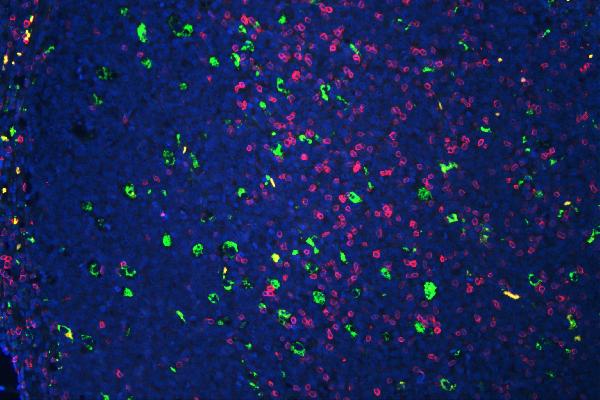
The catalog covers human, mouse, rat, and chicken reactivity (catalog reactivity). The figures show tissue IHC for six antibodies and human tonsil IF for PB9093 (IHC and IF captions).

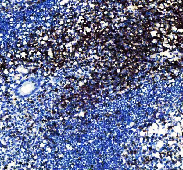
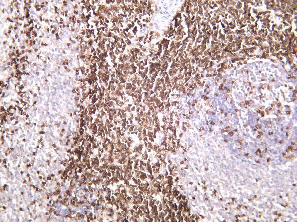
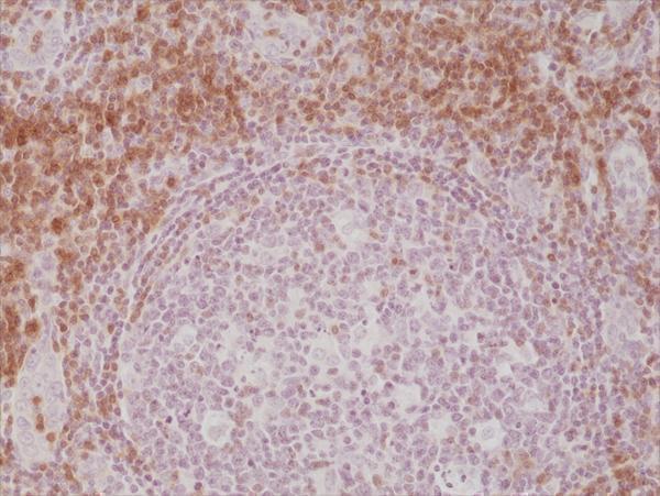
Real IHC data IHC analysis of CD3 Epsilon using anti-CD3 Epsilon antibody (PB9093). CD3 Epsilon was detected in paraffin-embedded section of human tonsil tissues. Heat mediated antigen retrieval was performed in citrate buffer (pH6, epitope retrieval solution) for 20 mins. The tissue section was blocked with 10% goat serum. The tissue section was then incubated with 1μg/ml rabbit anti-CD3 Epsilon Antibody (PB9093) overnight at 4°C. Biotinylated goat anti-rabbit IgG was used as secondary antibody and incubated for 30 minutes at 37°C. The tissue section was developed using Strepavidin-Biotin-Complex (SABC)(Catalog # SA1022) with DAB as the chromogen.
Anti-CD3 epsilon/CD3E Antibody ®
Cat # PB9093
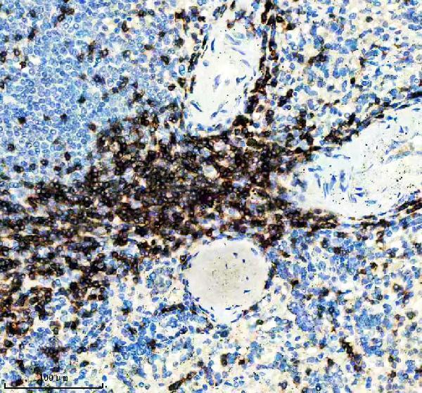
Real IHC data IHC analysis of CD3E using anti-CD3E antibody (M02675-1). CD3E was detected in a paraffin-embedded section of human appendix tissue. Heat mediated antigen retrieval was performed in EDTA buffer (pH 8.0, epitope retrieval solution). The tissue section was blocked with 10% goat serum. The tissue section was then incubated with a dilution of 1:50 rabbit anti-CD3E Antibody (M02675-1) overnight at 4°C. Peroxidase Conjugated Goat Anti-rabbit IgG was used as secondary antibody and incubated for 30 minutes at 37°C. The tissue section was developed using HRP Conjugated Rabbit IgG Super Vision Assay Kit (Catalog # SV0002) with DAB as the chromogen.
Anti-CD3 epsilon Rabbit Monoclonal Antibody
Cat # M02675-1
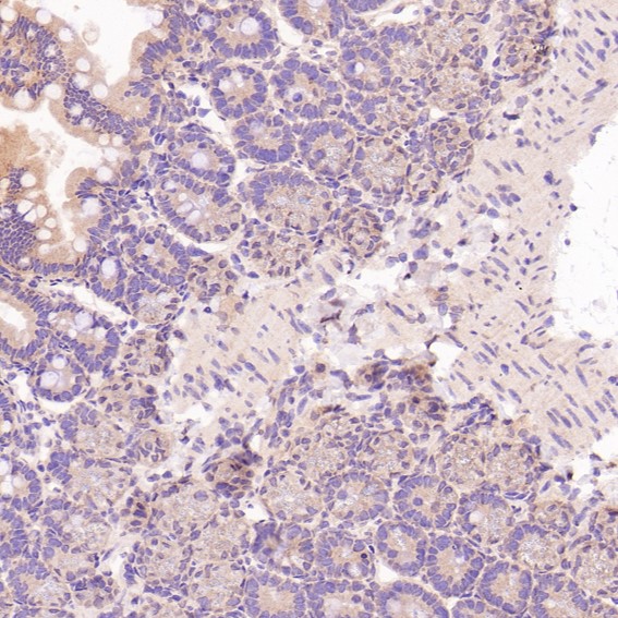
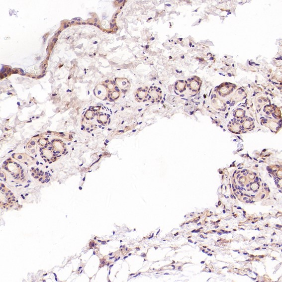
Real IHC data Immunohistochemical analysis of paraffin-embedded Mouse intestine, using the Antibody at 1:100 dilution.
Anti-CD3 epsilon Rabbit Monoclonal Antibody
Cat # M02675-2
Real IHC data Rat spleen was stained with anti-CD3 rabbit antibody
Anti-CD3 Rabbit Monoclonal Antibody
Cat # M02675-10
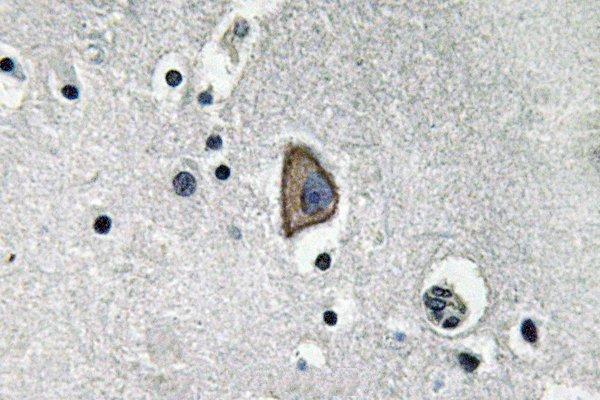
Real IHC data Immunohistochemistry (IHC) analyzes of CD3-ε (D71) pAb in paraffin-embedded human brain tissue.
Anti-CD3-Epsilon (D71) CD3E Antibody
Cat # A02675-2
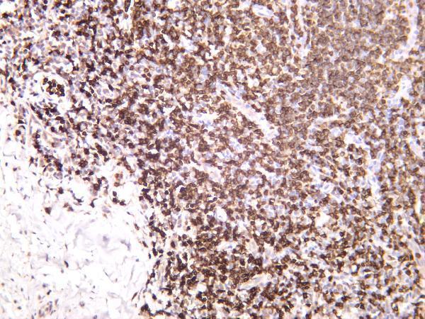
Real IHC data IHC result Immunohistochemical staining of formalin fixed and paraffin embedded human Tonsil tissue section using anti-CD3e rabbit monoclonal antibody (Clone RM344) at a 1:200 dilution.
Anti-CD3E Rabbit Monoclonal Antibody, Clone#RM344
Cat # M02675-3

PB9093 shows human tonsil paraffin IHC (PB9093 IHC caption); M02675-1 shows human appendix paraffin IHC (M02675-1 IHC caption); M02675-2 shows mouse intestine paraffin IHC, although its listed reactivity is human only (M02675-2 IHC caption; catalog reactivity). M02675-10 shows rat spleen IHC (M02675-10 IHC caption); A02675-2 shows human brain paraffin IHC (A02675-2 IHC caption); M02675-3 shows formalin-fixed, paraffin-embedded human tonsil IHC (M02675-3 IHC caption).

Which to pick: For human paraffin IHC, choose PB9093 when a documented starting condition helps: its tonsil image used citrate retrieval at pH 6 and 1 μg/ml primary antibody; the fixative is unreported (PB9093 IHC caption). For IF, PB9093 has human tonsil images; for ICC, M02675-1 lists that application, though its appendix paraffin IHC caption does not report the fixative (PB9093 IF captions; M02675-1 catalog applications and IHC caption). For cross-species IHC, PB9093 has human, mouse, and rat paraffin image captions plus listed chicken reactivity; those captions do not report the fixative (PB9093 IHC captions; catalog reactivity).

Each figure is that product's own IHC / IF validation image from its datasheet.

References

  1. UniProt Consortium. UniProt entry P07766 (CD3E_HUMAN, T-cell surface glycoprotein CD3 epsilon chain).
  2. Human Protein Atlas. CD3E tissue IHC expression (reliability: Enhanced).
  3. Human Protein Atlas. CD3E subcellular location (ICC-IF): Mainly localized to the plasma membrane and endoplasmic reticulum. In addition localized to the Golgi apparatus..
  4. Human Protein Atlas. CD3E antibody validation summary (5 antibodies).
  5. Assessing the role of STAT3 in DC differentiation and autologous DC immunotherapy in mouse models of GBM. PloS one 2014 — PMC4013007.
  6. Identification of immune-infiltrating cell-related biomarkers in hepatocellular carcinoma based on gene co-expression network analysis. Diagnostic pathology 2021 — PMC8255019.
  7. Genomic binding of PAX8-PPARG fusion protein regulates cancer-related pathways and alters the immune landscape of thyroid cancer. Oncotarget 2017 — PMC5351587.
  8. Aluminum adjuvant promotes liver inflammation and fibrosis in mice: A novel approach to establish a liver fibrosis animal model. Liver research (Beijing, China) 2025 — PMC12529491.
  9. PubMed PMID:3012357 — UniProt-cited evidence.
  10. PubMed PMID:3267235 — UniProt-cited evidence.
  11. PubMed PMID:14702039 — UniProt-cited evidence.