Click image to see more details
-

-
-
-
-
+9
Product Info Summary
| SKU: | PB9093 |
|---|---|
| Size: | 100 μg/vial |
| Reactive Species: | Chicken, Human, Mouse, Rat |
| Host: | Rabbit |
| Application: | IF, IHC, IHC-F, WB |
Customers Who Bought This Also Bought

Product info
Product Name
Anti-CD3 epsilon/CD3E Antibody Picoband®
SKU/Catalog Number
PB9093
Size
100 μg/vial
Form
Lyophilized
Description
Boster Bio Anti-CD3 epsilon/CD3E Antibody Picoband® catalog # PB9093. Tested in IF, IHC, IHC-F, ICC, WB applications. This antibody reacts with Chicken, Human, Mouse, Rat. The brand Picoband indicates this is a premium antibody that guarantees superior quality, high affinity, and strong signals with minimal background in Western blot applications. Only our best-performing antibodies are designated as Picoband, ensuring unmatched performance.
Storage & Handling
Store at -20˚C for one year from date of receipt. After reconstitution, at 4˚C for one month. It can also be aliquotted and stored frozen at -20˚C for six months. Avoid repeated freeze-thaw cycles.
Cite This Product
Anti-CD3 epsilon/CD3E Antibody Picoband® (Boster Biological Technology, Pleasanton CA, USA, Catalog # PB9093)
Host
Rabbit
Contents
Each vial contains antibody formulated with stabilizing components, 0.9 mg NaCl, 0.2 mg Na2HPO4, and 0.05 mg NaN3.
*This antibody is supplied in a stabilized formulation.
Compatibility with conjugation reactions depends on the chemistry of the conjugation method used.
For conjugation methods that are not compatible with the stabilizing components present in this formulation, a carrier-free antibody format is required.
Clonality
Polyclonal
Isotype
Rabbit IgG
Immunogen
E.coli-derived human CD3 epsilon recombinant protein (Position: D23-I207). Human CD3 epsilon shares 65% amino acid (aa) sequence identity with mouse CD3 epsilon.
Cross-reactivity
No cross-reactivity with other proteins
Reactive Species
PB9093 is reactive to CD3E in Chicken, Human, Mouse, Rat
Observed Molecular Weight
23 kDa
Calculated molecular weight
23.1 kDa
Background of CD3E
CD3e molecule, epsilon also known as CD3E is a polypeptide which in humans is encoded by the CD3E gene which resides on chromosome 11. It is mapped to 11q23.3. The protein encoded by this gene is the CD3-epsilon polypeptide, which together with CD3-gamma, -delta and -zeta, and the T-cell receptor alpha/beta and gamma/delta heterodimers, forms the T cell receptor-CD3 complex. This complex plays an important role in coupling antigen recognition to several intracellular signal-transduction pathways. The genes encoding the epsilon, gamma and delta polypeptides are located in the same cluster on chromosome 11. The epsilon polypeptide plays an essential role in T-cell development.
Antibody Validation
Boster validates all antibodies on WB, IHC, ICC, Immunofluorescence, and ELISA with known positive control and negative samples to ensure specificity and high affinity, including thorough antibody incubations.
Application & Images
Applications
PB9093 is guaranteed for IF, IHC, IHC-F, WB Boster Guarantee
Recommend Dilution
| Application | Dilution | Species |
|---|---|---|
| Western blot | 0.1-0.5μg/ml | Human |
| Immunohistochemistry (Paraffin-embedded Section) | 0.5-1μg/ml | Human, Mouse, Rat, Chicken |
| Immunohistochemistry (Frozen Section) | 0.5-1μg/ml | Mouse, Rat |
| Immunocytochemistry | 0.5-1μg/ml | Human |
| Immunofluorescence | 2μg/ml | Human, Mouse, Rat |
Tested application
Suggested blocking solution with 5% non-fat milk or BSA; (*)Recommended protein loading: 20-40 µg per lane
Use TE buffer pH 9.0 for antigen retrieval; (*) citrate buffer pH 6.0 is an alternative.
Validation Images & Assay Conditions
Click image to see more details
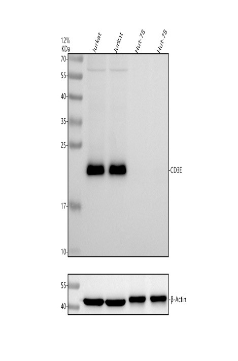
Western blot analysis of CD3E using anti-CD3E antibody (PB9093).
Electrophoresis was performed on a 5-20% SDS-PAGE gel at 70V (Stacking gel) / 90V (Resolving gel) for 2-3 hours. The sample well of each lane was loaded with 30 ug of sample under reducing conditions.
Lane 1: human Jurkat whole cell lysates,
Lane 2: human Jurkat whole cell lysates,
Lane 3: human Hut-78 whole cell lysates,
Lane 4: human Hut-78 whole cell lysates.
After electrophoresis, proteins were transferred to a nitrocellulose membrane at 150 mA for 50-90 minutes. Blocked the membrane with 5% non-fat milk/TBS for 1.5 hour at RT. The membrane was incubated with rabbit anti-CD3E antigen affinity purified polyclonal antibody (Catalog # PB9093) at 0.5 μg/mL overnight at 4°C, then washed with TBS-0.1%Tween 3 times with 5 minutes each and probed with a goat anti-rabbit IgG-HRP secondary antibody at a dilution of 1:5000 for 1.5 hour at RT. The signal is developed using an Enhanced Chemiluminescent detection (ECL) kit (Catalog # EK1002) with Tanon 5200 system. A specific band was detected for CD3E at approximately 23 kDa. The expected band size for CD3E is at 23 kDa.

Click image to see more details
IHC analysis of CD3 Epsilon using anti-CD3 Epsilon antibody (PB9093).
CD3 Epsilon was detected in paraffin-embedded section of rat spleen tissues. Heat mediated antigen retrieval was performed in citrate buffer (pH6, epitope retrieval solution) for 20 mins. The tissue section was blocked with 10% goat serum. The tissue section was then incubated with 1μg/ml rabbit anti-CD3 Epsilon Antibody (PB9093) overnight at 4°C. Biotinylated goat anti-rabbit IgG was used as secondary antibody and incubated for 30 minutes at 37°C. The tissue section was developed using Strepavidin-Biotin-Complex (SABC)(Catalog # SA1022) with DAB as the chromogen.

Click image to see more details
IHC analysis of CD3 Epsilon using anti-CD3 Epsilon antibody (PB9093).
CD3 Epsilon was detected in paraffin-embedded section of human tonsil tissues. Heat mediated antigen retrieval was performed in citrate buffer (pH6, epitope retrieval solution) for 20 mins. The tissue section was blocked with 10% goat serum. The tissue section was then incubated with 1μg/ml rabbit anti-CD3 Epsilon Antibody (PB9093) overnight at 4°C. Biotinylated goat anti-rabbit IgG was used as secondary antibody and incubated for 30 minutes at 37°C. The tissue section was developed using Strepavidin-Biotin-Complex (SABC)(Catalog # SA1022) with DAB as the chromogen.

Click image to see more details
IHC analysis of CD3 Epsilon using anti-CD3 Epsilon antibody (PB9093).
CD3 Epsilon was detected in paraffin-embedded section of mouse spleen tissues. Heat mediated antigen retrieval was performed in citrate buffer (pH6, epitope retrieval solution) for 20 mins. The tissue section was blocked with 10% goat serum. The tissue section was then incubated with 1μg/ml rabbit anti-CD3 Epsilon Antibody (PB9093) overnight at 4°C. Biotinylated goat anti-rabbit IgG was used as secondary antibody and incubated for 30 minutes at 37°C. The tissue section was developed using Strepavidin-Biotin-Complex (SABC)(Catalog # SA1022) with DAB as the chromogen.

Click image to see more details
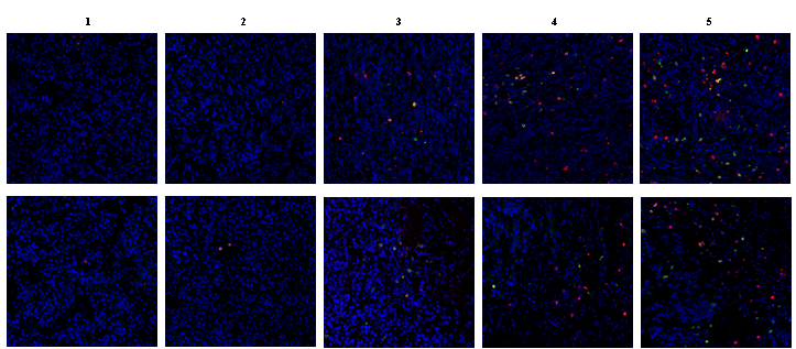
IF analysis of CD3E and CD20 using anti-CD3E antibody (PB9093) and anti-CD20 antibody (M03780-5).
CD3E and CD20 was detected in a paraffin-embedded section of human tonsil tissue. Heat mediated antigen retrieval was performed in EDTA buffer (pH 8.0, epitope retrieval solution). The tissue section was blocked with 10% goat serum. The tissue section was then incubated with 2 μg/mL rabbit anti-CD3E antibody (PB9093) and mouse anti-CD20 antibody (M03780-5) overnight at 4°C. DyLight®550 Conjugated Goat Anti-Rabbit IgG (BA1135), DyLight®488 Conjugated Goat Anti-Mouse IgG (BA1126) was used as secondary antibody at 1:100 dilution and incubated for 30 minutes at 37°C. The section was counterstained with DAPI. Visualize using a fluorescence microscope and filter sets appropriate for the label used.

Click image to see more details
IF analysis of CD3E using anti-CD3E antibody (PB9093).
CD3E was detected in a paraffin-embedded section of mouse 4T1 cell xenograft tumor tissue. Heat mediated antigen retrieval was performed in EDTA buffer (pH 8.0, epitope retrieval solution). The tissue section was blocked with 10% goat serum. The tissue section was then incubated with 1:200 rabbit anti-CD3E Antibody (PB9093) overnight at 4°C. DyLight 550-conjugated Goat Anti-Rabbit was used as secondary antibody incubated for 1 hour at RT. The section was counterstained with DAPI. Visualize using a fluorescence microscope and filter sets appropriate for the label used.

Click image to see more details
IHC analysis of CD3 Epsilon using anti-CD3 Epsilon antibody (PB9093).
CD3 Epsilon was detected in frozen section of rat spleen tissues. The tissue section was blocked with 10% goat serum. The tissue section was then incubated with 1μg/ml rabbit anti-CD3 Epsilon Antibody (PB9093) overnight at 4°C. Biotinylated goat anti-rabbit IgG was used as secondary antibody and incubated for 30 minutes at 37°C. The tissue section was developed using Strepavidin-Biotin-Complex (SABC)(Catalog # SA1022) with DAB as the chromogen.

Click image to see more details
IHC analysis of CD3 Epsilon using anti-CD3 Epsilon antibody (PB9093).
CD3 Epsilon was detected in frozen section of mouse spleen tissues. The tissue section was blocked with 10% goat serum. The tissue section was then incubated with 1μg/ml rabbit anti-CD3 Epsilon Antibody (PB9093) overnight at 4°C. Biotinylated goat anti-rabbit IgG was used as secondary antibody and incubated for 30 minutes at 37°C. The tissue section was developed using Strepavidin-Biotin-Complex (SABC)(Catalog # SA1022) with DAB as the chromogen.

Click image to see more details
IF analysis of CD3E and CD68 using anti-CD3E antibody (PB9093) and anti-CD68 antibody (M00602)
CD3E and CD68 was detected in paraffin-embedded section of human tonsil tissues. Heat mediated antigen retrieval was performed in citrate buffer (pH6, epitope retrieval solution ) for 20 mins. The tissue section was blocked with 10% goat serum. The tissue section was then incubated with 1μg/mL rabbit anti-CD3E Antibody (PB9093) and mouse anti CD68 Antibody(M00602) overnight at 4°C. DyLight®488 Conjugated Goat Anti-Mouse IgG (BA1126) and Biotin conjugated goat anti-rabbit IgG (BA1003) were used as secondary antibody at 1:100 dilution and incubated for 30 minutes at 37°C. The tissue section was developed using Cy3 Conjugated Avidin (BA1037). The section was counterstained with DAPI. Visualize using a fluorescence microscope and filter sets appropriate for the label used.

Click image to see more details
a Schematic diagram of BCP-GNCs-mediated macrophage polarization and DC maturation by releasing IL-4 and DXMS. b IF staining of Arg1 (red) and iNOS (green) of macrophage, CD83 (red) and CD40 (green) of DC and nuclei (blue) after macrophages or DCs seeded on the BCP with 690 nm far-red or 808 nm NIR stimulating the release of PBS (BCP-PBS), 690 nm far-red stimulating the release of IL-4 (BCP-IL4) or 808 nm NIR stimulating the release of DXMS (BCP-DXMS) for 24 h. Scale bar = 5 μm. c, d Semiquantification of positively stained cells in ( b ). n = 5, ** P < 0.01, *** P < 0.001. e Flowchart of time course for irradiating BCP-GNC in vivo. f–h IHC staining of M1, M2 macrophages (iNOS and Arg1), mature DCs (CD40), T cells (CD3), MSCs and osteoblasts (CD146, Runx2) under the BCP-GNCs implant in vivo. Scale bar = 100 μm. Red arrow, positive cells. M, material. i – k Semiquantification of positively stained cells in (E, F, G). n = 5, * P < 0.05, ** P < 0.01, **** P < 0.0001. l H&E and Masson staining of implant area of BCP-Con and BCP-GNCs in vivo after 4 weeks (4 W) (the red dash line shows the new bone formation area; NB, new bone; M, material). m Semiquantification of new bone area in ( k ). n = 5, **** P < 0.0001
Index in PubMed under a CC BY license. PMID: 34138183

Click image to see more details
a Implementation strategy of clodronate on macrophages depletion. b H&E and Masson staining of BCP implant area treating with PBS or clodronate in vivo after implant 4 weeks (the red dash line shows the new bone formation area; NB, new bone; M, material). Scale bar = 100 μm. c IHC staining of M2 macrophages (Arg1) and osteoblasts (Runx2, Col1a1) under the BCP implant treating with PBS or clodronate in vivo. Scale bar = 100 μm. Red arrow, positive cells. M, material. d – g Semiquantification of new bone area and positively stained cells in ( b, c ). n = 5, *** P < 0.001, **** P < 0.0001. h Implementation strategy of diphtheria toxin (DT) on DCs depletion. i H&E and Masson staining of BCP implant area treating with PBS or DT in vivo after implant 4 weeks (the red dash line shows the new bone formation area; NB, new bone; M, material). Scale bar = 100 μm. j IHC staining of T cells (CD3) and osteoblasts (Runx2, Col1a1) under the BCP implant treating with PBS or DT in vivo. Scale bar = 100 μm. Red arrow, positive cells. M, material. k – n Semiquantification of new bone area and positively stained cells in ( i, j ). n = 5, * P < 0.05, ** P < 0.01, *** P < 0.001, **** P < 0.0001
Index in PubMed under a CC BY license. PMID: 34138183

Click image to see more details
OSBP translocates from Golgi to PM via interacting with ORP4L. a Electron micrographs of ORP4L KI and wild-type T-cells, HepG2 and 293 T cells. Scale bar, 100 nm. Black triangles present in dividual data points for n = 20 cells from one or cell line. The central mark indicates the median, the bottom and top edges of the box indicate the interquartile range, and the whiskers represent the maximum and minimum point. One-way ANOVA test with a confidence interval of 95% was used to compute statistics. b Co-immunoprecipitation analysis of ORP4L binding to OSBP in ORP4L KI T-cells with or without anti-CD3 stimulation. c The localization of OSBP in ORP4L KI T-cells. Scale bar, 10 μm. d The localization of OSBP in ORP4L KI and wild-type T-cells with or without anti-CD3 stimulation. Scale bar, 10 μm. e Western blot analysis of OSBP protein levels in PM and Golgi of ORP4L KI or wild-type T-cells. f A schematic representation describing the BiFC technique. g Interactions between ORP4L/pVn-C1 and OSBP/pVc-C1 in ORP4L KI T-cells as determined by BiFC. Scar bar, 10 μm. h The localization of OSBP in ORP4L KI T-cells upon ORP4L knockdown. Scar bar, 10 μm. i Western blot analysis of OSBP protein levels in PM and Golgi of ORP4L KI T-cells upon ORP4L knockdown. j The localization of overexpressed wild-type OSBP or OSBP ( △ ORP4L) in ORP4L KI T-cells. Scar bar, 10 μm. k Western blot analysis of OSBP protein levels in PM and Golgi of ORP4L KI T-cells with or without VAPA knockdown. l Western blot analysis of overexpressed wild-type OSBP or OSBP( △ FFAT) protein levels in the PM and Golgi of ORP4L KI T-cells. Confocal microscopy and blot images are representative of n = 3 biological replicates with similar results. The same experiments were repeated twice in T-cells from two mice. In ( c , d , h and j ), the ration of OSBP fluorescence density from n = 10 cells from one between PM and Golgi are shown below. Source data are provided as a Source Data file.
Index in PubMed under a CC BY license. PMID: 35906240

Click image to see more details
Pathological findings of T-cell leukemia in ORP4L KI mice. a Kaplan–Meier comparative survival analysis of ORP4L KI and WT littermate mice ( n = 20 mice per group, Log-rank test). b White blood cell count on peripheral blood of ORP4L KI and WT littermate mice. Black triangles present in dividual data points for n = 15 (ORP4L KI) and n = 12 (WT) mice. c The percentage of indicated cells in peripheral blood of ORP4L KI and WT littermate mice. Black triangles present in dividual data points for n = 10 mice. d Percentage of CD25 + Foxp3 + or CD25 + CCR4 + cells in peripheral blood of ORP4L KI and WT littermate mice. Black triangles present in dividual data points for n = 8 (ORP4L KI) and n = 6 (WT) mice. e Flow cytometry analysis of Foxp3 and CCR4 expression in ORP4L KI and WT littermate mice. The same experiments were repeated twice in two mice. f Representative blood smears of ORP4L KI and WT littermate mice ( n = 3 mice). Scale bars, 10 μm. g , h Splenomegaly and hepatomegaly in ORP4L KI mice. The right panels illustrate the organ weights. Black triangles present in dividual data points for n = 10 (ORP4L KI) and n = 6 (WT) mice. Scale bars, 1 cm. i Representative H&E-staining of organs in ORP4L KI mice. Immunohistochemical anti-CD3 antibody staining (inserts) of the tissues is shown. Scale bars, 100 μm. j Gross and histological findings of splenomegaly in B-NDG mice after transplantation of spleen cells from ORP4L KI mice. Black triangles present in dividual data points for n = 6 mice. Scale bars, 1 cm. k Representative H&E and anti-CD3 antibody staining of spleen of j . Scale bars, 100 μm. Images of i and k are representative of n = 3 biological replicates with similar results. The same experiments were repeated twice in two mice. In each box plot, the central mark indicates the median, the bottom and top edges of the box indicate the interquartile range, and the whiskers represent the maximum and minimum point. Two-tailed unpaired t -test with a confidence interval of 95% was used to compute statistics. P -values are indicated in the figures. Source data are provided as a Source Data file.
Index in PubMed under a CC BY license. PMID: 35906240
Specific Publications For Anti-CD3 epsilon/CD3E Antibody Picoband® (PB9093)
Loading publications
Recommended Resources
Here are featured tools and databases that you might find useful.
- Boster's Pathways Library
- Protein Databases
- Bioscience Research Protocol Resources
- Data Processing & Analysis Software
- Photo Editing Software
- Scientific Literature Resources
- Research Paper Management Tools
- Molecular Biology Software
- Primer Design Tools
- Bioinformatics Tools
- Phylogenetic Tree Analysis
Customer Reviews
Have you used Anti-CD3 epsilon/CD3E Antibody Picoband®?
Share your experimental results or join a short interview to earn up to $1,000 in product credits or other rewards.
1 Reviews For Anti-CD3 epsilon/CD3E Antibody Picoband®
Accurate localization and good imaging performance.
Excellent
| SKU | PB9093 |
|---|---|
| Application | Immunofluorenscence |
| Sample | Mouse 4T1 cell xenograft tumor |
| Sample Processing Description | Fixed in 4% paraformaldehyde for 48 hours, followed by paraffin embedding and sectioning. |
| Primary Incubation | 1:200, overnight at 4 ℃ |
| Blocking Agent | Goat serum |
| Secondary Antibody | DyLight 550-conjugated goat anti-rabbit antibody. |
| Secondary Incubation | Incubate at room temperature for 1 hour |
| Detection | Laser confocal microscopy |
| Results Summary | The delivery time for antibodies is impressively fast — I usually receive the products within a week, which saves me a lot of valuable time. Both the pre-sales and after-sales support are efficient and reliable. Since my background is in chemistry, I often consulted Boster’s technical specialists about various details of biological experiments. They were always patient and thorough in their explanations, helping me avoid many detours during my experiments. Most importantly, these antibodies offer excellent value for money — they have strong binding performance, produce clear imaging results, and ensure a high experiment success rate, which has provided a solid foundation for my research. |
Yanhua Li,Shandong Normal University
Verified customer
Submitted 2025-10-16
Customer Q&As
Have a question?
Find answers in Q&As, reviews.
Can't find your answer?
Submit your question
6 Customer Q&As for Anti-CD3 epsilon/CD3E Antibody Picoband®
Question
Our lab used your anti-CD3 epsilon/CD3E antibody for WB on blood in a previous experiment. I am using mouse, and We want to use the antibody for IHC-P next. We are interested in examining blood as well as thymus in our next experiment. Could you please give me some suggestion on which antibody would work the best for IHC-P?
Verified Customer
Verified customer
Asked: 2020-04-09
Answer
I looked at the website and datasheets of our anti-CD3 epsilon/CD3E antibody and it seems that PB9093 has been tested on mouse in both WB and IHC-P. Thus PB9093 should work for your application. Our Boster satisfaction guarantee will cover this product for IHC-P in mouse even if the specific tissue type has not been validated. We do have a comprehensive range of products for IHC-P detection and you can check out our website bosterbio.com to find out more information about them.
Boster Scientific Support
Answered: 2020-04-09
Question
We are currently using anti-CD3 epsilon/CD3E antibody PB9093 for mouse tissue, and we are content with the IHC-F results. The species of reactivity given in the datasheet says chicken, human, mouse, rat. Is it likely that the antibody can work on monkey tissues as well?
Verified Customer
Verified customer
Asked: 2020-03-13
Answer
The anti-CD3 epsilon/CD3E antibody (PB9093) has not been tested for cross reactivity specifically with monkey tissues, but there is a good chance of cross reactivity. We have an innovator award program that if you test this antibody and show it works in monkey you can get your next antibody for free. Please contact me if I can help you with anything.
Boster Scientific Support
Answered: 2020-03-13
Question
We were content with the WB result of your anti-CD3 epsilon/CD3E antibody. However we have seen positive staining in leukemic t-cell cell membrane using this antibody. Is that expected? Could you tell me where is CD3E supposed to be expressed?
Verified Customer
Verified customer
Asked: 2020-03-11
Answer
From what I have seen in literature, leukemic t-cell does express CD3E. Generally CD3E expresses in cell membrane. Regarding which tissues have CD3E expression, here are a few articles citing expression in various tissues:
Blood, Pubmed ID: 3267235, 15489334
Leukemic T-cell, Pubmed ID: 15144186, 19690332
Thymus, Pubmed ID: 14702039
Boster Scientific Support
Answered: 2020-03-11
Question
We are currently using anti-CD3 epsilon/CD3E antibody PB9093 for mouse tissue, and we are content with the IHC-F results. The species of reactivity given in the datasheet says chicken, human, mouse, rat. Is it likely that the antibody can work on monkey tissues as well?
Verified Customer
Verified customer
Asked: 2019-06-28
Answer
The anti-CD3 epsilon/CD3E antibody (PB9093) has not been tested for cross reactivity specifically with monkey tissues, but there is a good chance of cross reactivity. We have an innovator award program that if you test this antibody and show it works in monkey you can get your next antibody for free. Please contact me if I can help you with anything.
Boster Scientific Support
Answered: 2019-06-28
Question
We have observed staining in chicken leukocyte. What should we do? Is anti-CD3 epsilon/CD3E antibody supposed to stain leukocyte positively?
Verified Customer
Verified customer
Asked: 2018-02-12
Answer
From what I have seen in literature leukocyte does express CD3E. From what I have seen in Uniprot.org, CD3E is expressed in leukocyte, blood, thymus, leukemic t-cell, among other tissues. Regarding which tissues have CD3E expression, here are a few articles citing expression in various tissues:
Blood, Pubmed ID: 3267235, 15489334
Leukemic T-cell, Pubmed ID: 15144186, 19690332
Thymus, Pubmed ID: 14702039
Boster Scientific Support
Answered: 2018-02-12
Question
I would like using your anti-CD3 epsilon/CD3E antibody for pd-1 signaling studies. Has this antibody been tested with western blotting on jurkat whole cell lysate? We would like to see some validation images before ordering.
J. Zhao
Verified customer
Asked: 2014-09-01
Answer
Thank you for your inquiry. This PB9093 anti-CD3 epsilon/CD3E antibody is tested on jurkat whole cell lysate, cem whole cell lysate. It is guaranteed to work for IF, IHC-P, IHC-F, ICC, WB in chicken, human, mouse, rat. Our Boster guarantee will cover your intended experiment even if the sample type has not been be directly tested.
Boster Scientific Support
Answered: 2014-09-01
