This website uses cookies to ensure you get the best experience on our website.
- Table of Contents
16 Q&As
16 Q&As
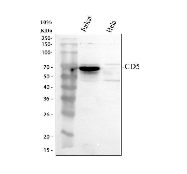
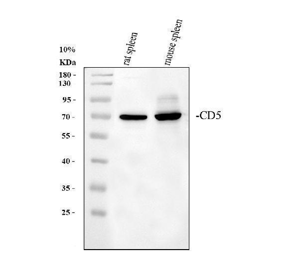
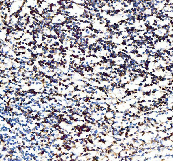
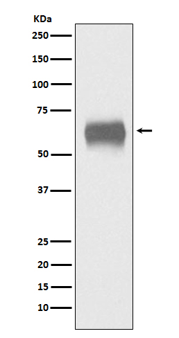
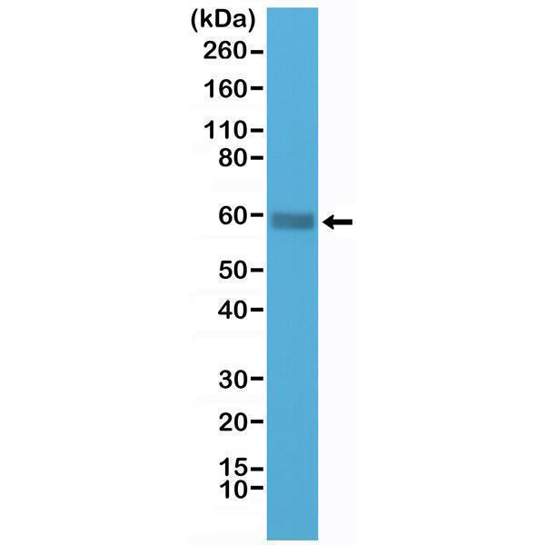
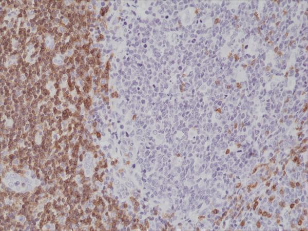
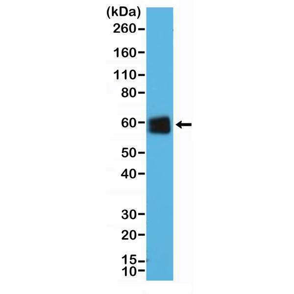
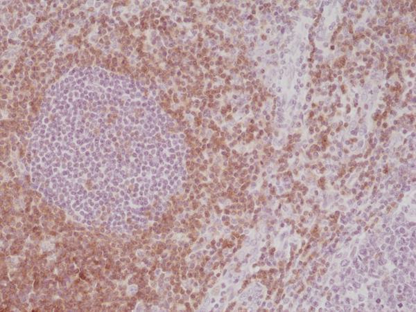
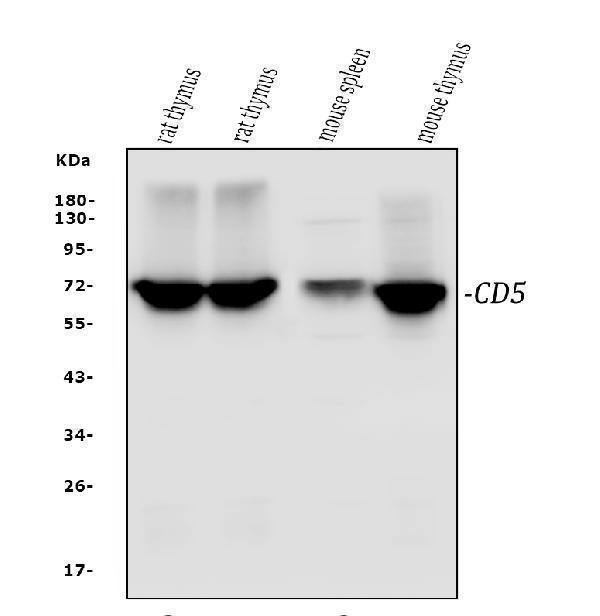
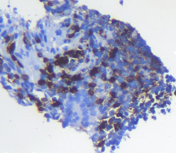
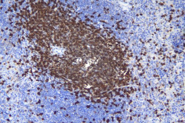
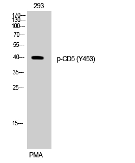
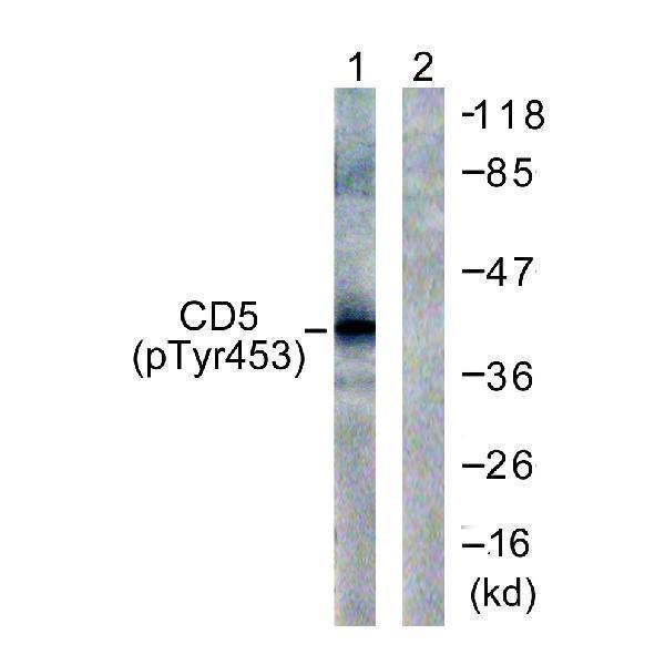
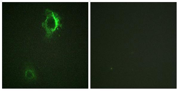
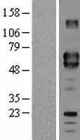
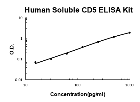
Facts about T-cell surface glycoprotein CD5.
Human | |
---|---|
Gene Name: | CD5 |
Uniprot: | P06127 |
Entrez: | 921 |
Belongs to: |
---|
No superfamily |
CD5 antigen (p56-62); CD5 antigen; CD5 molecule; CD5; LEU1T-cell surface glycoprotein CD5; Lymphocyte antigen T1/Leu-1; T1
Mass (kDA):
54.578 kDA
Human | |
---|---|
Location: | 11q12.2 |
Sequence: | 11; NC_000011.10 (61093963..61127852) |
Cell membrane; Single-pass type I membrane protein.
If you are interested in learning more about Boster Bio and the CD5 Marker, read on! We have included information on the CD5 marker, how to order a sample, and ordering options. In addition, we have included a few sample applications. You may also want to check out the sample application guide to see which flow procedures are best for your research. These guides will also include important information on how to optimize your experiments.
The Boster Bio: Best Uses Of The Human CD5 Marker provides a step-by-step immunohistochemistry protocol for the use of Human CD5 markers. Each step is clearly illustrated and includes recommended reagents and protocols. The guide also provides valuable immunostaining tips and troubleshooting information. You'll find detailed instructions for sample preparation and antibody-based immunohistochemistry.
CD5 is a lymphoid marker and is one of the earliest acquired during T-cell ontogeny. Its expression is elevated with the presence of the cell surface marker CD3.1, but disappears from the surfaces of some natural killer cells and lymphoid-committed progenitors. In addition to its role as a lymphoid marker, CD5 is also expressed on some B cells.
Although CD5 is normally expressed on T-cells, a subset of B-cells expresses it, which makes it an excellent tool for detecting non-Hodgkin B-cell lymphoproliferative disorders. However, the single immunohistochemical stain makes it difficult to compare different antigens, and CD5 positivity is largely amplified in B-cells.
In addition to its antigen-binding properties, CD5 also regulates the production of IL-10. These properties may explain the prolonged survival of human B-cell subsets. Human B-1 B cells are less well-characterized than murine B cells. They are found in adult and cord blood, and are enriched with cells that spontaneously produce polyreactive antibodies. Chronic lymphocytic leukemia is another disease that involves the overproduction of CD5+ cells.
The expression of CD5 is highly predictive of poor outcome in primary DLBCL. It did not influence survival, however, when compared with those with non-CD5-T-cell markers. It was Suzuki et al. who were the first to evaluate T-cell markers other than CD5 in DLBCL. Their results were encouraging, but additional studies are needed to confirm their findings. For now, CD5 remains a useful tool for prognostication in DLBCL.
The CD5 marker is a cell surface protein expressed on IgM secreting B cells. It is located on the gene 11q12.2. In humans, it is expressed more frequently on T cells than B cells. It was used for T-cell differentiation until monoclonal antibodies were developed that target CD3. The expression level of CD5 correlates with the strength of interaction of T cells with self-peptides.
If you're interested in a specific test for CD5 expression, then you should consider ordering it. Though CD5 expression was not significant in the test, it is an important factor in TCR signaling. The CD5 receptor binds to the CBL and UBASH3 molecules. They may interact in a negative feedback loop to inhibit the activity of Src kinase recruited to the synapse.
PMID: 3093892 by Jones N.H., et al. Isolation of complementary DNA clones encoding the human lymphocyte glycoprotein T1/Leu-1.
PMID: 8740779 by Calvo J., et al. Evolutionarily conserved transcription regulatory elements within the 5'-flanking region of the human CD5 gene.