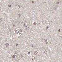
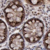
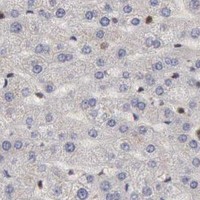
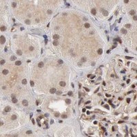
CDKN2A / Cyclin-dependent kinase inhibitor 2A · IHC design guide

Design Immunohistochemistry for CDKN2A

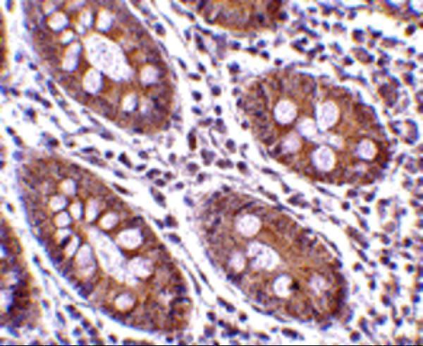
Plan chromogenic CDKN2A IHC-P using the catalog antibody’s human colon example (datasheet A00016-1 IHC-P). Expect mainly nuclear tissue staining, with high staining in colon glandular cells, and account for the reported splice/transcript discrepancy (HPA tissue IHC).

Evidence assembled Oct 2026 · For research use; verify linked source records and product datasheet before use
Immunohistochemistry protocol sheet for CDKN2A (IHC for CDKN2A): expected localisation Mainly nuclear in tissue (HPA tissue IHC); cytoplasmic location also annotated (UniProt), antibody A00016-1, validated IHC image, and IHC protocol steps
Printable CDKN2A IHC protocol sheet — expected localisation Mainly nuclear in tissue (HPA tissue IHC); cytoplasmic location also annotated (UniProt), antibody A00016-1, controls and protocol steps. Open the full CDKN2A IHC guide →

CDKN2A Immunohistochemistry Experimental Design Guide

Expected localisation, validated protocols, controls and antibodies — the at-a-glance facts below, then the full design guide.

Must know before staining
Expected localisation Mainly nuclear in tissue (HPA tissue IHC); cytoplasmic location also annotated (UniProt)
Staining pattern Mainly nuclear staining; high in colon glandular cells (HPA tissue IHC)
Antigen retrieval Citrate pH 6 HIER, heat-mediated (datasheet A00016-1)
Positive control ⓘ Appendix+4 more · see all
Negative control ⓘ Adipose tissue+4 more · see all
Important caveats
Reasons your staining may differ from the expected pattern.
Fixation Formaldehyde-fixed sections were used (datasheet A00016-1 IHC-P) (selected-SKU IHC image A00016-1)
Caveat Splice/transcript discrepancy complicates interpretation (HPA tissue IHC)
Regulation Staining intensity varies by cell type and tissue (HPA tissue IHC)
Isoform / epitope Six isoforms, including ARF; verify which isoform the epitope detects (UniProt)
Section 1

Recommended CDKN2A IHC & IF Protocols

Start with the catalog antibody’s IHC-P protocol (datasheet A00016-1), then compare the published melanoma protocol (PMC2948432: methods).

Recommended immunohistochemistry (IHC-P) protocol parameters
SampleFormaldehyde-fixed, paraffin-embedded Human Colon tissue (datasheet A00016-1)
FixationImage formalin-fixed; duration unreported (datasheet A00016-1); verify before use.
Sectioning4–5 µm sections on charged slides (standard)
DeparaffinisationXylene, graded ethanol series to water (standard)
Antigen retrievalHeat retrieval: Citrate pH 6 (datasheet A00016-1); 20 min, 95–100 °C (standard)
Peroxidase block3% H2O2, 10 min, room temperature (standard)
Blocking10% serum (datasheet A00016-1)
Primary antibodyRabbit anti-CDKN2A, 5 - 10 μg/mL (datasheet A00016-1)
Primary incubationOvernight at 4 °C (standard)
DetectionHRP-polymer secondary, DAB chromogen 5–10 min (standard)
CounterstainHematoxylin, blue, dehydrate and mount (standard)
Expected resultCDKN2A-positive staining in glandular cells of appendix (HPA tissue IHC: High). HPA tissue profile: Mainly nuclear expression in most tissues. No signal in the no-primary control.
💡Decision noteTry citrate pH 6 heat retrieval first (datasheet A00016-1); the melanoma excerpt does not specify retrieval (PMC2948432: methods).
Section 2

What Is the Expected CDKN2A Staining Pattern?

CDKN2A is a soluble protein with no transmembrane segment and is annotated in the nucleus and cytoplasm (UniProt P42771 topology and subcellular location). In tissue IHC, expect mainly nuclear staining, particularly in appendix and colon glandular cells and testicular elongated or late spermatids (HPA: mainly nuclear; High in these cells). HPA rates its tissue profile Enhanced but reports only medium agreement with RNA data and a splice or transcript discrepancy (HPA: reliability description).

What am I looking at on my slide?
Distinct nuclear staining in appendix or colon glandular cells, or in testicular elongated or late spermatids.This fits the reported high-expression cell types and mainly nuclear tissue pattern (HPA: High in these cells; mainly nuclear). Judge staining within the identified cells, since a tissue label alone does not identify which cells carry the signal (HPA: cell-level tissue IHC).
Strong staining confined to cell borders, lumens, or extracellular material, with no convincing nuclear signal.Treat this as a compartment mismatch requiring investigation: CDKN2A has no transmembrane segment, and HPA tissue staining is mainly nuclear (UniProt P42771 topology; HPA: tissue profile). Check morphology and the detection controls before assigning this signal to CDKN2A (general IHC practice).
Staining in cells reported as negative, such as skeletal-muscle myocytes or adipocytes.The pattern conflicts with those HPA observations and may reflect antibody cross-reactivity or endogenous detection activity (HPA: Not detected in skeletal-muscle myocytes and adipocytes; general IHC practice). A negative tissue observation is a comparator, not proof that every stained cell is false positive (HPA: tissue IHC profile).
Diffuse color across cells and surrounding tissue, without distinct nuclear boundaries.This does not reproduce the mainly nuclear profile (HPA: tissue IHC profile). Check background from the detection system and nonspecific binding using an appropriate negative control; score cells only where morphology and nuclear signal can be distinguished (general IHC practice).
No nuclear signal in appendix or colon glandular cells on an otherwise interpretable section.This disagrees with the reported high staining in those cell types (HPA: High in appendix and colon glandular cells). Review the sampled cells, antibody validation and run controls before calling the specimen negative; HPA also reports medium staining–RNA consistency (HPA: reliability description; general IHC practice).
💡Expected CDKN2A appearanceCall a positive result when identifiable appendix or colon glandular cells, or testicular elongated or late spermatids, show clear, predominantly nuclear staining consistent with HPA’s High ratings; isolated border, luminal, or diffuse staining without that pattern is suspect (HPA: tissue IHC profile; general IHC practice).
How each factor affects the staining
Compartment and topologyUse predominantly nuclear tissue staining as the primary interpretation anchor (HPA: mainly nuclear). Cytoplasmic CDKN2A is annotated, so a cytoplasmic component is possible, but a membrane-only pattern lacks support from its topology (UniProt P42771 subcellular location and topology).
Cell type and tissue selectionAppendix and colon glandular cells and testicular elongated or late spermatids provide reported high-staining comparators (HPA: High). Skeletal-muscle myocytes and adipocytes are reported as not detected; assess the named cells rather than all cells in either section (HPA: tissue IHC).
Transcript and isoform interpretationSix isoforms are listed, including ARF and smARF; isoform 3 is described as pancreas-specific (UniProt P42771 isoforms and tissue specificity). HPA flags a splice or transcript discrepancy, so tissue RNA abundance alone should not settle an unexpected IHC result (HPA: reliability description).
Antibody validationHPA lists CAB000445 as IHC Enhanced and CAB000093 and CAB018232 as IHC Approved (HPA: antibodies). These are antibody-specific statuses; apply a listed status only to the matching antibody, and use that antibody’s own validation when interpreting discordant staining (HPA: antibodies; general IHC practice).
IF/ICC Q: What location should be expected?A: Mainly nucleoplasm; HPA also lists cytosol and primary-cilium locations as uncertain (HPA: subcellular ICC-IF). Those ICC-IF observations support an imaging interpretation, but do not change the mainly nuclear tissue-IHC expectation (HPA: subcellular ICC-IF; tissue IHC).
Why is my staining missing, weak or wrong?
SituationLikely causeNext action
Expected glandular-cell nuclei are blank in appendix or colon.The sampled cells may be absent, or the IHC run may have failed; those cells are reported High (HPA: appendix and colon glandular cells; general IHC practice).Confirm glandular-cell morphology, inspect the run’s positive control, and review the IHC validation of the antibody used before interpreting absence as biological (general IHC practice; HPA: antibody statuses).
Only edge, luminal, or extracellular deposits appear.That distribution does not fit the mainly nuclear tissue pattern or the lack of a transmembrane segment (HPA: tissue profile; UniProt P42771 topology).Check the negative detection control and compare deposits with intact nuclei on the counterstained section; withhold a CDKN2A-positive call until a cellular pattern is resolved (general IHC practice).
A reported negative cell type stains strongly.Cross-reactivity or endogenous detection activity is possible, especially if the signal occurs in HPA-negative adipocytes or skeletal-muscle myocytes (HPA: Not detected in those cells; general IHC practice).Repeat with appropriate negative detection controls and confirm the exact stained cell type; compare antibody-specific IHC validation before treating the result as an exception (general IHC practice; HPA: antibody statuses).
The whole section looks uniformly colored.Diffuse detection background or nonspecific binding may obscure the reported mainly nuclear pattern (general IHC practice; HPA: tissue profile).Inspect a negative control, review blocking and detection steps, and evaluate whether counterstained nuclei remain distinguishable; do not score diffuse color as cellular positivity (general IHC practice).
Brain staining seems to conflict with the UniProt tissue note.UniProt says CDKN2A is not detected in brain, while HPA reports Medium staining in caudate glial cells and cerebellar molecular-layer cells (UniProt P42771 tissue specificity; HPA: caudate and cerebellum).Record the specific region, cell type, compartment and antibody, then report the source disagreement; HPA flags medium RNA–staining consistency and a transcript discrepancy (HPA: reliability description; general IHC practice).
ICC-IF shows a ciliary or cytosolic signal alongside nuclear staining.HPA supports nucleoplasmic localization but marks cytosol and primary-cilium locations uncertain (HPA: subcellular ICC-IF).Interpret those additional locations cautiously against the supported nucleoplasmic signal and imaging controls; use the tissue-IHC pattern when scoring paraffin sections (HPA: subcellular ICC-IF; tissue IHC; general IF practice).

Sample controls for CDKN2A IHC & IF

🧪Run colon first: its glandular cells should stain (HPA: High in colon glandular cells; selected-SKU caption: colon IHC). Use skeletal muscle as the negative tissue; its myocytes should lack staining (HPA: Not detected in skeletal-muscle myocytes). On the colon slide, treat non-glandular cells as an internal background comparison, not a validated negative population (HPA: positive call is specific to glandular cells).
Positive control tissue: Appendix (Glandular cells, HPA High)
Negative control tissue: Adipose tissue (HPA Not detected)
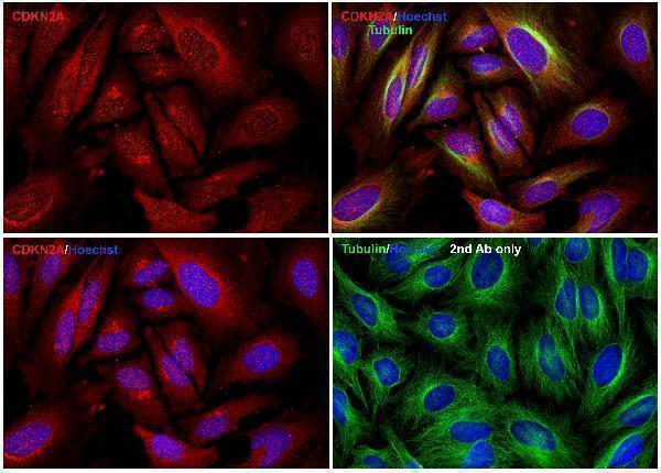
ICC-IF cell lines (HPA subcellular resource): HPA ICC-IF images show CDKN2A in A-431, U-251MG, U2OS, HEK293, SiHa, ASC52telo, RPTEC/TERT1, hTERT-RPE1 (serum starved), with annotated localisation: Nucleoplasm (supported) (HPA subcellular).
Technical controls: Include a no-primary, secondary-only slide; a nonimmune IgG control matched to the primary antibody’s host species, isotype, and clonality where known; and identically processed CDKN2A-knockout material as a biological negative. Quench endogenous peroxidase and check residual background in colon before interpreting HRP staining (selected-SKU caption: HRP secondary and colon tissue).
⚠️Feasibility: A target-specific fixation window or fixation effect is unreported; the caption describes formaldehyde fixation but gives no duration, while its structured fixative field says the fixative is unreported (selected-SKU caption; caption metadata: fixative not stated). The paraffin-section example uses heat retrieval in citrate buffer at pH 6, so verify retrieval in the local workflow; the supplied evidence does not establish whether frozen sections or IF are easier (selected-SKU caption: citrate heat retrieval; HPA: ICC-IF images). In colon, inspect gland lumina and tissue edges for background against the no-primary slide before scoring glandular staining (HPA: High in colon glandular cells).

HPA tissue IHC evidence for CDKN2A

Comprehensive Human Protein Atlas IHC scoring per tissue (reliability: Enhanced — Medium consistency between antibody staining and RNA expression data. Caution, Splice and/or transcript discrepancy exists.). Rows are taken directly from the HPA tissue chart — click any row's HPA link to view the source.

Positive expression · recommended positive controls

TissueCell typeLevelEvidenceSource
Appendix Glandular cells High Protein (IHC) HPA →
Colon Glandular cells High Protein (IHC) HPA →
Testis Elongated or late spermatids High Protein (IHC) HPA →
Adrenal gland Glandular cells Medium Protein (IHC) HPA →
Breast Glandular cells Medium Protein (IHC) HPA →

Undetected expression · recommended negative controls

TissueCell typeLevelEvidenceSource
Adipose tissue Adipocytes Not detected Protein (IHC) HPA →
Fallopian tube Ciliated cells (cell body) Not detected Protein (IHC) HPA →
Skeletal muscle Myocytes Not detected Protein (IHC) HPA →
Soft tissue Fibroblasts Not detected Protein (IHC) HPA →
Vagina Squamous epithelial cells Not detected Protein (IHC) HPA →
Section 3

Advanced CDKN2A IHC Tips

Troubleshoot CDKN2A staining in paraffin section IHC by checking retrieval, cellular localisation, antibody specificity and scoring before interpreting biological differences.

What should I change when CDKN2A staining is weak after antigen retrieval?
Start with heat-mediated retrieval in citrate buffer at pH 6 (datasheet A00016-1). The selected paraffin-section example used this retrieval with 10 µg/ml primary antibody incubated overnight at 4°C (A00016-1 tissue-IHC caption). Compare a freshly processed positive control, such as colon glandular cells, across retrieval runs before changing antibody concentration (HPA: High in colon glandular cells; standard IHC practice). If staining remains weak, test an alternative retrieval buffer as a controlled fallback, recording tissue preservation and background alongside nuclear signal (standard IHC practice; HPA: mainly nuclear tissue expression).
How can I assess whether fixation is causing weak CDKN2A IHC?
Target-specific sensitivity of CDKN2A staining to fixation duration or fixative type is unknown from the supplied evidence; assess it experimentally rather than assigning a cause from staining alone. Compare similarly handled sections with documented fixation and processing histories, keeping citrate pH 6 retrieval and the 10 µg/ml primary concentration consistent (datasheet A00016-1; A00016-1 tissue-IHC caption; standard IHC practice). Include a positive control and examine morphology, nuclear staining and background in each run (HPA: mainly nuclear tissue expression; standard IHC practice). A change confined to poorly preserved areas supports a processing problem, but does not establish CDKN2A-specific fixation sensitivity (standard IHC practice).
Should CDKN2A staining be nuclear, cytoplasmic or both?
Score nuclear staining as the principal tissue-IHC pattern because HPA reports mainly nuclear expression across tissues (HPA tissue IHC). Nucleoplasmic localisation is supported in cell imaging, whereas cytosolic localisation is uncertain; UniProt also lists nucleus and cytoplasm (HPA subcellular; UniProt P42771 subcellular). Record cytoplasmic signal separately from nuclear signal, especially if it varies with retrieval or detection conditions (standard IHC practice). If staining is exclusively diffuse cytoplasmic, compare an expected positive tissue compartment and a no-primary control before treating it as CDKN2A expression (HPA: High in colon glandular cells; standard IHC practice).
Can this antibody distinguish p16 from other CDKN2A isoforms?
Do not assign staining to p16 alone until the catalog antibody’s immunogen or epitope is mapped against the relevant CDKN2A products (UniProt P42771: 6 isoforms; standard IHC interpretation). The record includes p16-related isoforms and tumor suppressor ARF, so an unspecified epitope leaves isoform recognition unresolved (UniProt P42771 isoforms). Request an epitope map or compare validated isoform-selective reagents under matched IHC conditions before interpreting a staining difference as isoform-specific (standard IHC practice). HPA flags splice or transcript discrepancy in its tissue assessment, which further limits conclusions drawn from RNA and this stain alone (HPA tissue IHC: Enhanced reliability description).
How should I adapt CDKN2A localisation checks for multiplex IF?
For an IF/ICC cross-check, pair CDKN2A with a marker identifying the expected cell population and compare nuclear signal within that marked population (HPA: High in colon glandular cells; HPA: mainly nuclear tissue expression; standard IF practice). Choose a fluorophore channel with low measured tissue autofluorescence, and inspect single-stain controls before interpreting overlap (standard IF practice). Because CDKN2A has no transmembrane segment and its supported location is nucleoplasmic, use permeabilisation appropriate for access to an intracellular nuclear epitope, guided by the antibody’s epitope information (UniProt P42771 topology; HPA subcellular; standard IF practice). Report uncertain cytosolic or ciliary signals separately from supported nucleoplasmic staining (HPA subcellular).
How do I reduce diffuse or misleading CDKN2A chromogenic background?
First compare the stain with a no-primary control and inspect whether colour follows tissue edges, damaged areas or endogenous peroxidase-rich regions (standard IHC practice). Include a peroxidase block for HRP detection, then adjust blocking, washing or chromogen development one variable at a time (standard IHC practice). The selected example used 10% serum for 1 hour at room temperature, an HRP secondary at 1:250 and hematoxylin counterstain (A00016-1 tissue-IHC caption). Preserve discernible nuclear staining in an expected positive compartment while reducing diffuse colour; HPA reports High staining in colon glandular cells and mainly nuclear tissue expression (HPA tissue IHC).
What is a defensible way to score CDKN2A IHC across samples? ⚠ ANSWER MARKED FOR VERIFICATION
Define the cell population and nuclear positivity threshold before scoring, because HPA describes mainly nuclear tissue expression (HPA tissue IHC; standard IHC practice). Report the percentage of positive nuclei and, where intensity is reproducible, an H-score based on nuclear intensity and its percentage; for spatial questions, report positive-cell density per mm² of evaluated tissue (standard IHC practice). Normalise counts to the number of evaluable cells or the measured viable tissue area in the same compartment, excluding folds and necrosis by a prespecified rule (standard IHC practice). Record cytoplasmic staining separately and use matched retrieval and detection runs for comparisons (UniProt P42771 subcellular; standard IHC practice).
How can I distinguish true CDKN2A positivity from IHC artefact?
A convincing result has cellular staining in the expected compartment and passes control checks: HPA reports mainly nuclear tissue expression and High staining in colon glandular cells (HPA tissue IHC; standard IHC practice). Question signal restricted to tissue edges, folds or necrotic areas, and compare it with a no-primary control for nonspecific detection (standard IHC practice). Investigate chromogen in inappropriate cells or diffuse colour after HRP detection with a peroxidase-block control before calling those cells positive (standard IHC practice). Interpret disagreements with RNA cautiously because HPA rates tissue staining Enhanced while noting medium staining–RNA consistency and splice or transcript discrepancy (HPA tissue IHC).
Boster reagents

Best CDKN2A / Cyclin-dependent kinase inhibitor 2A IHC Antibodies

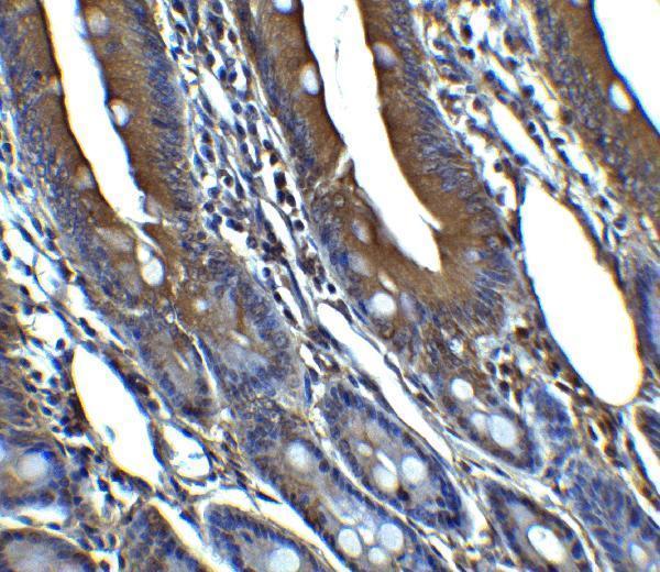
The catalog shows IHC data from human, rat and mouse tissues (catalog IHC captions), plus IF images from human cells and human and rat colon (A00016-1 IF captions).

Real IHC data Immunohistochemistry Validation of CDKN2A in Human Colon Tissue Immunohistochemical analysis of paraffin-embedded Human Colon Tissue using anti- CDKN2A antibody (A00016-1) at 10 μg/ml. Tissue was fixed with formaldehyde and blocked with 10% serum for 1 h at RT; antigen retrieval was by heat mediation with a citrate buffer (pH6). Samples were incubated with primary antibody overnight at 4˚C. A goat anti-rabbit IgG H&L (HRP) at 1/250 was used as secondary. Counter stained with Hematoxylin.
Anti-CDKN2A/CDKN2A/p16INK4a Antibody
Cat # A00016-1
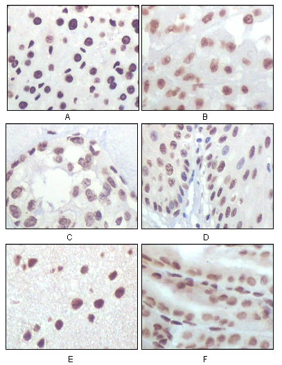
Real IHC data Immunohistochemistry analysis of paraffin-embedded rat liver tissue (A) , human brain tumor (B) , breast cancer (C) , esophageal epithelium tissue (D) , mouse brain tissue (E) and stomach tisue (F) , showing nuclear localization with DAB staining using p16 Mon
Anti-p16 CDKN2A Monoclonal Antibody
Cat # A00016-3
Real IHC data Immunohistochemical analysis of paraffin-embedded human gastric, using p16 INK Antibody.
Anti-p16 INK Monoclonal Antibody
Cat # M00016

A00016-1 has paraffin-section IHC images from human and rat colon and IF images from human cells and human and rat colon (A00016-1 image captions). A00016-3 has paraffin-section IHC images from rat, human and mouse tissues; M00016 has a paraffin-section IHC image from human gastric tissue (A00016-3 and M00016 IHC captions).

Which to pick: For tissue IHC, start with A00016-1: its human and rat colon captions report paraffin sections fixed with formaldehyde and stained at 10 μg/mL and 5 μg/mL, respectively (A00016-1 IHC captions). For broader cross-species tissue examples, consider mouse monoclonal A00016-3; its caption shows rat, human and mouse paraffin sections but does not report a fixative (A00016-3 catalog entry and IHC caption). For human IF/ICC, rabbit monoclonal M00016 lists both applications, while A00016-1 offers specific IF images of human cells and colon; M00016’s gastric IHC caption reports paraffin sections but no fixative (M00016 catalog entry and IHC caption; A00016-1 IF captions).

Each figure is that product's own IHC / IF validation image from its datasheet.

References

  1. UniProt Consortium. UniProt entry P42771 (CDN2A_HUMAN, Cyclin-dependent kinase inhibitor 2A).
  2. Human Protein Atlas. CDKN2A tissue IHC expression (reliability: Enhanced).
  3. Human Protein Atlas. CDKN2A subcellular location (ICC-IF): Mainly localized to the nucleoplasm. In addition localized to the cytosol, primary cilium, primary cilium tip and primary cilium transition zone..
  4. Human Protein Atlas. CDKN2A antibody validation summary (4 antibodies).
  5. p16 Immunohistochemistry as a Screening Tool for Homozygous CDKN2A Deletions in CNS Tumors. The American journal of surgical pathology 2024 — PMC10723769.
  6. Deletion at chromosome arm 9p in relation to BRAF/NRAS mutations and prognostic significance for primary melanoma. Genes, chromosomes & cancer 2010 — PMC2948432.
  7. p16 Immunohistochemical Expression as a Surrogate Assessment of CDKN2A Alteration in Gliomas Leading to Prognostic Significances. Cancers 2023 — PMC10000516.
  8. Deletions of CDKN2A and MTAP Detected by Copy-Number Variation Array Are Associated with Loss of p16 and MTAP Protein in Pleural Mesothelioma. Cancers 2023 — PMC10605896.
  9. PubMed PMID:8259215 — UniProt-cited evidence.
  10. PubMed PMID:10445844 — UniProt-cited evidence.
  11. PubMed PMID:12228235 — UniProt-cited evidence.