Click image to see more details
-

-
-
-
-
+8
Product Info Summary
| SKU: | A00016-1 |
|---|---|
| Size: | 0.1 mg |
| Reactive Species: | Human, Mouse, Rat |
| Host: | Rabbit |
| Application: | ELISA, IF, IHC-P, WB |
Customers Who Bought This Also Bought

Product info
Product Name
Anti-CDKN2A Antibody
SKU/Catalog Number
A00016-1
Size
0.1 mg
Form
Liquid
Description
Boster Bio Anti-CDKN2A Antibody (Catalog # A00016-1). Tested in ELISA, WB, IHC-P, IF applications. This antibody reacts with Human, Mouse, Rat.
Storage & Handling
Antibody can be stored at 4°C up to one year. Antibodies should not be exposed to prolonged high temperatures.
Cite This Product
Anti-CDKN2A Antibody (Boster Biological Technology, Pleasanton CA, USA, Catalog # A00016-1)
Host
Rabbit
Contents
CDKN2A Antibody is supplied in PBS containing 0.02% sodium azide.
Clonality
Polyclonal
Isotype
IgG
Immunogen
Anti-CDKN2A antibody was raised against a peptide corresponding to 18 amino acids near the amino terminus of human CDKN2A. The immunogen is located within the first 50 amino acids of CDKN2A.
Reactive Species
A00016-1 is reactive to CDKN2A in Human, Mouse, Rat
Observed Molecular Weight
68 kDa
Calculated molecular weight
16.5 kDa
Background of CDKN2A
The CDKN2A locus gives rise to 2 distinct transcripts from different promoters. The transcripts have been designated p16(INK4A) and p14(ARF). This chromosomal region undergoes a number of inversions, translocations, heterozygous deletions, and homozygous deletions in a variety of malignant cell lines including those from glioma, non-small cell lung cancer, leukemia, and melanoma. Deletion of the region containing CDKN2A is found in more than half of all melanoma cell lines. Conversely, transfection of CDKN2A suppressed the growth of two independent mesothelioma cell lines, suggesting that inactivation of the CDKN2 gene is an essential step in the etiology of malignant mesotheliomas. CDKN2A induces a G1 cell cycle arrest by inhibiting the phosphorylation of the Rb protein by the cyclin-dependent kinases CDK4 and CDK6. CDKN2A is expressed as at least three distinct isoforms.
Antibody Validation
Boster validates all antibodies on WB, IHC, ICC, Immunofluorescence, and ELISA with known positive control and negative samples to ensure specificity and high affinity, including thorough antibody incubations.
Application & Images
Applications
A00016-1 is guaranteed for ELISA, IF, IHC-P, WB Boster Guarantee
Assay Dilutions Recommendation
The recommendations below provide a starting point for assay optimization. The actual working concentration varies and should be decided by the user.
WB: 1 - 4 μg/mL; IHC-P: 5 - 10 μg/mL; IF: 20 μg/mL.
Antibody validated: Western Blot in human and mouse samples; Immunohistochemistry in human and rat samples; Immunofluorescence in human and rat samples. All other applications and species not yet tested. Optimal dilutions for each application should be determined by the researcher.
Validation Images & Assay Conditions
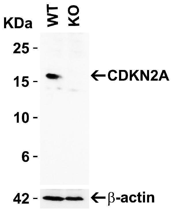
Click image to see more details
KO Validation of CDKN2A in 293 Cells
Loading: 10 μg of 293 WT cell lysates or CDKN2A KO cell lysates. Antibodies: CDKN2A, A00016-1 (2 μg/mL) and beta-actin, 3779 (1 μg/mL), 1 h incubation at RT in 5% NFDM/TBST.
Secondary: Goat Anti-Rabbit IgG HRP conjugate at 1:10000 dilution.

Click image to see more details
Immunofluorescence Validation of CDKN2A In HeLa Cells
Immunofluorescent analysis of methanol-fixed HeLa cells labeling CDKN2A with A00016-1 at 20 μg/mL, followed by goat anti-rabbit IgG secondary antibody at 1/1000 dilution (red) and Hoechst staining (blue). Alpha tubulin was stained with anti-alpha tubulin antibody following by goat anti-mouse IgG secondary antibody (green). Images were captured with confocal microscopy.

Click image to see more details
Independent Antibody Validation (IAV) via Protein Expression Profile in Cell Lines
Loading: 15 μg of lysates per lane.
Antibodies: CDKN2A A00016-1 (4 μg/mL), CDKN2A 24-023 (4 μg/mL), and beta-actin (1 μg/mL), overnight incubation at 4˚C in 5% NFDM/TBST.
Secondary: Goat anti-rabbit IgG HRP conjugate at 1:10000 dilution.

Click image to see more details
Recombinant Protein Test
Loading: 15 μg of lysates per lane.
Antibodies: CDKN2A A00016-1 (2 μg/mL), overnight incubation at 4˚C in 5% NFDM/TBST.
Secondary: Goat anti-rabbit IgG HRP conjugate at 1:10000 dilution.
Lane 1: Human prostate (benign hyperplasia)
Lane 2: Human CDKN2A recombinant protein (arrow: monomer and dimer)

Click image to see more details
Western Blot Validation in Human Cell Lines
Loading: 15 μg of lysates per lane.
Antibodies: CDKN2A, A00016-1 (2 μg/mL), 1h incubation at RT in 5% NFDM/TBST.
Secondary: Goat anti-rabbit IgG HRP conjugate at 1:10000 dilution.

Click image to see more details
Western Blot Validation in Human Normal and Cancer Tissue
Loading: 15 μg of lysates per lane.
Antibodies CDKN2A A00016-1 (2 μg/mL), overnight incubation at 4˚C in 5% NFDM/TBST.
Secondary: Goat anti-rabbit IgG HRP conjugate at 1:10000 dilution.
Lane 1: Human breast
Lane 2: Human lung tumor
Lane 3: Human colon cancer

Click image to see more details
Western Blot Validation in Mouse Colon Tissue
Loading: 15 μg of lysates per lane.
Antibodies: CDKN2A A00016-1 (A: 1 μg/mL, B: 2 μg/mL), 1h incubation at RT in 5% NFDM/TBST.
Secondary: Goat anti-rabbit IgG HRP conjugate at 1:10000 dilution.

Click image to see more details
Immunofluorescence Validation of CDKN2A in Human A431 Cells
Immunofluorescent analysis of 4% paraformaldehyde-fixed A431 cells labeling CDKN2A with A00016-1 at 20 μg/mL, followed by goat anti-rabbit IgG secondary antibody at 1/500 dilution (green) and DAPI staining (blue).

Click image to see more details
Immunofluorescence Validation of CDKN2A in Human Colon Tissue
Immunofluorescent analysis of 4% paraformaldehyde-fixed human colon tissue labeling CDKN2A with A00016-1 at 20 μg/mL, followed by goat anti-rabbit IgG secondary antibody at 1/500 dilution (green) and DAPI staining (blue).

Click image to see more details
Immunofluorescence Validation of CDKN2A in Rat Colon Tissue
Immunofluorescent analysis of 4% paraformaldehyde-fixed Rat Colon Tissue labeling CDKN2A with A00016-1 at 20 μg/mL, followed by goat anti-rabbit IgG secondary antibody at 1/500 dilution (red) and DAPI staining (blue).

Click image to see more details
Immunohistochemistry Validation of CDKN2A in Human Colon Tissue
Immunohistochemical analysis of paraffin-embedded Human Colon Tissue using anti- CDKN2A antibody (A00016-1) at 10 μg/ml. Tissue was fixed with formaldehyde and blocked with 10% serum for 1 h at RT; antigen retrieval was by heat mediation with a citrate buffer (pH6). Samples were incubated with primary antibody overnight at 4˚C. A goat anti-rabbit IgG H&L (HRP) at 1/250 was used as secondary. Counter stained with Hematoxylin.

Click image to see more details
Immunohistochemistry Validation of CDKN2A in Rat Colon Tissue
Immunohistochemical analysis of paraffin-embedded Rat Colon Tissue using anti- CDKN2A antibody (A00016-1) at 5 μg/ml. Tissue was fixed with formaldehyde and blocked with 10% serum for 1 h at RT; antigen retrieval was by heat mediation with a citrate buffer (pH6). Samples were incubated with primary antibody overnight at 4˚C. A goat anti-rabbit IgG H&L (HRP) at 1/250 was used as secondary. Counter stained with Hematoxylin.
Specific Publications For Anti-CDKN2A Antibody (A00016-1)
Loading publications
Recommended Resources
Here are featured tools and databases that you might find useful.
- Boster's Pathways Library
- Protein Databases
- Bioscience Research Protocol Resources
- Data Processing & Analysis Software
- Photo Editing Software
- Scientific Literature Resources
- Research Paper Management Tools
- Molecular Biology Software
- Primer Design Tools
- Bioinformatics Tools
- Phylogenetic Tree Analysis
Customer Reviews
Have you used Anti-CDKN2A Antibody?
Share your experimental results or join a short interview to earn up to $1,000 in product credits or other rewards.
0 Reviews For Anti-CDKN2A Antibody
Customer Q&As
Have a question?
Find answers in Q&As, reviews.
Can't find your answer?
Submit your question
