CDX2 / Homeobox protein CDX-2 · IHC design guide

Design Immunohistochemistry for CDX2

Plan chromogenic CDX2 IHC on paraffin sections around the nuclear staining reported in intestinal endocrine cells (HPA tissue IHC). This guide covers the catalog antibody’s 1:50 starting dilution and helps you choose intestinal controls and score nuclear staining (datasheet M00877-1; HPA tissue IHC).

Evidence assembled Oct 2026 · For research use; verify linked source records and product datasheet before use
Immunohistochemistry protocol sheet for CDX2 (IHC for CDX2): expected localisation Nuclear staining in intestinal tissue (HPA tissue IHC), antibody M00877-1, validated IHC image, and IHC protocol steps
Printable CDX2 IHC protocol sheet — expected localisation Nuclear staining in intestinal tissue (HPA tissue IHC), antibody M00877-1, controls and protocol steps. Open the full CDX2 IHC guide →

CDX2 Immunohistochemistry Experimental Design Guide

Expected localisation, validated protocols, controls and antibodies — the at-a-glance facts below, then the full design guide.

Must know before staining
Expected localisation Nuclear staining in intestinal tissue (HPA tissue IHC)
Staining pattern Intestinal endocrine-cell nuclei stain (HPA tissue IHC)
Antigen retrieval EDTA pH 8.0 HIER, heat-mediated (datasheet M00877-1)
Positive control ⓘ Appendix+4 more · see all
Negative control ⓘ Adipose tissue+4 more · see all
Important caveats
Reasons your staining may differ from the expected pattern.
Fixation Keep fixation consistent across paraffin sections (standard IHC practice; not target-specific)
Caveat Small-intestine endocrine staining is medium (HPA tissue IHC)
Regulation Intestine-enriched RNA (HPA tissue RNA)
Isoform / epitope No isoforms; a single 1–313 chain (UniProt)
Section 1

Recommended CDX2 IHC & IF Protocols

The catalog antibody’s IHC-P protocol is followed by published CDX2 staining protocols for colon tumors, odontogenic tumors, and malignant peripheral nerve sheath tumors (PMC11119288; PMC13535953; PMC9097546).

Recommended immunohistochemistry (IHC-P) protocol parameters
SampleParaffin-embedded human colon tissue; fixative not specified (datasheet M00877-1)
FixationImage fixative and duration unreported (datasheet M00877-1); verify before use.
Sectioning4–5 µm sections on charged slides (standard)
DeparaffinisationXylene, graded ethanol series to water (standard)
Antigen retrievalHeat retrieval: EDTA pH 8.0 (datasheet M00877-1); 20 min, 95–100 °C (standard)
Peroxidase block3% H2O2, 10 min, room temperature (standard)
Blocking10% goat serum (datasheet M00877-1)
Primary antibodyRabbit monoclonal (clone AFA-3) anti-CDX2, 1:50 (datasheet M00877-1)
Primary incubationOvernight at 4 °C (datasheet M00877-1)
DetectionHRP-conjugated secondary, DAB chromogen (datasheet M00877-1)
CounterstainHematoxylin, blue, dehydrate and mount (standard)
Expected resultCDX2-positive staining in endocrine cells of appendix (HPA tissue IHC: High). HPA tissue profile: Nuclear expression in intestinal tract. No signal in the no-primary control.
💡Decision noteStart with heat-mediated EDTA retrieval at pH 8.0 (datasheet: M00877-1); optimize against the published retrieval condition when reproducing PMC9097546.
Section 2

What Is the Expected CDX2 Staining Pattern?

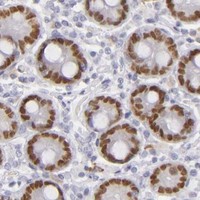
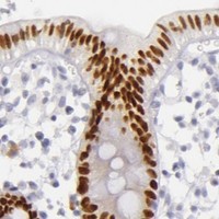
CDX2 is a nuclear transcription factor without a transmembrane segment (UniProt Q99626: nucleus, topology). In paraffin-section IHC, expect nuclear staining in intestinal-tract endocrine cells: high in appendix, colon, duodenum and rectum, and medium in small intestine (HPA: tissue IHC). HPA rates the tissue pattern Enhanced because antibody staining is highly consistent with RNA expression (HPA: tissue IHC reliability).

What am I looking at on my slide?
Distinct nuclear signal in intestinal-tract endocrine cells, with little surrounding haze.This matches the reported compartment and cell population (HPA: nuclear expression in intestinal tract; endocrine-cell staining). Compare intensity within the named tissue and cell type: HPA reports High in appendix, colon, duodenum and rectum, but Medium in small intestine (HPA: tissue IHC).
Cytoplasmic or vesicular signal dominates the paraffin-section IHC slide.Recheck localisation before scoring this as the expected IHC result (HPA: tissue IHC nuclear profile). HPA also reports cytosol and vesicles as additional ICC-IF locations, so those observations alone do not prove an artefact across both applications (HPA: subcellular ICC-IF).
Signal appears in respiratory epithelial cells of bronchus or glial cells of caudate.Those named cell populations were Not detected in the HPA tissue survey (HPA: bronchus respiratory epithelial cells; caudate glial cells). Review staining specificity and endogenous detection activity as possible explanations (general IHC practice); do not infer that every cell in either tissue must be negative.
A broad, poorly defined deposit obscures nuclei and tissue boundaries.Diffuse background cannot establish CDX2-positive cells or nuclear localisation (general IHC interpretation). Check a no-primary control and the detection and blocking steps to locate nonspecific signal (general IHC practice); interpret the slide only after discrete nuclei can be distinguished.
No nuclear signal is visible in a designated positive intestinal section.First confirm that the examined section contains the relevant endocrine cells (HPA: intestinal-tract endocrine-cell staining). An absent internal positive signal makes a negative test result difficult to interpret; review section integrity, retrieval and detection controls (general IHC practice).
💡Expected CDX2 appearanceCall the IHC result positive when intestinal-tract endocrine cells show distinct nuclear chromogen signal, with intensity assessed against the relevant HPA tissue level; diffuse deposits or signal confined to unexpected cells do not establish that pattern (HPA: tissue IHC; general IHC interpretation).
How each factor affects the staining
Which tissue and cells provide a reference?Use HPA's named endocrine-cell populations, not an assumption that all intestinal cells stain equally: appendix, colon, duodenum and rectum are High; small intestine is Medium (HPA: tissue IHC).
How strong is the IHC pattern evidence?The tissue profile has Enhanced reliability from staining–RNA consistency, and CAB002221 has an Enhanced IHC validation status (HPA: tissue IHC reliability; HPA: antibody validation). Neither designation substitutes for controls on the current slide (general IHC practice).
IF/ICC Q: Should cytosolic or vesicular signal count as the expected result here?A: This page's IHC call is nuclear (HPA: tissue IHC). HPA's ICC-IF record places CDX2 mainly in nucleoplasm and additionally in cytosol and vesicles; interpret IF/ICC with its own guide (HPA: subcellular ICC-IF).
Does the protein record suggest a membrane or shed-product pattern?UniProt reports no transmembrane segment or signal peptide and describes one chain spanning residues 1–313 (UniProt Q99626: topology, processing). Those annotations support assessing nuclear localisation; they do not establish how fixation affects staining.
Why is my staining missing, weak or wrong?
SituationLikely causeNext action
The positive intestinal control has no nuclear stain.The section may lack the scored endocrine-cell population (HPA: tissue IHC), or a staining step may have failed (general IHC practice).Verify the cells on the section, then check retrieval, primary-antibody and detection controls; repeat with a verified positive section if needed (general IHC practice).
Nuclear staining is faint in small intestine.HPA reports Medium staining there, compared with High in the listed appendix, colon, duodenum and rectum endocrine cells (HPA: tissue IHC).Judge signal against the matching tissue and cell type; compare a verified positive control before changing staining conditions (HPA: tissue IHC; general IHC practice).
Bronchus respiratory epithelial cells show a strong signal.HPA reports this cell population as Not detected; cross-reactivity or endogenous detection activity is possible (HPA: bronchus; general IHC practice).Check a no-primary control and inspect whether the deposit is nuclear and cell-specific; confirm unexpected staining with an independently validated antibody (general IHC practice).
Most visible signal lies outside nuclei.The expected paraffin-section IHC profile is nuclear, although HPA reports additional cytosol and vesicle locations in ICC-IF (HPA: tissue IHC; subcellular ICC-IF).Compare signal with the nuclear counterstain and positive control, then reassess specificity before assigning an IHC positive call (general IHC practice).
Diffuse chromogen covers both cells and background.Nonspecific detection or inadequate blocking can obscure cell boundaries (general IHC practice).Review the no-primary control, blocking and detection steps; adjust the general workflow until discrete nuclei can be assessed (general IHC practice).
A test section appears negative while its positive control stains correctly.The test may lack detectable signal in the cells examined; HPA levels apply to specific tissue and cell populations (HPA: tissue IHC).Confirm the test section's cell identity and morphology, score only interpretable nuclei, and report the tested population rather than extending the result to the whole tissue (general IHC practice).

Sample controls for CDX2 IHC & IF

🧪Run colon first: its endocrine cells show High CDX2 staining (HPA: colon endocrine cells, High), and the selected antibody has a paraffin-section colon IHC caption (catalog antibody: M00877-1 caption). Use adipose tissue as a negative tissue (HPA: adipocytes, Not detected); on the colon slide, compare stained nuclei with neighboring cells that lack nuclear signal, without assuming every other cell type is CDX2-negative (UniProt Q99626: nucleus; HPA: colon endocrine cells, High).
Positive control tissue: Appendix (Endocrine cells, HPA High)
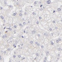
Negative control tissue: Adipose tissue (HPA Not detected)
ICC-IF cell lines (HPA subcellular resource): HPA ICC-IF images show CDX2 in CACO-2, HEK293, U2OS, JURKAT, NIH 3T3, with annotated localisation: Nucleoplasm (approved) (HPA subcellular).
Technical controls: Include a no-primary, secondary-only control and an isotype control matched to the primary antibody’s host species, immunoglobulin class, and monoclonal or polyclonal format; use a CDX2 knockout specimen, if available, as a biological specificity control (standard IHC practice). For chromogenic colon IHC, quench endogenous peroxidase and block endogenous biotin if using avidin–biotin detection; for IF, assess colon autofluorescence with an unstained control (standard IHC/IF practice).
⚠️Feasibility: A target-specific fixation window and fixation effect are unreported in the supplied evidence, and the selected M00877-1 paraffin-section colon caption does not state a fixative (catalog antibody: M00877-1 caption). Retrieval dependence is unreported, so optimize antigen retrieval for paraffin IHC empirically (standard IHC practice); the supplied evidence does not establish whether frozen sections or IF are easier, although HPA provides ICC-IF images for CDX2 (HPA: subcellular images). In colon, luminal material can complicate background assessment, so score nuclear staining in the identified cells against the negative controls (standard IHC practice; UniProt Q99626: nucleus).

HPA tissue IHC evidence for CDX2

Comprehensive Human Protein Atlas IHC scoring per tissue (reliability: Enhanced — High consistency between antibody staining and RNA expression data.). Rows are taken directly from the HPA tissue chart — click any row's HPA link to view the source.

Positive expression · recommended positive controls

TissueCell typeLevelEvidenceSource
Appendix Endocrine cells High Protein (IHC) HPA →
Colon Endocrine cells High Protein (IHC) HPA →
Duodenum Endocrine cells High Protein (IHC) HPA →
Rectum Endocrine cells High Protein (IHC) HPA →
Small intestine Endocrine cells Medium Protein (IHC) HPA →

Undetected expression · recommended negative controls

TissueCell typeLevelEvidenceSource
Adipose tissue Adipocytes Not detected Protein (IHC) HPA →
Adrenal gland Glandular cells Not detected Protein (IHC) HPA →
Bone marrow Hematopoietic cells Not detected Protein (IHC) HPA →
Breast Adipocytes Not detected Protein (IHC) HPA →
Bronchus Respiratory epithelial cells Not detected Protein (IHC) HPA →
Section 3

Advanced CDX2 IHC Tips

CDX2 IHC is read primarily as nuclear staining in intestinal tissue (UniProt Q99626; HPA tissue IHC). Use compartment and cell context when troubleshooting.

What should I change when CDX2 nuclear staining is weak in paraffin sections?
Start with heat-mediated EDTA at pH 8.0 for antigen retrieval (datasheet M00877-1). Keep section thickness, heating time, cooling, antibody concentration and detection constant while comparing retrieval conditions on adjacent sections (standard IHC practice). Include human colon as a positive tissue control because the selected antibody has a paraffin-section image there and CDX2 has an intestinal nuclear profile (caption M00877-1; HPA tissue IHC). If staining remains weak, test a different retrieval buffer on a separate section as a documented fallback, checking whether nuclear signal improves without increased background (standard IHC practice).
Could fixation explain weak CDX2 staining in my paraffin sections?
Target-specific fixation sensitivity is unknown from the supplied evidence; the selected paraffin-section caption does not state a fixative (caption M00877-1). Record the actual fixative, fixation duration and processing history for each specimen before changing the assay (standard IHC practice). Compare similarly processed positive-control sections in the same staining run, then vary retrieval or antibody concentration one factor at a time to locate the source of signal loss (standard IHC practice). Do not infer a CDX2-specific fixation effect from its nuclear location, phosphorylation sites or tissue distribution; those observations do not test fixation sensitivity (UniProt Q99626; HPA tissue IHC).
How should I assess CDX2 staining outside the nucleus?
Score crisp nuclear staining as the primary IHC signal because CDX2 is annotated as nuclear and tissue IHC shows an intestinal nuclear profile (UniProt Q99626; HPA tissue IHC). HPA subcellular imaging also reports nucleoplasmic staining with additional cytosolic and vesicular localisation, so record reproducible extranuclear signal separately rather than folding it into nuclear positivity (HPA subcellular). Compare the signal with morphology, a matched positive section and an appropriate reagent control before assigning biological meaning (standard IHC practice). If diffuse cytoplasmic color rises as nuclear contrast falls, review retrieval strength, antibody concentration and chromogen development for background (standard IHC practice).
Could an isoform or inaccessible epitope cause discordant CDX2 IHC results?
The supplied record lists 0 isoforms and a single CDX2 chain spanning residues 1–313, so it does not support an isoform-specific explanation (UniProt Q99626). The record does not identify this antibody’s epitope, and the two listed phosphoserines at residues 60 and 283 do not establish phospho-dependent recognition (UniProt Q99626; datasheet M00877-1). Check the antibody documentation for its immunogen or mapped epitope before making an epitope-based claim (standard IHC practice). For discordant paraffin sections, first compare retrieval, processing, nuclear morphology and control staining across runs (standard IHC practice).
How can I adapt the CDX2 readout for multiplex immunofluorescence?
Treat IF as a separate assay and confirm that the chosen antibody and fixation conditions work in that format before interpreting a multiplex result (standard IF practice). Pair CDX2 with a validated marker for the expected intestinal cell population, then assess colocalisation by cell and nucleus rather than by overlapping tissue regions (HPA tissue IHC; standard IF practice). Choose spectrally separated fluorophores after checking tissue autofluorescence in an unstained section, placing the weaker signal in the cleaner channel (standard IF practice). CDX2 is a nuclear protein without a transmembrane segment, so optimise permeabilisation for nuclear epitope access after fixation and check that nuclear morphology remains intact (UniProt Q99626; standard IF practice).
What causes widespread brown signal despite poor CDX2 nuclear contrast?
First compare a no-primary control with the stained section to separate detection-system color from antibody-dependent staining (standard IHC practice). Use an appropriate peroxidase block before chromogenic detection and inspect whether DAB development has obscured cellular boundaries; these are general IHC workflow steps, not CDX2-specific requirements (standard IHC practice). Then titrate the primary antibody and assess blocking and washing while holding retrieval and development constant (standard IHC practice). Judge any residual signal against the expected intestinal nuclear pattern, since broad staining of cytoplasm or unrelated structures is insufficient evidence of CDX2 positivity (HPA tissue IHC; UniProt Q99626).
Which measurement best captures variable CDX2 nuclear staining? ⚠ ANSWER MARKED FOR VERIFICATION
Define the tissue compartment and eligible cell population before scoring, using nuclear staining as the CDX2-positive event (UniProt Q99626; HPA tissue IHC). Report the percentage of positive nuclei and, when intensity matters, an H-score calculated from the percentages at each prespecified intensity grade (standard IHC practice). Normalise counts to the number of evaluable nuclei in the same compartment; use positive nuclei per mm² only when area-based density answers the question (standard IHC practice). Exclude folds, necrosis and poorly preserved regions consistently, and apply identical thresholds and imaging conditions across specimens (standard IHC practice).
How do I distinguish genuine CDX2 positivity from staining artefact?
Give greatest weight to well-preserved nuclei in an appropriate intestinal cellular context, consistent with the reported nuclear intestinal profile (HPA tissue IHC; UniProt Q99626). Human colon is an available paraffin-section reference for the selected antibody, but its caption does not identify the fixative (caption M00877-1). Treat isolated cytoplasmic color, staining confined to section edges or necrotic areas, and color persisting in a no-primary control as reasons to investigate artefact before calling a cell positive (standard IHC practice). Check endogenous peroxidase blocking and DAB development when unexpected brown deposits appear, then repeat scoring on intact regions (standard IHC practice).
Boster reagents

Best CDX2 / Homeobox protein CDX-2 IHC Antibodies

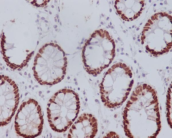
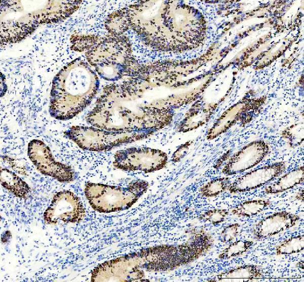
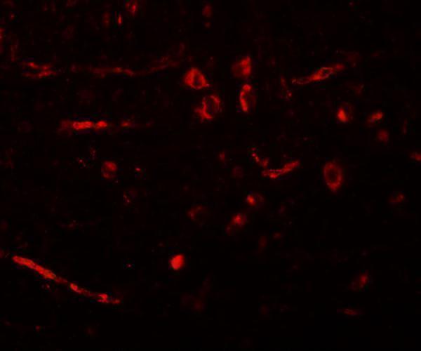
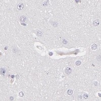
Both anti-CDX2 antibodies have IHC images: human and mouse colon for M00877-1, and rat brain for A00877-1; A00877-1 also has a rat-brain IF image (catalog image captions).

Real IHC data Immunohistochemical analysis of paraffin-embedded human colon, using CDX2 Antibody.
Anti-CDX2 Rabbit Monoclonal Antibody
Cat # M00877-1
Real IHC data Immunohistochemistry of CDX2 in rat brain tissue with CDX2 antibody at 5 μg/mL.
Anti-Homeobox protein CDX-2 CDX2 Antibody
Cat # A00877-1

M00877-1 lists IHC and ICC/IF, with IHC images of paraffin-embedded human and mouse colon (catalog applications and IHC captions: M00877-1). A00877-1 lists IHC-P and IF, with IHC and IF images of rat brain (catalog applications and image captions: A00877-1).

Which to pick: For paraffin-section IHC, choose M00877-1 when human or mouse colon image evidence is useful; its captions describe EDTA retrieval at pH 8.0, and the fixative is unreported (catalog IHC captions: M00877-1). For ICC, choose monoclonal M00877-1, which lists ICC/IF; for tissue IF with image evidence, choose A00877-1, whose IF image shows rat brain (catalog clone and applications: M00877-1; catalog IF caption: A00877-1). Both list human, mouse and rat reactivity for cross-species planning; the pictured samples are limited to human and mouse colon for M00877-1 and rat brain for A00877-1 (catalog reactivity and image captions).

Each figure is that product's own IHC / IF validation image from its datasheet.

References

  1. UniProt Consortium. UniProt entry Q99626 (CDX2_HUMAN, Homeobox protein CDX-2).
  2. Human Protein Atlas. CDX2 tissue IHC expression (reliability: Enhanced).
  3. Human Protein Atlas. CDX2 subcellular location (ICC-IF): Mainly localized to the nucleoplasm. In addition localized to vesicles and cytosol..
  4. Human Protein Atlas. CDX2 antibody validation summary (3 antibodies).
  5. Evaluation of the Immunohistochemical Scoring System of CDX2 Expression as a Prognostic Biomarker in Colon Cancer. Diagnostics (Basel, Switzerland) 2024 — PMC11119288.
  6. Special AT-rich sequence binding protein 2 (SATB2), β-catenin, and CD10 immunohistochemistry provides insights into the histopathologic features and cellular differentiation of adenomatoid odontogenic tumor. Journal of dental sciences 2026 — PMC13535953.
  7. CDX2 expression in malignant peripheral nerve sheath tumour: a potential diagnostic pitfall associated with PRC2 inactivation. Histopathology 2022 — PMC9097546.
  8. Diagnostic and Prognostic Roles of CDX2 Immunohistochemical Expression in Colorectal Cancers. Diagnostics (Basel, Switzerland) 2022 — PMC8947721.
  9. PubMed PMID:9459001 — UniProt-cited evidence.
  10. PubMed PMID:9036867 — UniProt-cited evidence.
  11. PubMed PMID:11161380 — UniProt-cited evidence.