Click image to see more details
-

-
-
-
-
+2
Product Info Summary
| SKU: | M00877-1 |
|---|---|
| Size: | 100 μl |
| Reactive Species: | Human, Mouse, Rat |
| Host: | Rabbit |
| Application: | Flow Cytometry, IF, IHC, ICC, WB |
Customers Who Bought This Also Bought

Product info
Product Name
Anti-CDX2 Rabbit Monoclonal Antibody
SKU/Catalog Number
M00877-1
BM4014 is an alternative SKU for this antibody, used in previous lots.
Size
100 μl
Form
Liquid
Description
Boster Bio Anti-CDX2 Rabbit Monoclonal Antibody catalog # M00877-1. Tested in WB, IHC, ICC/IF, Flow Cytometry applications. This antibody reacts with Human, Mouse, Rat.
Storage & Handling
Store at -20°C for one year. For short term storage and frequent use, store at 4°C for up to one month. Avoid repeated freeze-thaw cycles.
Cite This Product
Anti-CDX2 Rabbit Monoclonal Antibody (Boster Biological Technology, Pleasanton CA, USA, Catalog # M00877-1)
Host
Rabbit
Contents
Rabbit IgG in stabilizing components, phosphate buffered saline, pH 7.4, 150mM NaCl, 0.02% sodium azide and 50% glycerol.
*This antibody is supplied in a stabilized formulation.
Compatibility with conjugation reactions depends on the chemistry of the conjugation method used.
For conjugation methods that are not compatible with the stabilizing components present in this formulation, a carrier-free antibody format is required.
Clonality
Monoclonal
Clone Number
AFA-3
Isotype
Rabbit IgG
Immunogen
A synthesized peptide derived from human CDX2
Reactive Species
M00877-1 is reactive to CDX2 in Human, Mouse, Rat
Observed Molecular Weight
37 kDa
Calculated molecular weight
33.5 kDa
Antibody Validation
Boster validates all antibodies on WB, IHC, ICC, Immunofluorescence, and ELISA with known positive control and negative samples to ensure specificity and high affinity, including thorough antibody incubations.
Application & Images
Applications
M00877-1 is guaranteed for Flow Cytometry, IF, IHC, ICC, WB Boster Guarantee
Assay Dilutions Recommendation
The recommendations below provide a starting point for assay optimization. The actual working concentration varies and should be decided by the user.
WB 1:500-2000
IHC 1:50-200
ICC/IF 1:50-200
FC 1:50
Positive Control
IHC: human colon tissue, human colon cancer tissue, mouse colon tissue
Validation Images & Assay Conditions
Click image to see more details
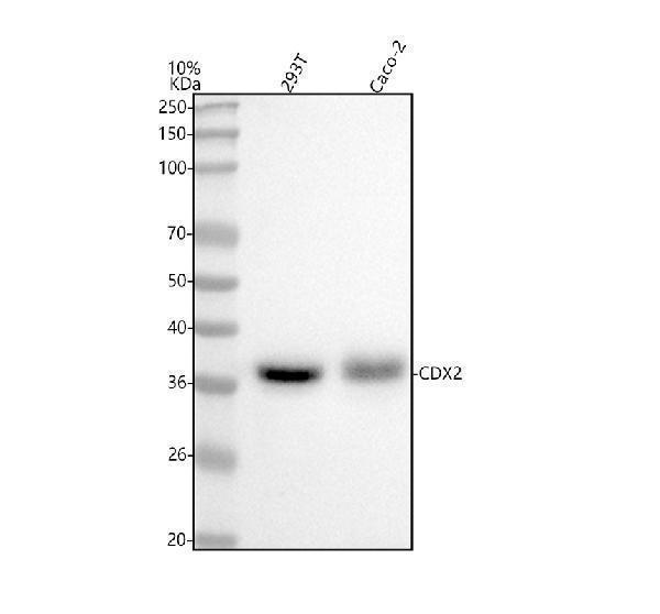
Western blot analysis of CDX2 using anti-CDX2 antibody (M00877-1).
Electrophoresis was performed on a 5-20% SDS-PAGE gel at 70V (Stacking gel) / 90V (Resolving gel) for 2-3 hours. The sample well of each lane was loaded with 30 ug of sample under reducing conditions.
Lane 1: human 293T whole cell lysates,
Lane 2: human Caco-2 whole cell lysates.
After electrophoresis, proteins were transferred to a nitrocellulose membrane at 150 mA for 50-90 minutes. Blocked the membrane with 5% non-fat milk/TBS for 1.5 hour at RT. The membrane was incubated with rabbit anti-CDX2 antigen affinity purified monoclonal antibody (Catalog # M00877-1) at 1:1000 overnight at 4°C, then washed with TBS-0.1%Tween 3 times with 5 minutes each and probed with a goat anti-rabbit IgG-HRP secondary antibody at a dilution of 1:500 for 1.5 hour at RT. The signal is developed using an Enhanced Chemiluminescent detection (ECL) kit (Catalog # EK1002) with Tanon 5200 system. A specific band was detected for CDX2 at approximately 37 kDa. The expected band size for CDX2 is at 34 kDa.

Click image to see more details
All lanes use the Antibody at 1:3K dilution for 1 hour at room temperature.

Click image to see more details
IHC analysis of CDX2 using anti-CDX2 antibody (M00877-1).
CDX2 was detected in a paraffin-embedded section of human colon cancer tissue. Heat mediated antigen retrieval was performed in EDTA buffer (pH 8.0, epitope retrieval solution). The tissue section was blocked with 10% goat serum. The tissue section was then incubated with 1:50 rabbit anti-CDX2 Antibody (M00877-1) overnight at 4°C. Peroxidase Conjugated Goat Anti-rabbit IgG was used as secondary antibody and incubated for 30 minutes at 37°C. The tissue section was developed using HRP Conjugated Rabbit IgG Super Vision Assay Kit (Catalog # SV0002) with DAB as the chromogen.

Click image to see more details
The distribution of Lgr5 + ISCs in the intestinalmucosa and the subcellular localization and relative expression level detection of epithelial function proteins CDX2 and villin in the intestinal mucosa of IBD at 7 days after termination of DSS administration. (A) The Lgr5 + ISCs (brown) in the small intestinal mucosa: (A1) the normal group, the villi and the crypts were arranged compactly, and Lgr5 + ISCs were observed in the crypts; (A2) the DSS group, the villi and the crypts were scattered, with few Lgr5 + ISCs; (A3) the DSS + B. subtilis -fermented milk group, there were more Lgr5 + ISCs in villi and crypts compared with those in the DSS group. (B) The Lgr5 + ISCs (brown) in the colonic mucosa: (B1) the normal group, the glands were arranged compactly, and there were large amounts of Lgr5 + ISCs at the bottom of the glands; (B2) the DSS group: the ulcers were replaced by scars. No Lgr5 + ISCs were observed in the scars; (B3) the DSS + B. subtilis -fermented milk group: the colonic epithelium was integrated, with some regenerated glands. A number of Lgr5 + ISCs were observed at the bottom of the regenerated glands. (C) The CDX2 was localized in the epithelial cellular nuclei (brown) by immunohistochemistry staining in the small intestinal mucosa: (C1) the normal group: the villi and the crypts were arranged compactly, and CDX2 + epithelial cells were observed on the surface of the villi and the crypts; (C2) the DSS group: the villi and the crypts were scattered, and few CDX2 + epithelial cells were observed on the surface of the crypt and the villi; (C3) the DSS + B. subtilis -fermented milk group: more villi and crypts were observed in comparison with the DSS group, and there were more CDX2 + epithelial cells covering the villi and crypts. (D) The CDX2 was localized in the epithelial cellular nuclei (brown) by immunohistochemistry staining in the colonic mucosa: (D1) the normal group: the colonic glands were arranged compactly, and CDX2 + epithelial cells were observed on the surface of the glands; (D2) the DSS group: the glands were scattered, and few CDX2 + epithelial cells were observed in the scar; (D3) the DSS + B. subtilis -fermented milk group: more colonic glands were observed in comparison with the DSS group, and there were more CDX2 + epithelial cells in the glands. (E) The Mucin2 was localized in the cytoplasm of the goblet cells (brown) by immunohistochemistry staining in the small intestinal mucosa: (E1) the normal group, a number of Mucin2 + goblet cells observed in the epithelium; (E2) the DSS group: only few Mucin2 + goblet cells were observed in the remaining villi and crypts; (E3) the DSS + B. subtilis -fermented milk group: more Mucin2 + goblet cells were observed in the recovered mucosa. (F) The Mucin2 was localized in the cytoplasm of the goblet cells (brown) by immunohistochemistry staining in the colonic mucosa: (F1) the normal group, large amounts of Mucin2 + goblet cells were observed in the mucosa; (F2) the DSS group: only few Mucin2 + goblet cells were observed in the scars; (F3) the DSS + B. subtilis -fermented milk group: more Mucin2 + goblet cells were observed in the recovered colonic mucosa. (G,H) Western blotting was applied for detection of the relative expression level of Lgr5, CDX2, and Mucin2 in the samples containing equivalent ileum and colon. The expression level of Lgr5, CDX2, and Mucin2 in the DSS group was significantly lower than that of the normal (control) group. The expression level of Lgr5, CDX2, and Mucin2 in the DSS + B. subtilis -fermented milk (FM) group was significantly higher than that of the DSS group ( n = 5, ** represents p < 0.01).
Index in PubMed under a CC BY license. PMID: 33519783

Click image to see more details
IHC analysis of CDX2 using anti-CDX2 antibody (M00877-1).
CDX2 was detected in a paraffin-embedded section of mouse colon tissue. Heat mediated antigen retrieval was performed in EDTA buffer (pH 8.0, epitope retrieval solution). The tissue section was blocked with 10% goat serum. The tissue section was then incubated with 1:50 rabbit anti-CDX2 Antibody (M00877-1) overnight at 4°C. Peroxidase Conjugated Goat Anti-rabbit IgG was used as secondary antibody and incubated for 30 minutes at 37°C. The tissue section was developed using HRP Conjugated Rabbit IgG Super Vision Assay Kit (Catalog # SV0002) with DAB as the chromogen.

Click image to see more details
Immunohistochemical analysis of paraffin-embedded human colon, using CDX2 Antibody.
Specific Publications For Anti-CDX2 Rabbit Monoclonal Antibody (M00877-1)
Loading publications
Recommended Resources
Here are featured tools and databases that you might find useful.
- Boster's Pathways Library
- Protein Databases
- Bioscience Research Protocol Resources
- Data Processing & Analysis Software
- Photo Editing Software
- Scientific Literature Resources
- Research Paper Management Tools
- Molecular Biology Software
- Primer Design Tools
- Bioinformatics Tools
- Phylogenetic Tree Analysis
Customer Reviews
Have you used Anti-CDX2 Rabbit Monoclonal Antibody?
Share your experimental results or join a short interview to earn up to $1,000 in product credits or other rewards.
0 Reviews For Anti-CDX2 Rabbit Monoclonal Antibody
Customer Q&As
Have a question?
Find answers in Q&As, reviews.
Can't find your answer?
Submit your question
4 Customer Q&As for Anti-CDX2 Rabbit Monoclonal Antibody
Question
Will M00877-1 anti-CDX2 Rabbit Monoclonal antibody work on parafin embedded sections? If so, which fixation method do you recommend we use (PFA, paraformaldehyde, other)?
Verified Customer
Verified customer
Asked: 2020-01-27
Answer
As indicated on the product datasheet, M00877-1 anti-CDX2 Rabbit Monoclonal antibody as been validated on IHC. It is best to use PFA for fixation because it has better tissue penetration ability. PFA needs to be prepared fresh before use. Long term stored PFA turns into formalin, as the PFA molecules congregate and become formalin.
Boster Scientific Support
Answered: 2020-01-27
Question
I was wanting to use your anti-CDX2 Rabbit Monoclonal antibody for IHC for human mucosa of transverse colon on frozen tissues, but I want to know if it has been tested for this particular application. Has this antibody been tested and is this antibody a good choice for human mucosa of transverse colon identification?
Verified Customer
Verified customer
Asked: 2019-12-04
Answer
It shows on the product datasheet, M00877-1 anti-CDX2 Rabbit Monoclonal antibody has been tested for IF, IHC, WB on human tissues. We have an innovator award program that if you test this antibody and show it works in human mucosa of transverse colon in IHC-frozen, you can get your next antibody for free.
Boster Scientific Support
Answered: 2019-12-04
Question
We need to test anti-CDX2 Rabbit Monoclonal antibody M00877-1 on human mucosa of transverse colon for research purposes, then I may be interested in using anti-CDX2 Rabbit Monoclonal antibody M00877-1 for diagnostic purposes as well. Is the antibody suitable for diagnostic purposes?
Verified Customer
Verified customer
Asked: 2019-04-15
Answer
The products we sell, including anti-CDX2 Rabbit Monoclonal antibody M00877-1, are only intended for research use. They would not be suitable for use in diagnostic work. If you have the means to develop a product into diagnostic use, and are interested in collaborating with us and develop our product into an IVD product, please contact us for more discussions.
Boster Scientific Support
Answered: 2019-04-15
Question
We are currently using anti-CDX2 Rabbit Monoclonal antibody M00877-1 for human tissue, and we are happy with the IHC results. The species of reactivity given in the datasheet says human. Is it possible that the antibody can work on bovine tissues as well?
Verified Customer
Verified customer
Asked: 2018-08-23
Answer
The anti-CDX2 Rabbit Monoclonal antibody (M00877-1) has not been validated for cross reactivity specifically with bovine tissues, though there is a good chance of cross reactivity. We have an innovator award program that if you test this antibody and show it works in bovine you can get your next antibody for free. Please contact me if I can help you with anything.
Boster Scientific Support
Answered: 2018-08-23
