CHEK1 / Serine/threonine-protein kinase Chk1 · Western blot design guide

Design a Western Blot for CHEK1

Real validated CHEK1 Western blot protocols, expected-band and isoform facts, troubleshooting for weak or shifted signal, and recommended anti-CHEK1 WB antibodies. Everything you need to plan the experiment before you commit precious samples.

Evidence assembled September 2026 · For research use; verify linked source records and product datasheet before use
Western blot protocol sheet for CHEK1: expected band ~54.4 kDa, hero antibody A01060, catalog values and labelled standard workflow; separate PMC comparisons on the guide
Printable CHEK1 Western blot protocol sheet — expected band ~54.4 kDa, antibody A01060, controls and PMC citations. Open the full CHEK1 WB guide →

CHEK1 Western Blot Experimental Design Guide

Expected bands, validated protocols, controls and antibodies — the at-a-glance facts below, then the full design guide.

Must know before running
Expected band ~54.4 kDa
Observed band ~54 kDa
Gel 5–20% (catalog A01060)
Negative control ⓘ Suggested KO / knockdown lysate
Important caveats
Reasons your observed band may differ from the expected size.
PTM Phosphorylated + Ubl conjugation
Caveat Modification-state controls
Gene-set association MSigDB Hallmark membership
Isoform 3 isoform(s)
Section 1

Real Curated CHEK1 Western Blot Protocols

The A01060 protocol combines labelled catalog values with standard starting conditions. Published comparisons retain their own sample, reagent and detection scope.

Recommended Western blot protocol parameters
Sample / lysatehuman 293T, human K562 (catalog A01060)
Gel %5–20% (catalog A01060)
Load30 ug; reducing conditions (catalog A01060)
Transfera nitrocellulose membrane at 150 mA for 50-90 minutes (catalog A01060)
Membranenitrocellulose membrane (catalog A01060)
Blocking5% non-fat milk/TBS for 1.5 hour at RT (catalog A01060)
Primary antibodyA01060 · 0.5 μg/mL (catalog A01060)
Primary incubationovernight at 4°C (catalog A01060)
Secondary antibodygoat anti-rabbit IgG-HRP, 1:5000 (catalog A01060)
Secondary incubation1.5 hour at RT (catalog A01060)
WashTBS-0.1%Tween 3 times with 5 minutes each (catalog A01060)
DetectionECL (catalog A01060)
Section 2

What Is the Expected CHEK1 Western Blot Band Size?

CHEK1 is predicted at 54.4 kDa and observed at ~54 kDa; the small difference has no established cause, and extra bands require identity controls.

What am I looking at on my blot?
Band at ~54 kDaMatches the empirical CHEK1 band and its 54.4 kDa predicted mass; confirm identity with appropriate controls
Band near 54 kDa in a nuclear fractionConsistent with CHEK1 nuclear localization
Several bands at different positionsIsoforms 1, 2, and 3 are annotated, but distinct migration is unverified
Doublet near 54 kDaCHEK1 has phosphorylation sites, but a phosphorylation-dependent shift is unverified
💡Expected CHEK1 appearanceCHEK1 has a predicted mass of 54.4 kDa and an empirical band at ~54 kDa; confirm band identity with a positive control or CHEK1 depletion.
How each factor affects band size
Predicted CHEK1 mass54.4 kDa predicted; the empirical band is ~54 kDa
Isoform 1Its individual mass and migration are not supplied
Isoform 2Its size relative to isoform 1 is not supplied
Isoform 3Its size relative to the other isoforms is not supplied
Why is my band missing or off?
SituationLikely causeNext action
No band in lysateCHEK1 recovery or detection may be insufficientCheck a positive-control lysate and nuclear protein recovery
Band higher than expectedAn isoform or phosphorylation could affect migration, but no shift is establishedCheck CHEK1 depletion and compare fractionated samples
Band lower than expectedAn alternative isoform or unrelated band is possible; isoform sizes are unknownConfirm the band with CHEK1 depletion and a second antibody
Multiple bandsAnnotated isoforms or phosphorylated forms are possible, but distinct bands are unverifiedUse CHEK1 depletion to identify specific bands
Weak or no signalCHEK1 may be poorly recovered from a nuclear or chromosomal fractionCheck fraction recovery and include a positive-control lysate

Sample controls for CHEK1 Western blot

🧪For positive controls for CHEK1 in Western blot, you can use no HPA-supported tissue or cell line from the supplied evidence.
Positive control: No high/medium HPA tissue identified
Negative control: Suggested KO / knockdown lysate
Loading controls: Run GAPDH, β-actin, and a total-protein stain such as Ponceau alongside the samples.
⚠️Feasibility: With no HPA expression data, a positive tissue control cannot be selected from the supplied evidence.

HPA tissue expression evidence for CHEK1

Comprehensive Human Protein Atlas IHC scoring per tissue. Rows are taken directly from the HPA tissue chart — click any row's HPA link to view the source.

Higher expression tissues · candidate positive controls from IHC

TissueCell typeLevelEvidenceSource
No high/medium HPA tissues identified in the supplied evidence.

Lower expression tissues · IHC evidence, not confirmed WB-negative controls

TissueCell typeLevelEvidenceSource
No lower-expression tissue rows available in the supplied evidence.
Section 3

Advanced CHEK1 Western Blot Tips

Deeper troubleshooting and optimisation questions for CHEK1, answered from its protein features.

How should CHEK1 band migration be interpreted?
Band shift · Use the separately labelled calculated mass and catalog-observed evidence above. A sequence annotation does not establish an observed migration shift. Verify target identity with orthogonal controls.
Could CHEK1 isoforms produce additional bands?
Isoforms · UniProt lists three isoforms. Relative to isoform 1, isoform 2 lacks residues 1–94 and changes residues 95–97 from RIE to MEK; isoform 3 lacks residues 412–445. These sequence differences could affect band size. Check whether the antibody recognizes a region retained in the isoform under study.
Does CHEK1 phosphorylation explain a shifted band?
PTM · Canonical CHEK1 has a predicted mass of 54.4 kDa, consistent with the observed band near 54 kDa. UniProt lists nine phosphoserines, but their presence alone does not establish a visible shift or explain an apparent mass difference.

UniProt identifies phosphoserines at residues 317 and 345 as modified by ATM and ATR. It also lists phosphoserines at 280, 286, 296, 301, 331, 467, and 468 without assigning those enzymes. These are UniProt coordinates; verify the numbering convention used by a phosphosite antibody before interpreting its signal.

DNA damage is a relevant condition for comparison because CHEK1 is linked to DNA damage and repair, and UniProt assigns phosphorylation at residues 317 and 345 to ATM and ATR. Compare treated and matched untreated samples with a site-specific antibody; the listed features do not establish that either signal will increase under every condition.

Measure a site-specific phospho-CHEK1 signal alongside total CHEK1 in matched samples. UniProt lists multiple phosphoserines, so a change at one site should not be taken as a change at all sites or in total protein. Identify the site and use UniProt coordinates when reporting results.
Does this guide establish induction of CHEK1?
Induction · No general induction response is established by this guide. A pathway or gene-set association is not evidence of induction in a particular specimen. Verify the relevant treatment and control in a target-specific experiment.
How should transfer be checked for CHEK1?
Transfer · Standard workflow guidance: verify transfer efficiency for the intended target size before interpreting a weak signal. Use total-protein assessment and optimize transfer for the membrane, gel and apparatus; the labelled catalog values take precedence.
How should blocking be optimized?
Blocking · Standard workflow guidance: follow the A01060 datasheet where specified. Otherwise compare 5% milk or 5% BSA in TBST; for a phospho-specific assay start with BSA. Optimize background and specific signal with matched controls.
How should CHEK1 be quantified?
Quantitation · Standard workflow guidance: quantify only a validated target band within the linear exposure range. Use consistent sample preparation and loading, retain biological replicates, and avoid interpreting saturation or loading differences as regulation.
How can unexpected CHEK1 bands be interpreted?
Interpretation · First compare them with the approximately 54 kDa observed band and the antibody’s target region. Isoform 2 lacks residues 1–94 and isoform 3 lacks 412–445, which may affect detection or apparent size. Phosphorylation is documented, but the feature list alone cannot assign an unexpected band to phosphorylation or an isoform.
Boster reagents

CHEK1 Western Blot Antibodies

Catalog antibodies with Western blot application and product-specific WB images. Evaluate suitability with the reported sample, controls and experimental conditions.

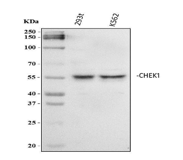
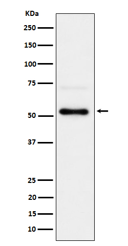
Real WB data Western blot analysis of Chk1/CHEK1 using anti-Chk1/CHEK1 antibody (A01060). Electrophoresis was performed on a 5-20% SDS-PAGE gel at 70V (Stacking gel) / 90V (Resolving gel) for 2-3 hours. The sample well of each lane was loaded with 30 ug of sample under reducing conditions. Lane 1: human 293T whole cell lysates, Lane 2: human K562 whole cell lysates. After electrophoresis, proteins were transferred to a nitrocellulose membrane at 150 mA for 50-90 minutes. Blocked the membrane with 5% non-fat milk/TBS for 1.5 hour at RT. The membrane was incubated with rabbit anti-Chk1/CHEK1 antigen affinity purified polyclonal antibody (Catalog # A01060) at 0.5 μg/mL overnight at 4°C, then washed with TBS-0.1%Tween 3 times with 5 minutes each and probed with a goat anti-rabbit IgG-HRP secondary antibody at a dilution of 1:5000 for 1.5 hour at RT. The signal is developed using an Enhanced Chemiluminescent detection (ECL) kit (Catalog # EK1002) with Tanon 5200 system. A specific band was detected for Chk1/CHEK1 at approximately 54 kDa. The expected band size for Chk1/CHEK1 is at 54 kDa.
Anti-Chk1/CHEK1 Antibody Picoband®
Cat # A01060
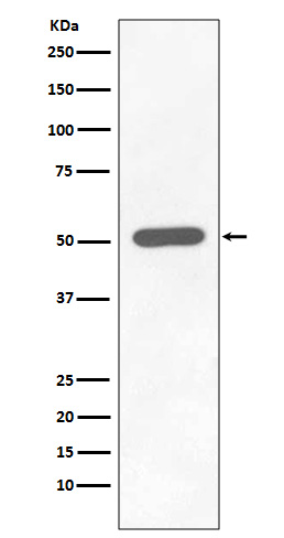
Real WB data Western blot analysis of Phospho-Chk1 (S296) expression in HEK293 cell lysate Treated with Calyculin.
Anti-Phospho-Chk1 (S296) CHEK1 Rabbit Monoclonal Antibody
Cat # P01060
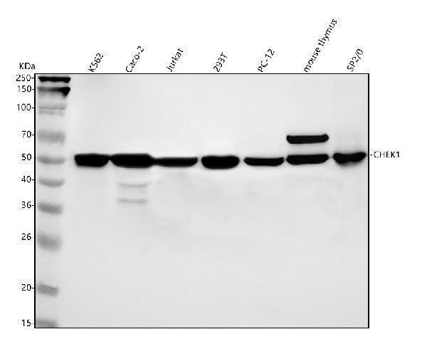
Real WB data Western blot analysis of Chk1 using anti-Chk1 antibody (M01060-1). Electrophoresis was performed on a 5-20% SDS-PAGE gel at 70V (Stacking gel) / 90V (Resolving gel) for 2-3 hours. The sample well of each lane was loaded with 30 ug of sample under reducing conditions. Lane 1: human K562 whole cell lysates, Lane 2: human CACO-2 whole cell lysates, Lane 3: human Jurkat whole cell lysates, Lane 4: human 293T whole cell lysates, Lane 5: rat PC-12 whole cell lysates, Lane 6: mouse thymus tissue lysates, Lane 7: mouse SP2/0 whole cell lysates. After electrophoresis, proteins were transferred to a nitrocellulose membrane at 150 mA for 50-90 minutes. Blocked the membrane with 5% non-fat milk/TBS for 1.5 hour at RT. The membrane was incubated with rabbit anti-Chk1 antigen affinity purified monoclonal antibody (Catalog # M01060-1) at 1:500 overnight at 4°C, then washed with TBS-0.1%Tween 3 times with 5 minutes each and probed with a goat anti-rabbit IgG-HRP secondary antibody at a dilution of 1:500 for 1.5 hour at RT. The signal is developed using an Enhanced Chemiluminescent detection (ECL) kit (Catalog # EK1002) with Tanon 5200 system. A specific band was detected for Chk1 at approximately 54 kDa. The expected band size for Chk1 is at 54 kDa.
Anti-Chk1 CHEK1 Rabbit Monoclonal Antibody
Cat # M01060-1
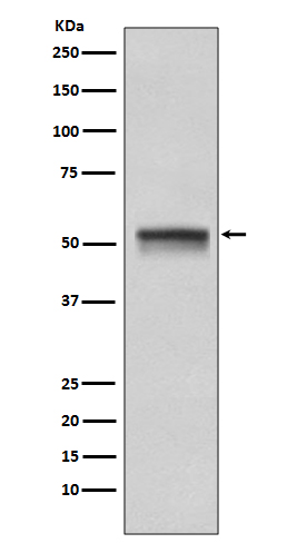
Real WB data Western blot analysis of Chk1 expression in HeLa cell lysate.
Anti-Chk1 CHEK1 Rabbit Monoclonal Antibody
Cat # M01060
Real WB data Western blot analysis of Phospho-Chk1 (S280) expression in 293T treated with Calyculin A cell lysate.
Anti-Phospho-Chk1 (S280) Rabbit Monoclonal Antibody
Cat # M01060S280

The listed CHEK1 antibodies include total Chk1 and phospho-S296 or phospho-S280 options. Each has a WB image. Captions show selected cell or tissue lysates; these examples establish tested contexts, not universal performance across samples or species.

Which to pick: For total CHEK1, M01060-1 has a WB image with human, rat, and mouse samples; A01060 shows human 293T and K562, while M01060 shows HeLa. For site-specific detection, choose P01060 for S296 or M01060S280 for S280; both show treated human cell lysates.

Source: BosterBio CHEK1 gene-info card — filtered to Western-blot-capable antibodies; each card shows that product's actual WB validation figure.