This website uses cookies to ensure you get the best experience on our website.


- Table of Contents
Plan chromogenic paraffin IHC with the IHC-validated antibody at 2–5 μg/mL (datasheet A01585-2). Use skin granular-layer cells as a high-staining reference and colon glandular cells as an undetected comparator (HPA tissue IHC).

Expected localisation, validated protocols, controls and antibodies — the at-a-glance facts below, then the full design guide.
| Expected localisation | Membranous tissue staining (HPA tissue IHC) | |
| Staining pattern | Membranous staining, most abundant in squamous epithelium (HPA tissue IHC) | |
| Antigen retrieval | EDTA pH 8.0 HIER, heat-mediated (datasheet A01585-2) | |
| Positive control | Kidney+4 more · see all | |
| Negative control | Adipose tissue+4 more · see all |
| Fixation | Keep fixation conditions consistent across paraffin sections. (standard IHC practice; not target-specific) | |
| Caveat | Antibody staining and RNA show medium consistency (HPA tissue IHC) | |
| Regulation | RNA is tissue enhanced in liver and skin (HPA tissue RNA) | |
| Isoform / epitope | No annotated isoforms; map extracellular vs cytoplasmic epitope (UniProt) |
Start with the catalog antibody’s IHC-P protocol (datasheet A01585-2). These four published CLDN1 IHC protocols provide conditions for breast, colorectal, mouse colonic, and paraffin-embedded tissue sections (PMC1064136; PMC4202803; PMC7571313; PMC12624041).
| Sample | Paraffin-embedded human lung cancer tissue; fixative not specified (datasheet A01585-2) |
| Fixation | Image fixative and duration unreported (datasheet A01585-2); verify before use. |
| Sectioning | 4–5 µm sections on charged slides (standard) |
| Deparaffinisation | Xylene, graded ethanol series to water (standard) |
| Antigen retrieval | Heat retrieval: EDTA pH 8.0 (datasheet A01585-2); 20 min, 95–100 °C (standard) |
| Peroxidase block | 3% H2O2, 10 min, room temperature (standard) |
| Blocking | 10% goat serum (datasheet A01585-2) |
| Primary antibody | Rabbit anti-CLDN1, 2μg/ml (datasheet A01585-2) |
| Primary incubation | Overnight at 4 °C (datasheet A01585-2) |
| Detection | Streptavidin-biotin complex (SABC), DAB chromogen (datasheet A01585-2) |
| Counterstain | Hematoxylin, blue, dehydrate and mount (standard) |
| Expected result | CLDN1-positive staining in bowman's capsule of kidney (HPA tissue IHC: High). HPA tissue profile: Membranous expression in several tissues, most abundant in squamous epithelium. No signal in the no-primary control. |
CLDN1 is a four-pass membrane protein found at tight junctions and cell membranes, with a reported basolateral location in its CD81 complex (UniProt O95832 topology and subcellular location). In paraffin-section IHC, expect membranous staining in epithelial cells, especially skin granular-layer cells, tonsillar squamous epithelium, and kidney Bowman’s capsule (HPA: High in each). HPA rates its tissue IHC profile Enhanced, with medium consistency between staining and RNA expression (HPA tissue IHC).
| Membranous outlines in skin granular-layer cells, tonsillar squamous epithelium, or Bowman’s capsule. | This matches the expected compartment and High staining in these cells (UniProt O95832 subcellular location; HPA tissue IHC). Judge cell identity and membrane pattern together: a dark chromogenic deposit alone does not establish a correct CLDN1 result (general IHC practice). |
| Predominantly nuclear staining, or widespread cytoplasmic staining without clear cell borders. | This does not fit the annotated junctional and membrane location (UniProt O95832 subcellular location). Review morphology, detection controls, and antibody conditions before assigning it to CLDN1; the supplied sources do not establish a normal nuclear pattern (general IHC interpretation). |
| Strong staining in adipocytes, bone-marrow hematopoietic cells, or colon glandular cells. | HPA reports CLDN1 as not detected in these cell populations (HPA tissue IHC). If staining persists after confirming the cell type, consider nonspecific antibody binding or endogenous detection activity; a discordant result alone cannot distinguish them (general IHC practice). |
| Diffuse color across tissue, stroma, and slide, with little separation between cells. | A diffuse deposit cannot be scored as the epithelial membrane pattern described by HPA (HPA tissue IHC). Uneven reagent coverage, nonspecific binding, or detection background are general IHC possibilities; inspect a no-primary control and compare matched processing before interpreting tissue expression (general IHC practice). |
| No signal in a well-preserved skin granular layer, tonsillar squamous epithelium, or Bowman’s capsule. | These are expected High cell-specific positive references (HPA tissue IHC). First verify that the named cells are present and the section is interpretable; then review the catalog antibody’s IHC-P instructions, reagent activity, and detection controls. Absence in one section alone does not establish biological loss (general IHC practice). |
| Membrane topology and epitope location | CLDN1 has 4 transmembrane segments, two extracellular regions, and cytoplasmic termini and loop (UniProt O95832 topology). The antibody epitope is unspecified here, so topology cannot justify a CLDN1-specific retrieval, permeabilisation, or fixation-sensitivity claim. |
| Cell-specific reference pattern | Skin granular-layer cells, tonsillar squamous cells, and Bowman’s capsule are High; breast glandular and bronchial respiratory epithelial cells are Medium (HPA tissue IHC). Compare the named cells, not whole-tissue color, when choosing positive references. |
| Strength of tissue evidence | HPA labels the tissue profile Enhanced but notes medium consistency with RNA expression; its listed antibodies have IHC ratings Enhanced and Supported (HPA tissue IHC; HPA antibodies). Treat an unexpected pattern as a finding to verify, rather than proof of absence or ectopic expression. |
| IF/ICC question: should the same pattern be expected? | Membrane localization is biologically expected (UniProt O95832 subcellular location; HPA subcellular summary: Membrane). HPA supplies no ICC-IF cell-line images or main-location assignment here, so this record cannot validate an IF/ICC staining pattern or supply an IF protocol (HPA subcellular). |
| Situation | Likely cause | Next action |
|---|---|---|
| Expected positive epithelium is blank. | The target cells may be absent from the section, or the IHC detection run may have failed; HPA reports High staining in the specified skin, tonsil, and kidney cells (HPA tissue IHC; general IHC practice). | Confirm cell identity and section integrity, then check the catalog antibody’s IHC-P instructions, reagent activity, and a run control. Change retrieval conditions only as a general optimization step; no CLDN1-specific retrieval effect is supplied (general IHC practice). |
| Signal is mainly nuclear or diffuse cytoplasmic. | The compartment conflicts with CLDN1’s junctional and membrane annotation (UniProt O95832 subcellular location). A mismatch can reflect staining or interpretation problems but does not identify one specific cause (general IHC practice). | Recheck focus and cell boundaries, compare a no-primary control, and repeat using the catalog antibody’s validated IHC-P conditions. Score membrane signal separately from any other deposit (general IHC practice). |
| HPA-negative cells appear strongly positive. | Adipocytes, bone-marrow hematopoietic cells, and colon glandular cells are reported not detected (HPA tissue IHC). Nonspecific binding or endogenous detection activity may produce misleading color (general IHC practice). | Verify morphology and compare no-primary and detection controls. If the deposit remains antibody dependent, reassess antibody conditions and seek independent confirmation before reporting CLDN1 in those cells (general IHC practice). |
| The entire section has a colored haze. | Broad background obscures the membranous epithelial pattern reported by HPA (HPA tissue IHC). Blocking, washing, or detection conditions can contribute to background in chromogenic IHC (general IHC practice). | Check a no-primary control and inspect reagent coverage. Review blocking, wash steps, primary-antibody concentration, and detection timing under the catalog IHC-P procedure; retain conditions that preserve interpretable positive-cell borders (general IHC practice). |
| A moderate-staining tissue is treated as a failed positive control. | HPA rates breast glandular and bronchial respiratory epithelial cells Medium, below its High examples (HPA tissue IHC). A weaker result in these named cells alone does not show that the run failed. | Compare like cell populations and include a High reference such as skin granular-layer cells, tonsillar squamous epithelium, or Bowman’s capsule when available (HPA tissue IHC). Interpret control intensity with morphology and run controls (general IHC practice). |


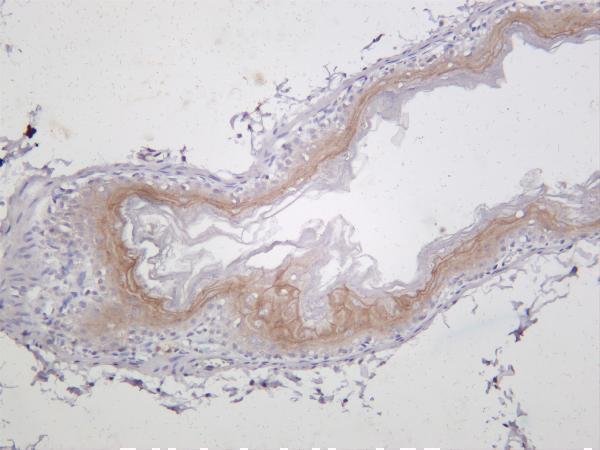
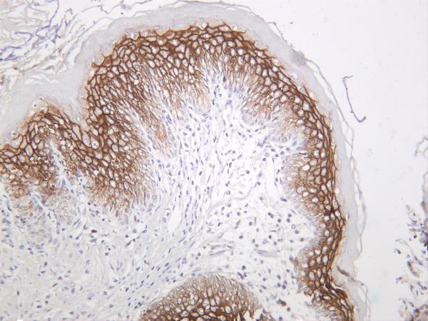
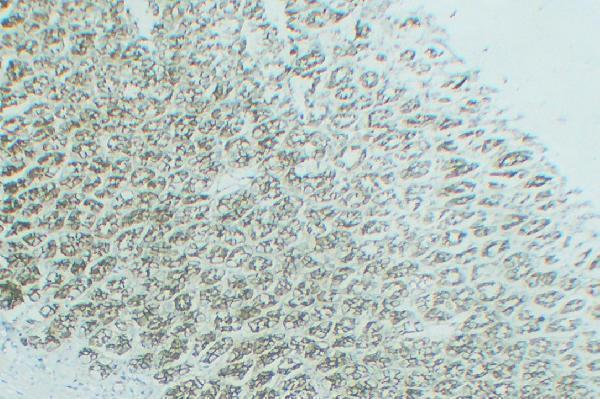
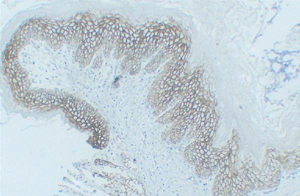
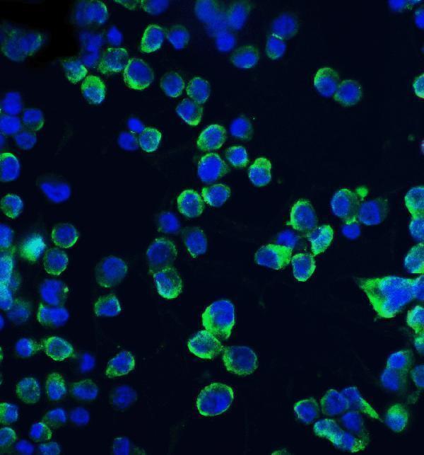


Comprehensive Human Protein Atlas IHC scoring per tissue (reliability: Enhanced — Medium consistency between antibody staining and RNA expression data.). Rows are taken directly from the HPA tissue chart — click any row's HPA link to view the source.
| Tissue | Cell type | Level | Evidence | Source |
|---|---|---|---|---|
| Kidney | Bowman's capsule | High | Protein (IHC) | HPA → |
| Skin | Cells in granular layer | High | Protein (IHC) | HPA → |
| Tonsil | Squamous epithelial cells | High | Protein (IHC) | HPA → |
| Breast | Glandular cells | Medium | Protein (IHC) | HPA → |
| Bronchus | Respiratory epithelial cells | Medium | Protein (IHC) | HPA → |
| Tissue | Cell type | Level | Evidence | Source |
|---|---|---|---|---|
| Adipose tissue | Adipocytes | Not detected | Protein (IHC) | HPA → |
| Bone marrow | Hematopoietic cells | Not detected | Protein (IHC) | HPA → |
| Caudate | Glial cells | Not detected | Protein (IHC) | HPA → |
| Cerebellum | Cells in granular layer | Not detected | Protein (IHC) | HPA → |
| Cerebral cortex | Endothelial cells | Not detected | Protein (IHC) | HPA → |
Troubleshoot CLDN1 staining by checking membrane localisation, cell identity and controls alongside retrieval and detection conditions (UniProt O95832; HPA tissue IHC).
Anti-CLDN1 antibodies have IHC images from human, mouse, and rat tissues and IF images from human cells and tissue (catalog image captions).
A01585-2 has IHC images from paraffin sections of human lung and rectal cancers; M01585-3 has IHC images from mouse and rat stomach and human breast (catalog IHC image captions). M01585-2 has IHC images from rat stomach and esophagus; A01585-1 has an IF image from human HepG2 cells (catalog image captions).
Which to pick: For human paraffin-section IHC, choose A01585-2: its IHC captions document EDTA pH 8.0 retrieval and 2 μg/mL primary antibody, but do not report the fixative (A01585-2 IHC image captions). For IHC across human, mouse, and rat samples, consider M01585-3, a monoclonal antibody listed as reactive with all three species and validated for IHC (M01585-3 catalog); its IHC captions do not report fixation or section processing (M01585-3 IHC image captions). For IF/ICC, choose A01585-1, which lists both applications and has an IF image in human HepG2 cells (A01585-1 catalog and IF image caption).