Click image to see more details
-

-
-
-
-
+1
Product Info Summary
| SKU: | A01585-1 |
|---|---|
| Size: | 0.1 mg |
| Reactive Species: | Human, Rat |
| Host: | Rabbit |
| Application: | ELISA, IF, ICC, WB |
Customers Who Bought This Also Bought

Product info
Product Name
Anti-Claudin-1 CLDN1 Antibody
SKU/Catalog Number
A01585-1
Size
0.1 mg
Form
Liquid
Description
Boster Bio Anti-Claudin-1 CLDN1 Antibody (Catalog # A01585-1). Tested in ELISA, WB, ICC, IF applications. This antibody reacts with Human, Rat.
Storage & Handling
CLDN1 antibody can be stored at 4°C for three months and -20°C, stable for up to one year. Avoid repeated freeze-thaw cycles. Antibodies should not be exposed to prolonged high temperatures.
Cite This Product
Anti-Claudin-1 CLDN1 Antibody (Boster Biological Technology, Pleasanton CA, USA, Catalog # A01585-1)
Host
Rabbit
Contents
CLDN1 Antibody is supplied in PBS containing 0.02% sodium azide.
Clonality
Polyclonal
Isotype
IgG
Immunogen
Anti-CLDN1 antibody was raised against a peptide corresponding to 20 amino acids near the carboxy terminus of human CLDN1. The immunogen is located within the last 50 amino acids of CLDN1.
Reactive Species
A01585-1 is reactive to CLDN1 in Human, Rat
Observed Molecular Weight
68 kDa
Calculated molecular weight
22.7 kDa
Background of CLDN1
Claudin1 (CLDN1), a member of the claudin family, is an integral membrane protein and a component of tight junction strands. Tight junctions are specialized regions of cell to cell contact consisting of networking strands that act as a molecular gasket for preventing the leakage of ions, water, etc., between cells. They are abundant in luminal epithelial sheets where they maintain epithelial cell polarity. Different tissues exhibit different Claudin composition and CLDN1 expression is often cell type and tissue dependent. Loss of function mutations result in neonatal ichthyosis-sclerosing cholangitis syndrome. CLDN1 and CLDN2 were found to be overexpressed in colonal cancer tissues and may be useful as tumor markers and targets for the treatment of colorectal cancer. Characterization of Claudins expression in human tumors can be an additional diagnostic tool. Recent studies show that CLDN1 has gastric tumor suppressive activity and is a direct transcriptional target of RUNX3. Along with SCARB1, LDL-R, and the tetraspanin superfamily member CD81, CLDN1 has been reported to be an entry factor for the Hepatitis C virus.
Antibody Validation
Boster validates all antibodies on WB, IHC, ICC, Immunofluorescence, and ELISA with known positive control and negative samples to ensure specificity and high affinity, including thorough antibody incubations.
Application & Images
Applications
A01585-1 is guaranteed for ELISA, IF, ICC, WB Boster Guarantee
Recommend Dilution
WB: 1-2 μg/mL; ICC: 5 μg/mL; IF: 20 μg/mL.
Antibody validated: Western Blot in human and rat samples; Immunocytochemistry in human samples; Immunofluorescence in human samples. All other applications and species not yet tested. Optimal dilutions for each application should be determined by the researcher.
Validation Images & Assay Conditions
Click image to see more details
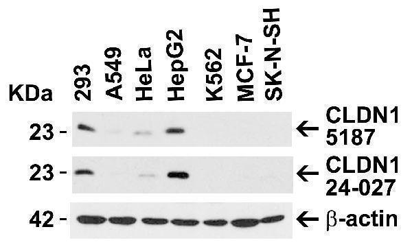
Independent Antibody Validation (IAV) via Protein Expression Profile in Human Cell Lines
Loading: 15 μg of lysates per lane.
Antibodies: CLDN1, A01585-1 (1 μg/mL), CLDN1, 24-027 (2 μg/mL), and beta-actin (1 μg/mL), 1h incubation at RT in 5% NFDM/TBST.
Secondary: Goat anti-rabbit IgG HRP conjugate at 1:10000 dilution.

Click image to see more details
Western Blot Validation in Human Tissue Lysates
Loading: 15 μg of lysates per lane.
Antibodies: CLDN1, A01585-1 (1 μg/mL) , 1h incubation at RT in 5% NFDM/TBST.
Secondary: Goat anti-rabbit IgG HRP conjugate at 1:10000 dilution.

Click image to see more details
Western Blot Validation in Human HepG2 Cell Lysate
Loading: 15 μg of lysates per lane.
Antibodies: CLDN1, A01585-1 (A: 1 μg/mL and B: 2 μg/mL), 1h incubation at RT in 5% NFDM/TBST.
Secondary: Goat anti-rabbit IgG HRP conjugate at 1:10000 dilution.

Click image to see more details
Western Blot Validation in Human Tissue Lysates
Loading: 15 μg of lysates per lane.
Antibodies: CLDN1, A01585-1 (1 μg/mL) , 1h incubation at RT in 5% NFDM/TBST.
Secondary: Goat anti-rabbit IgG HRP conjugate at 1:10000 dilution.

Click image to see more details
Immunofluorescence Validation of CLDN1 in HepG2 Cells
Immunofluorescent analysis of 4% paraformaldehyde-fixed HepG2 cells labeling CLDN1 with A01585-1 at 20 μg/mL, followed by goat anti-rabbit IgG secondary antibody at 1/500 dilution (green) and DAPI staining (blue).
Specific Publications For Anti-Claudin-1 CLDN1 Antibody (A01585-1)
Loading publications
Recommended Resources
Here are featured tools and databases that you might find useful.
- Boster's Pathways Library
- Protein Databases
- Bioscience Research Protocol Resources
- Data Processing & Analysis Software
- Photo Editing Software
- Scientific Literature Resources
- Research Paper Management Tools
- Molecular Biology Software
- Primer Design Tools
- Bioinformatics Tools
- Phylogenetic Tree Analysis
Customer Reviews
Have you used Anti-Claudin-1 CLDN1 Antibody?
Share your experimental results or join a short interview to earn up to $1,000 in product credits or other rewards.
0 Reviews For Anti-Claudin-1 CLDN1 Antibody
Customer Q&As
Have a question?
Find answers in Q&As, reviews.
Can't find your answer?
Submit your question
