This website uses cookies to ensure you get the best experience on our website.
- Table of Contents
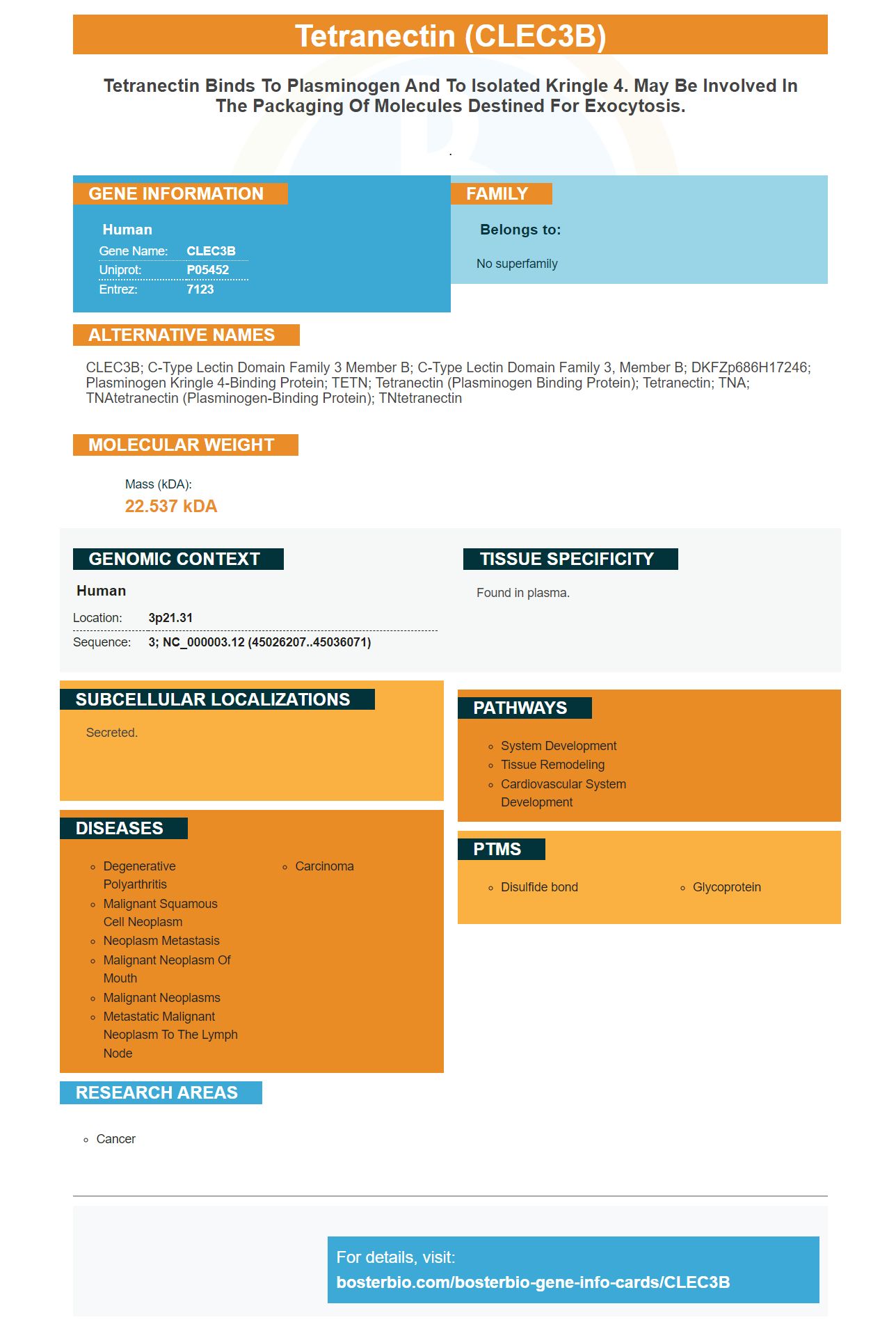
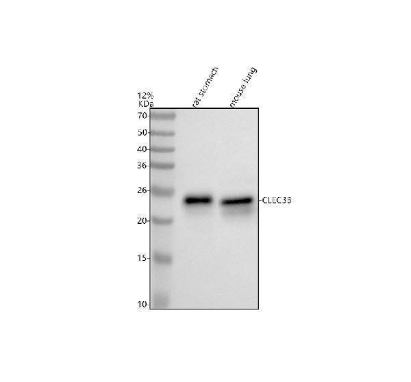
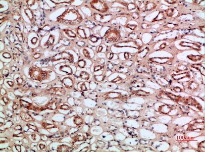
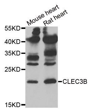
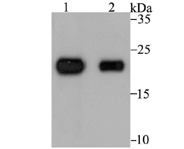
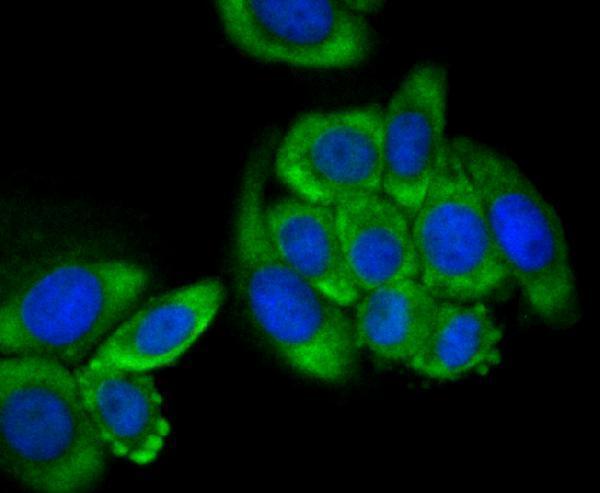
Facts about Tetranectin.
.
Human | |
---|---|
Gene Name: | CLEC3B |
Uniprot: | P05452 |
Entrez: | 7123 |
Belongs to: |
---|
No superfamily |
CLEC3B; C-type lectin domain family 3 member B; C-type lectin domain family 3, member B; DKFZp686H17246; Plasminogen kringle 4-binding protein; TETN; tetranectin (plasminogen binding protein); Tetranectin; TNA; TNAtetranectin (plasminogen-binding protein); TNtetranectin
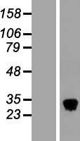
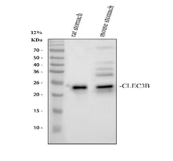
Mass (kDA):
22.537 kDA
Human | |
---|---|
Location: | 3p21.31 |
Sequence: | 3; NC_000003.12 (45026207..45036071) |
Found in plasma.
Secreted.
If you're looking for a high-affinity, reliable CLEC3B antibody, Boster's main antibodies are the solution. Having been validated in ELISA, immunohistochemistry, and Western Blotting, Boster antibodies have been extensively cited in the research community. Make use of Boster's CLEC3B antibodies for your next experiment and begin seeing results faster!
This study examined the expression of CLEC3B in lung cancer tissues. It included both small-cell and large-cell varieties. The samples were examined using PCR and immunohistochemistry. The results of these studies were analyzed using a Chi square test. This allowed us to determine the relationship between CLEC3B protein expression and prognosis. We also utilized a receiver-operating-characteristic (ROC) curve and TIMER database. We also performed gene set enrichment analysis in order to determine the biological processes associated with tumors that are related to CLEC3B.
The lung cancer tissue microarray LAC-1403 found that there were 18 immunomarkers. The 18 biomarkers detected in the lung cancer tissue microarray LAC-1403 were synaptophysin and the chromogranin. Each biomarker was evaluated for its prognostic significance. Our findings suggest that CLEC3B could play a part in the regulation of the lung's immune system carcinoma.
The expression of DcR3 protein was also observed to be associated with presence and nature of lung cancer tissues. Non-small-cell lung cancers had higher levels of DcR3 than small-cell ones. Furthermore, the expression of DcR3 was correlated with the size of the tumor TNM stage, tumor size, and distant metastasis. These studies support lung cancer screening and early stage diagnosis.
This study has shown that DcR3 protein expression was detected by immunohistochemistry. Tissue microarrays were taken from patients during surgical resection and deidentified slides are obtained from the Mayo Clinic SPORE study in Pancreatic Cancer. All patients provided written informed consent to participate in the study. The study was approved by Mayo Clinic's Institutional Review Board. The images were captured using the Zeiss Axioscope A1 microscopy that was specifically designed for histological investigations. Zen 2 software was used to capture the images. Zen 2 software was used to capture the images.
MB complements IHC in lung cancer . It can be utilized together with immunohistochemistry to identify molecular targets in cancer samples. The results of immunohistochemistry may be used to develop new therapies that are effective for patients suffering from lung cancer. However, tissues cannot always be used to evaluate all molecular targets. Different strategies are possible dependent on the particulars of the patient.
Numerous new MB techniques are being used in tumor biopsies that contain few tumor cells. These techniques can identify EGFR mutation status in a tissue sample within a matter of hours. These techniques can detect more mutations than IHC. This is great news for patients suffering from lung cancer and for scientists interested in detecting it. If you're interested in using immunohistochemistry as a screening tool this study might be worth your consideration.
Recent research has revealed an association between CLEC3B gene expression and the level of lung cancer's immune infiltrate. The authors utilized data from the TCGA database to identify immune-infiltrating cells within tumor tissues. They also identified two patient groups: lower-score and high-score. CLEC3B levels were higher in patients with high-score SCC but not in patients with low-score ADC. CLEC3B levels in patients with low-score ADC did not correlate well with immune infiltration.
These data were used for investigating the mechanisms that underlie the relationship between CLEC3B expression levels and the infiltrates of the immune system in lung cancer. To verify CLEC3B mRNA expression, the TCGA data were classified into low and high-expression groups and the GO gene set GMT file C5 was used for analysis. After analysing the data, the top 20 enrichment results were identified. The P value was 0.05 and the FDR was 0.25.
This study was designed to find out if a genetic variant CLEC3B could affect lung cancer survival. It found a significant correlation between the expression level of the gene and the immune infiltration levels in lung cancer. Researchers found that patients who had high levels of CLEC3B expression were more likely than those with lower levels to develop advanced stage cancers or to spread far away.
Although there has been limited research in the area of immunohistochemistry for lung cancer these results are significant and could aid the development of more effective biomarkers. These biomarkers can help physicians optimize patient care and increase survival rates. They are currently being developed by researchers from the United States and China. They provide a novel method to determine the most effective therapies and ensure that patients are receiving the most effective treatment available.
There was a time when only a few studies investigated the significance of SIDT1 in tumors. The research has revealed that it plays an important role in pancreatic and breast cancer, but there is little information about its role in lung cancer. The authors of this study wanted to explore the role played by SIDT1 and explore its prognostic value.
They also assessed the degree of infiltration by the immune system and found that patients with high SIDT1 expression had worse prospects of survival. These findings could be the basis for targeted therapy and immunotherapy. Yubin Tian, Yong Zhou and co-first authors of this study carried out the study. They concluded that an association between SIDT1 expression and immune infiltration levels for lung cancer could improve prognosis.
CLEC3B expression has been linked to poor overall survival rates as well as a reduced chance of developing cancer. Several studies have linked low CLEC3B expression with poor prognosis in patients with HCC. In particular, low CLEC3B expression has been associated with poor survival among patients with gastric and ovarian cancer. Additionally the low CLEC3B expression was associated with low overall survival and relapse-free rates in breast cancer.
Studies of CLEC3B expression have also shown an association between the protein's expression and the immune cells population. Tumors that have high levels of CLEC3B expression are more likely to have T cells that are CD8+ than tumors that do not have it. Cluster I demonstrated a stronger association for LUAD-positive tumors.
Seven genes were studied to determine whether there was a link between CLEC3B expression and OS. The expression of CAV1 caveolin1, complement factor D flavin-containing monooxygenase-2 and HIC-5 and FMO2 was significantly. However, in a recent study, CLEC3B expression in tumor samples was associated negatively with OS, while the expression of six other genes correlated with OS.
The study utilized a vast data set of lung adenocarcinomas from two groups and identified five biomarkers that have independently prognostic value. Additionally, the CAV1 gene was found to have a positive association with overall survival, which is the most potent prognostic biomarker currently available. The other two markers, CFD and FMO2, were associated with a significant degree of immunosuppression.
The cancer microenvironment also includes resident host cells and immune cells. These cells are crucial for the progression of cancer, and they exhibit a variety of anti-inflammatory and pro-inflammatory functions. Infiltrating T-cells contribute to tumor growth, but they also exhibit immunosuppressive capabilities. This is the primary function of regulatory T cells in the tumor microenvironment.
It is unknown if CLEC3B will be used as a cancer immunotherapy target. The study highlights the importance the m6ARNA methylation process in the tumor microenvironments. Additionally, m6A RNA methylation has been associated with poor prognosis, and that is the reason why it is essential to study the connection between M6A RNA methylation and the immune microenvironment in LUAD patients.
CLEC3B even though it has a modest impact on the tumor microenvironment, is a promising candidate for immunotherapy. A phase II study demonstrated that treatment of a patient with lung cancer that has CLEC3B resulted in a significant increase number of lymphocytes. The clinical value of CLEC3B for the treatment of tumors is increasing. A positive correlation was also observed between CLEC3B and immune infiltration.
Recent studies have shown that the expression of REXO4 is linked to poor survival overall and progression-free in patients with HCC. It is also associated with infiltration and activation of the immune system. Its involvement in immune infiltration and survival is vital in HCC. CLEC3B immunotherapy can enhance the survival rate of cancer patients. CLEC3B is a powerful treatment for cancer and a promising biomarker that could be used to develop cancer immunotherapy.
PMID: 1354271 by Wewer U.M., et al. Tetranectin, a plasminogen kringle 4-binding protein. Cloning and gene expression pattern in human colon cancer.
PMID: 1511740 by Berglund L., et al. The gene structure of tetranectin, a plasminogen binding protein.