This website uses cookies to ensure you get the best experience on our website.


- Table of Contents
Plan chromogenic IHC in paraffin sections around CREB1’s widespread nuclear staining (HPA tissue IHC). This guide covers fixation, antibody selection and interpretation of total versus phospho-CREB1 staining (UniProt; datasheet A00577).

Expected localisation, validated protocols, controls and antibodies — the at-a-glance facts below, then the full design guide.
| Expected localisation | Nuclear staining in tissue sections (HPA tissue IHC) | |
| Staining pattern | Widespread nuclear staining across tissue cell types (HPA tissue IHC) | |
| Antigen retrieval | Tris-EDTA pH 9.0 HIER, 95–98 °C, 20 min (rule: nuclear antigen) | |
| Positive control | Adipose tissue+4 more · see all | |
| Negative control | None in HPA (detected in all 45 tissues); use no-primary + isotype controls |
| Fixation | Fix promptly in 10% NBF; prolonged fixation may mask epitopes (standard IHC practice) | |
| Caveat | Phospho-specific staining may differ from total CREB1 (datasheet A00577) | |
| Regulation | Phosphorylation can change phospho-CREB signal (UniProt) | |
| Isoform / epitope | 3 isoforms; check antibody epitope coverage across splice forms (UniProt) |
Compare the catalog antibody’s IHC-P protocol with four published CREB1 or phospho-CREB1 IHC methods (PMC8421817; PMC3050929; PMC4496382; PMC13109986).
| Sample | FFPE tissue sections (standard) |
| Fixation | 10% neutral-buffered formalin, 24 h, standard processing (standard) |
| Sectioning | 4–5 µm sections on charged slides (standard) |
| Deparaffinisation | Xylene, graded ethanol series to water (standard) |
| Antigen retrieval | Heat-induced epitope retrieval in Tris-EDTA buffer, pH 9.0, 20 min at 95–98 °C (standard rule: nuclear antigen) |
| Peroxidase block | 3% H2O2, 10 min, room temperature (standard) |
| Blocking | 10% normal serum of the secondary host, 30 min, room temperature (standard) |
| Primary antibody | Rabbit anti-CREB1, 1:2000-1:10000 (datasheet M00577-1) |
| Primary incubation | Overnight at 4 °C (standard) |
| Detection | HRP-polymer secondary, DAB chromogen 5–10 min (standard) |
| Counterstain | Hematoxylin, blue, dehydrate and mount (standard) |
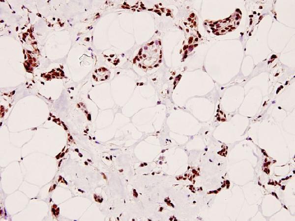
| Expected result | CREB1-positive staining in adipocytes of adipose tissue (HPA tissue IHC: High). HPA tissue profile: Ubiquitous nuclear expression. No signal in the no-primary control. |
CREB1 is nuclear and has no transmembrane segment (UniProt P16220). Expect nuclear staining across many cell types, including adipocytes, glandular cells and hematopoietic cells (HPA tissue IHC: Supported; ubiquitous nuclear expression).
| Nuclei stain across many cell types (HPA tissue IHC). | Expected CREB1 pattern (HPA tissue IHC: ubiquitous nuclear expression). |
| Cytoplasm stains more strongly than nuclei (HPA tissue IHC: nuclear pattern). | Suspect nonspecific staining or an IHC artefact; check controls (standard IHC practice). |
| Signal appears only in an unexpected cell population (HPA tissue IHC: ubiquitous nuclear pattern). | Check cross-reactivity or endogenous detection activity (standard IHC practice). |
| Diffuse colour obscures nuclei (HPA tissue IHC: nuclear pattern). | Background may reflect excess antibody or incomplete blocking (standard IHC practice). |
| Adipocyte nuclei show no signal (HPA tissue IHC: High in adipocytes). | Check retrieval, antibody incubation and detection controls (standard IHC practice). |
| Fixation and cross-linking (standard IHC practice) | Overfixation can mask nuclear epitopes and weaken signal (standard IHC practice). |
| Antigen retrieval (standard paraffin IHC practice) | Insufficient retrieval can reduce nuclear staining (standard IHC practice). |
| Isoforms (UniProt P16220: 3 isoforms) | Coverage may vary if the antibody epitope differs among isoforms (UniProt P16220; IHC practice). |
| Protein processing (UniProt P16220: chain 1–327) | No signal peptide or propeptide is annotated (UniProt P16220). |
| Endogenous peroxidase (standard chromogenic IHC practice) | Incomplete quenching can create misleading colour (standard chromogenic IHC practice). |
| Situation | Likely cause | Next action |
|---|---|---|
| No nuclear signal in adipocytes (HPA tissue IHC: High). | Epitope masking after fixation (standard IHC practice). | Optimise retrieval using the catalog IHC-P protocol (standard IHC practice). |
| Nuclear signal is uniformly faint (HPA tissue IHC: nuclear pattern). | Weak antibody or detection conditions (standard IHC practice). | Check incubation and detection controls (standard IHC practice). |
| Cytoplasmic colour dominates (HPA tissue IHC: nuclear pattern). | Nonspecific staining or background (standard IHC practice). | Review blocking and antibody concentration (standard IHC practice). |
| Diffuse colour covers the section (HPA tissue IHC: nuclear pattern). | Excess antibody or incomplete washing (standard IHC practice). | Optimise antibody dilution and washes (standard IHC practice). |
| Colour persists in a no-primary control (standard IHC practice). | Endogenous peroxidase or detection background (standard chromogenic IHC practice). | Check peroxidase quenching and detection reagents (standard IHC practice). |
| IF/ICC signal is mainly nucleoplasmic (HPA subcellular ICC-IF). | Expected localisation (HPA subcellular ICC-IF: nucleoplasm enhanced). | Assess IF/ICC using its separate guide (HPA subcellular ICC-IF). |




Comprehensive Human Protein Atlas IHC scoring per tissue (reliability: Supported — High consistency between antibody staining and RNA expression data.). Rows are taken directly from the HPA tissue chart — click any row's HPA link to view the source.
| Tissue | Cell type | Level | Evidence | Source |
|---|---|---|---|---|
| Adipose tissue | Adipocytes | High | Protein (IHC) | HPA → |
| Adrenal gland | Glandular cells | High | Protein (IHC) | HPA → |
| Appendix | Glandular cells | High | Protein (IHC) | HPA → |
| Bone marrow | Hematopoietic cells | High | Protein (IHC) | HPA → |
| Breast | Glandular cells | High | Protein (IHC) | HPA → |
| Tissue | Cell type | Level | Evidence | Source |
|---|---|---|---|---|
| None in HPA: CREB1 is detected in all 45 scored tissues. Use a no-primary (secondary-only) and an isotype control instead. | ||||
Troubleshoot CREB1 staining by checking nuclear signal, matched processing and epitope specificity before comparing cells or conditions.
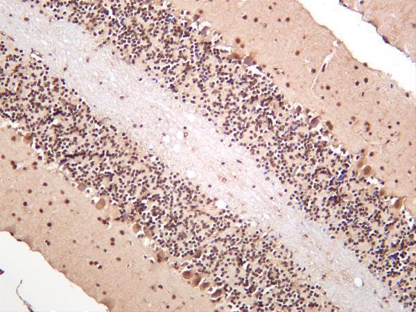
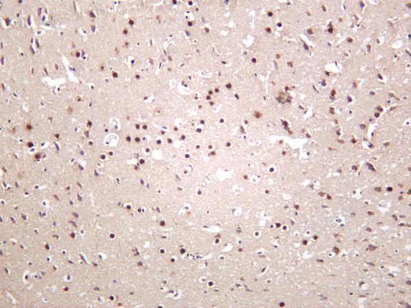
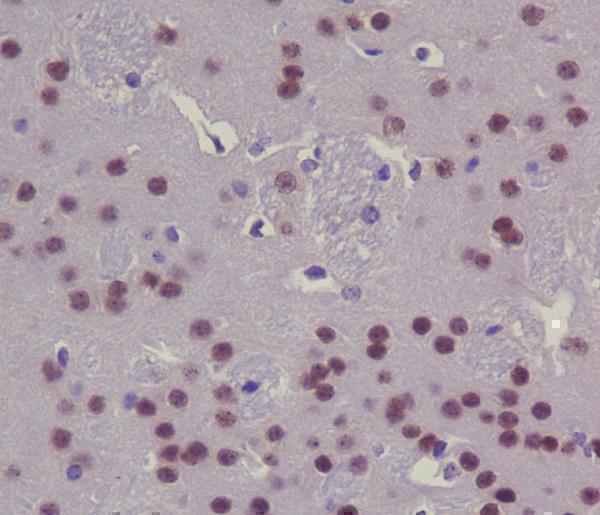
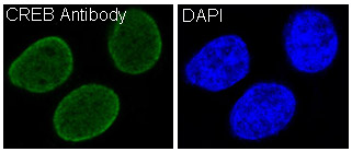
The catalog includes CREB1 IHC images from human, mouse, and rat samples (catalog IHC images), plus a HeLa IF image (catalog IF image: M00577).
M00577-1 has IHC images of human, mouse, and rat brain and human thyroid carcinoma (catalog IHC images); A00577 has human hepatocarcinoma and colon carcinoma IHC images (catalog IHC images); A00577S129 has a human breast carcinoma IHC image (catalog IHC image). M00577 has a mouse brain IHC image and a HeLa IF image (catalog images); P00577-1 has a human gastric carcinoma IHC image (catalog IHC image).
Which to pick: For FFPE IHC, choose M00577-1 for total CREB1 in human, mouse, or rat samples (catalog title, reactivity, and IHC images); choose P00577-1 when the target is phospho-S133 CREB1 (catalog title and IHC application). For IF/ICC, M00577 lists both applications and has a HeLa IF image (catalog applications and IF image). For IHC across species, M00577-1 is monoclonal and has brain IHC images from human, mouse, and rat (catalog description and IHC images).