This website uses cookies to ensure you get the best experience on our website.


- Table of Contents
Plan CUEDC2 paraffin IHC with the IHC-validated antibody at 0.5–1 µg/mL (datasheet: PA1962). Assess cytoplasmic staining in lung macrophages or glomerular cells (HPA tissue IHC), and interpret results cautiously because tissue IHC reliability is uncertain (HPA tissue IHC).

Expected localisation, validated protocols, controls and antibodies — the at-a-glance facts below, then the full design guide.
| Expected localisation | Cytoplasmic tissue staining (HPA tissue IHC); nuclear location annotated (UniProt) | |
| Staining pattern | Cytoplasmic staining in lung macrophages and glomerular cells (HPA tissue IHC) | |
| Antigen retrieval | Tris-EDTA pH 9.0 HIER, 95–98 °C, 20 min (rule: nuclear antigen) | |
| Positive control | Bone marrow+3 more · see all | |
| Negative control | Adipose tissue+4 more · see all |
| Fixation | Keep fixation consistent across sections. (standard IHC practice; not target-specific) | |
| Caveat | Tissue staining has low RNA concordance; verify independently (HPA tissue IHC) | |
| Regulation | Higher in breast tumors; inverse to ESR1 (UniProt) | |
| Isoform / epitope | No isoforms or precursor cleavage reported (UniProt) |
The catalog antibody’s IHC-P protocol appears alongside published CUEDC2 protocols for colorectal cancer, rat brain, and serous ovarian cancer (PMC6381460; PMC7906146; PMC7521062).
| Sample | Tissue sections; selected-image fixative not specified (standard IHC workflow) |
| Fixation | Image fixative and duration unreported (datasheet PA1962); verify before use. |
| Sectioning | 4–5 µm sections on charged slides (standard) |
| Deparaffinisation | Xylene, graded ethanol series to water (standard) |
| Antigen retrieval | Heat-induced epitope retrieval in Tris-EDTA buffer, pH 9.0, 20 min at 95–98 °C (standard rule: nuclear antigen) |
| Peroxidase block | 3% H2O2, 10 min, room temperature (standard) |
| Blocking | 10% normal serum of the secondary host, 30 min, room temperature (standard) |
| Primary antibody | Rabbit anti-CUEDC2, 0.5-1μg/ml (datasheet PA1962) |
| Primary incubation | Overnight at 4 °C (standard) |
| Detection | HRP-polymer secondary, DAB chromogen 5–10 min (standard) |
| Counterstain | Hematoxylin, blue, dehydrate and mount (standard) |
| Expected result | CUEDC2-positive staining in hematopoietic cells of bone marrow (HPA tissue IHC: Medium). HPA tissue profile: Cytoplasmic expression in a few tissues. No signal in the no-primary control. |
In paraffin-section IHC, expect CUEDC2 staining mainly in the cytoplasm of selected cells, including bone marrow hematopoietic cells, glomerular cells, lung macrophages and smooth muscle cells (HPA tissue IHC: Medium; cytoplasmic expression in a few tissues). Nuclear staining is biologically plausible (UniProt Q9H467: cytoplasm and nucleus). CUEDC2 has no transmembrane segment (UniProt Q9H467 topology). Treat the tissue pattern as provisional: HPA rates its IHC reliability Uncertain because staining and RNA expression show low consistency.
| Cytoplasmic staining in lung macrophages or smooth muscle cells, with other cells less conspicuous. | This matches reported Medium staining in those cell populations (HPA tissue IHC). Compare the cell pattern with a control section; HPA calls the tissue IHC profile Uncertain, so agreement alone does not establish antibody specificity. |
| Nuclear staining accompanies cytoplasmic staining in a plausible positive cell population. | Nuclear localization is consistent with UniProt Q9H467 and supported nucleoplasmic ICC-IF localization (HPA subcellular). HPA describes tissue IHC chiefly as cytoplasmic; assess prominent nuclear IHC against controls before calling it a confirmed tissue pattern. |
| Strong extracellular or luminal deposits dominate while expected intracellular staining is absent. | That distribution does not fit the reported cytoplasmic and nuclear localization (UniProt Q9H467; HPA tissue IHC). Consider precipitated chromogen, trapped reagent or nonspecific staining; inspect the no-primary control and tissue morphology before interpreting it as CUEDC2. |
| Adipocytes or bronchial respiratory epithelial cells stain strongly. | Both populations are reported as Not detected in the listed HPA tissue IHC observations. Suspect cross-reactivity or endogenous detection activity, then check matched controls. An HPA negative observation is a reference pattern, not proof that every specimen must be negative. |
| No intracellular signal appears in a section containing an expected positive cell population. | Bone marrow hematopoietic cells, glomerular cells, lung macrophages and smooth muscle cells each have reported Medium staining (HPA tissue IHC). Check cell identity and controls first; the profile's Uncertain reliability prevents treating any one section as an infallible positive control. |
| Which cells provide a provisional positive reference? | Bone marrow hematopoietic cells, kidney glomerular cells, lung macrophages and smooth muscle cells show Medium staining (HPA tissue IHC). Confirm that the relevant cells are present in the actual section before judging assay sensitivity. |
| How much confidence does the tissue IHC map carry? | HPA rates it Uncertain: antibody staining and RNA expression have low consistency, with external verification pending (HPA tissue IHC). HPA036544 also has an Uncertain IHC validation status (HPA antibodies). Interpret distribution alongside controls. |
| Does protein architecture predict membrane staining or an epitope effect? | CUEDC2 lacks a transmembrane segment, has no reported signal peptide or propeptide, and contains a CUE domain at residues 144–187 (UniProt Q9H467). No antibody epitope or target-specific fixation effect is supplied; architecture cannot determine retrieval conditions. |
| IF/ICC Q&A: What localization should I expect on its separate guide page? | Main signals are nucleoplasmic and cytosolic; nuclear membrane localization is also supported, while plasma membrane and primary cilium tip assignments are uncertain (HPA subcellular). Both listed antibodies have Supported ICC status (HPA antibodies). These ICC-IF observations do not validate an IHC protocol. |
| Situation | Likely cause | Next action |
|---|---|---|
| A putative positive section has no signal. | The expected cells may be absent, or a general IHC step may have failed; HPA's positive pattern is provisional (HPA tissue IHC: Uncertain). | Locate the relevant cells on the counterstained section. As general IHC practice, verify the validated control, retrieval, primary incubation and detection reagents before changing conditions. |
| Most tissue compartments show a uniform brown haze. | Diffuse staining obscures the restricted cytoplasmic pattern reported by HPA (HPA tissue IHC). General IHC causes include excess primary or detection reagent and inadequate blocking or washing. | Compare a no-primary control; as general IHC practice, review blocking, washing and reagent concentration while retaining the same tissue and detection controls. |
| Adipocytes or bronchial respiratory epithelium stain strongly. | Those cells are listed as Not detected (HPA tissue IHC). Cross-reactivity or endogenous enzyme activity may explain an unexpected chromogenic signal. | Inspect a no-primary control and the detection-system controls. If signal persists, address endogenous activity using standard IHC controls; if primary-dependent, reassess antibody specificity. |
| Signal appears chiefly in lumina or extracellular deposits. | This conflicts with the intracellular localization record (UniProt Q9H467; HPA tissue IHC). Chromogen deposits or retained reagent are possible general IHC explanations. | Inspect section morphology and no-primary control; repeat the affected staining step with clean reagents if deposits are visible. Do not score deposits as positive cells. |
| Only intense nuclear staining appears in a tissue section. | Nuclear localization is plausible (UniProt Q9H467; HPA subcellular), but HPA's tissue IHC profile is mainly cytoplasmic and rated Uncertain. | Record nuclear and cytoplasmic compartments separately. Compare expected cell types and controls before assigning the nuclear-only IHC pattern to CUEDC2. |
| A tissue result disagrees with an ICC-IF image. | HPA supports nucleoplasm and cytosol in ICC-IF, while its tissue IHC profile is Uncertain (HPA subcellular; HPA tissue IHC). The preparations report different observations. | Interpret the paraffin section against tissue IHC controls and cell identity. Use ICC-IF localization as context, without treating it as validation of the chromogenic tissue result. |



Comprehensive Human Protein Atlas IHC scoring per tissue (reliability: Uncertain — Low consistency between antibody staining and RNA expression data. Pending external verification.). Rows are taken directly from the HPA tissue chart — click any row's HPA link to view the source.
| Tissue | Cell type | Level | Evidence | Source |
|---|---|---|---|---|
| Bone marrow | Hematopoietic cells | Medium | Protein (IHC) | HPA → |
| Kidney | Cells in glomeruli | Medium | Protein (IHC) | HPA → |
| Lung | Macrophages | Medium | Protein (IHC) | HPA → |
| Smooth muscle | Smooth muscle cells | Medium | Protein (IHC) | HPA → |
| Tissue | Cell type | Level | Evidence | Source |
|---|---|---|---|---|
| Adipose tissue | Adipocytes | Not detected | Protein (IHC) | HPA → |
| Adrenal gland | Glandular cells | Not detected | Protein (IHC) | HPA → |
| Appendix | Glandular cells | Not detected | Protein (IHC) | HPA → |
| Bronchus | Respiratory epithelial cells | Not detected | Protein (IHC) | HPA → |
| Caudate | Glial cells | Not detected | Protein (IHC) | HPA → |
Troubleshoot CUEDC2 staining in paraffin sections by checking retrieval, cellular compartment, controls and scoring before interpreting a chromogenic signal.
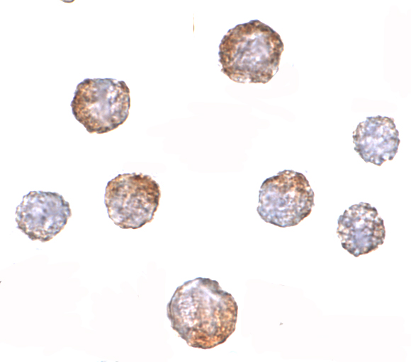
CUEDC2 reagents have illustrated paraffin-section IHC in human thyroid cancer tissue (PA1962 image caption) and ICC in HeLa cells (A08885 image caption); both list Human, Mouse and Rat reactivity (catalog).
PA1962 lists IHC and shows staining of human thyroid cancer tissue in a paraffin section (catalog applications; PA1962 image caption). A08885 lists ICC and shows staining of HeLa cells at 5 μg/mL (catalog applications; A08885 image caption).
Which to pick: Choose PA1962 for paraffin-section IHC: its image caption documents IHC(P) in human thyroid cancer tissue; the fixative is unreported (PA1962 image caption). Choose A08885 for cultured-cell IF/ICC: its ICC image shows HeLa cells at 5 μg/mL (A08885 image caption). Both list Human, Mouse and Rat reactivity, so the catalog gives neither a cross-species advantage; both are rabbit antibodies with clone type unreported (catalog reactivity, host and clone fields).