CXCL12 / Stromal cell-derived factor 1 · IHC design guide

Design Immunohistochemistry for CXCL12

Plan paraffin-section CXCL12 IHC around cytoplasmic and nuclear staining, with additional plasma positivity (HPA tissue IHC). The IHC-validated antibody has a stated working range of 0.5–1 μg/mL (datasheet A00053-2).

Evidence assembled Oct 2026 · For research use; verify linked source records and product datasheet before use
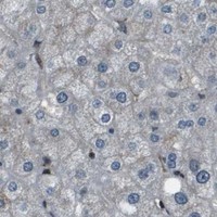
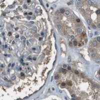
Immunohistochemistry protocol sheet for CXCL12 (IHC for CXCL12): expected localisation Cytoplasmic and nuclear staining, with plasma positivity (HPA tissue IHC), antibody A00053-2, validated IHC image, and IHC protocol steps
Printable CXCL12 IHC protocol sheet — expected localisation Cytoplasmic and nuclear staining, with plasma positivity (HPA tissue IHC), antibody A00053-2, controls and protocol steps. Open the full CXCL12 IHC guide →

CXCL12 Immunohistochemistry Experimental Design Guide

Expected localisation, validated protocols, controls and antibodies — the at-a-glance facts below, then the full design guide.

Must know before staining
Expected localisation Cytoplasmic and nuclear staining, with plasma positivity (HPA tissue IHC)
Staining pattern Most tissues: cytoplasmic/nuclear; Leydig cells medium (HPA tissue IHC)
Antigen retrieval Citrate pH 6 HIER, heat-mediated (datasheet A00053-2)
Positive control ⓘ Testis
Negative control ⓘ Adipose tissue+4 more · see all
Important caveats
Reasons your staining may differ from the expected pattern.
Fixation Keep fixation consistent across sections (standard IHC practice; not target-specific); Matched tissue-IHC evidence does not establish the fixation claim. Validate the specimen-specific method before use. (selected-SKU IHC image A00053-2)
Caveat Secreted CXCL12: tissue RNA and protein may differ (HPA tissue IHC)
Regulation Isoform expression varies by tissue (UniProt)
Isoform / epitope 7 isoforms; N-terminal epitope coverage is unclear (UniProt)
Section 1

Recommended CXCL12 IHC & IF Protocols

The catalog antibody’s IHC-P protocol (datasheet: citrate pH 6 HIER) is accompanied by four published CXCL12 IHC protocols (PMC10028106; PMC4012992; PMC11955448; PMC3085597).

Recommended immunohistochemistry (IHC-P) protocol parameters
SampleParaffin-embedded human endometrial carcinoma tissue; fixative not specified (datasheet A00053-2)
FixationImage fixative and duration unreported (datasheet A00053-2); verify before use.
Sectioning4–5 µm sections on charged slides (standard)
DeparaffinisationXylene, graded ethanol series to water (standard)
Antigen retrievalHeat retrieval: Citrate pH 6, 20 min (datasheet A00053-2)
Peroxidase block3% H2O2, 10 min, room temperature (standard)
Blocking10% goat serum (datasheet A00053-2)
Primary antibodyRabbit anti-CXCL12, 0.5-1μg/ml (datasheet A00053-2)
Primary incubationOvernight at 4 °C (datasheet A00053-2)
DetectionStreptavidin-biotin complex (SABC), DAB chromogen (datasheet A00053-2)
CounterstainHematoxylin, blue, dehydrate and mount (standard)
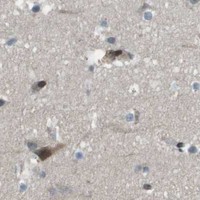
Expected resultCXCL12-positive staining in leydig cells of testis (HPA tissue IHC: Medium). HPA tissue profile: Cytoplasmic and nuclear expression in most tissues with additional plasma positivity. No signal in the no-primary control.
💡Decision noteStart with citrate pH 6 heat retrieval (datasheet: citrate pH 6 HIER); one published protocol boils sections for 10 minutes, then cools them for 20 minutes (PMC3085597).
Section 2

What Is the Expected CXCL12 Staining Pattern?

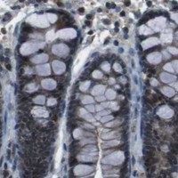
CXCL12 is secreted and has no transmembrane segment (UniProt P48061 topology). In paraffin tissue IHC, HPA reports cytoplasmic and nuclear staining in most tissues, with additional plasma positivity (HPA: Approved tissue profile). Leydig cells in testis provide a medium staining example (HPA: Testis, Leydig cells). Because the protein is secreted, the cells containing stain need not match the cells expressing its RNA (HPA: reliability description).

What am I looking at on my slide?
Cytoplasmic and nuclear signal, with possible plasma staining, in tissue IHC.This fits the reported distribution (HPA: Approved tissue profile). Judge a positive result by its cell context and controls as well as its compartment; secreted CXCL12 may be detected away from its producing cells (UniProt P48061 subcellular location; HPA: reliability description).
Leydig cells show moderate chromogenic staining in a testis section.This is a practical positive reference: HPA scores Leydig cells Medium (HPA: Testis, Leydig cells). Match the cell type before comparing intensity; the HPA score describes an observed example, not a required threshold for every specimen.
Signal forms an exclusive, sharp membrane rim with no cytoplasmic or nuclear staining.Treat this as suspect because CXCL12 has no transmembrane segment (UniProt P48061 topology) and HPA reports cytoplasmic and nuclear tissue staining (HPA: Approved tissue profile). Check specificity controls before calling the rim CXCL12.
Strong stain appears in cells HPA lists as undetected, such as adipocytes.HPA reports adipocytes as Not detected in adipose tissue and breast (HPA: tissue IHC). Unexpected stain warrants checks for antibody cross-reactivity or endogenous chromogenic activity (general IHC practice); the reference observation alone cannot prove either cause.
Color spreads across tissue and blank areas, obscuring cell boundaries.This is background until controls support a cell-specific signal (general IHC practice). Because HPA also reports plasma positivity (HPA: Approved tissue profile), assess vascular color separately from diffuse reagent or detection background.
💡Expected CXCL12 appearanceCall a section positive when interpretable cytoplasmic and nuclear staining, possibly with plasma positivity, follows the HPA tissue profile and a testis reference shows medium Leydig cell staining; isolated membrane rims or staining in blank controls are suspect (HPA: Approved tissue profile; HPA: Testis, Leydig cells; UniProt P48061 topology; general IHC practice).
How each factor affects the staining
Secretion and tissue contextCXCL12 is secreted (UniProt P48061 subcellular location). HPA cautions that protein and RNA locations may differ (HPA: reliability description), so an IHC-positive cell need not be identified as the producing cell solely from its stain.
Processing and isoformsUniProt lists a 1–21 signal peptide, processed chains beginning at residue 22 or 24, and 7 isoforms (UniProt P48061 processing; isoforms). Interpret antibody coverage only if its documented epitope is known; the supplied record gives none.
Reference tissue and validationHPA reports Medium Leydig cell staining and marks tissue IHC Approved (HPA: Testis, Leydig cells; tissue reliability). CAB017564 is IHC Approved (HPA: antibodies); this does not establish enhanced agreement between independent antibodies.
Antigen retrieval choiceNo CXCL12-specific retrieval condition or fixation sensitivity is supplied (HPA: tissue IHC; UniProt P48061). For paraffin IHC, follow the IHC-validated antibody's documented retrieval instructions and compare controls if optimizing (general IHC practice).
IF/ICC: is the same pattern established?No cell lines with ICC-IF images or subcellular image location are provided (HPA: subcellular ICC-IF). HPA labels CXCL12 secreted (HPA: subcellular summary); its tissue IHC pattern should not be treated as an independently demonstrated IF/ICC image pattern.
Why is my staining missing, weak or wrong?
SituationLikely causeNext action
No signal appears in the testis reference or study section.A failed staining run or an unsuitable antibody workflow is possible (general IHC practice); HPA reports Medium Leydig cell staining as a reference (HPA: Testis, Leydig cells).Check that Leydig cells are present, then verify primary antibody, documented retrieval, detection reagents and a run control (general IHC practice). Do not infer specimen negativity from a failed positive reference.
Only a sharp membrane outline is visible.Membrane-only staining conflicts with the absence of a transmembrane segment (UniProt P48061 topology) and the reported cytoplasmic and nuclear profile (HPA: Approved tissue profile).Review the primary-omission control and compare adjacent cells; if the rim persists without specific intracellular staining, withhold a CXCL12-positive call (general IHC practice).
Adipocytes stain strongly in a comparison section.HPA lists adipocytes as Not detected in adipose tissue and breast (HPA: tissue IHC); cross-reactivity or endogenous detection activity is possible (general IHC practice).Inspect cell identity and a primary-omission control, including any endogenous enzyme signal, before interpreting the stain (general IHC practice).
The whole section or blank spaces develop color.Nonspecific background from blocking, washing or chromogenic detection may obscure localized signal (general IHC practice). Plasma positivity can also occur in tissue (HPA: Approved tissue profile).Use a primary-omission control and review blocking, washing and detection exposure; distinguish vascular plasma color from color in cell-free blank areas (general IHC practice; HPA: Approved tissue profile).
The observed distribution differs from CXCL12 RNA maps.Secreted protein can occupy a different tissue location from its RNA (HPA: reliability description; UniProt P48061 subcellular location).Score protein staining in identified cells and tissue spaces using IHC controls; do not assign the protein's source solely from colocalization with RNA (general IHC practice; HPA: reliability description).
A specimen has little or no stain despite a working positive reference.HPA records Not detected in several specified cell populations, including bone marrow hematopoietic cells (HPA: tissue IHC); its examples do not establish that every cell or specimen must stain.Record the scored cell type and control performance, compare with the matching HPA cell-level observation, and report an interpretable negative without generalizing it to the entire tissue (HPA: tissue IHC; general IHC practice).

Sample controls for CXCL12 IHC & IF

🧪Run testis first and assess staining in Leydig cells (HPA: Medium in Leydig cells). Use adipose tissue adipocytes as a negative comparison (HPA: Not detected in adipocytes); on the testis slide, assess non-Leydig areas for background staining without assuming those cells are CXCL12-negative.
Positive control tissue: Testis (Leydig cells, HPA Medium)
Negative control tissue: Adipose tissue (HPA Not detected)
ICC-IF cell lines (HPA subcellular resource): HPA carries no ICC-IF cell line for CXCL12; derive a cell-line control from the positive tissue's cell type (Leydig cells) and confirm it by RNA or western blot first.
Technical controls: Include a no-primary (secondary-only) control, a concentration-matched rabbit isotype control (caption: rabbit primary antibody), and CXCL12-knockout tissue or a validated peptide-block control. Quench endogenous peroxidase and check for endogenous biotin because the reported IHC detection uses a biotin-based system with DAB (selected-SKU IHC caption).
⚠️Feasibility: A target-specific fixation window or fixation effect is unreported, and the selected-SKU paraffin-section caption does not state a fixative. The reported IHC example used citrate retrieval at pH 6 for 20 minutes (selected-SKU IHC caption); whether retrieval is essential is unreported. The supplied evidence does not establish that frozen sections or IF are easier, and reports no testis-specific artefact (HPA: no ICC-IF images; selected-SKU IHC caption).

HPA tissue IHC evidence for CXCL12

Comprehensive Human Protein Atlas IHC scoring per tissue (reliability: Approved — Secreted protein, tissue location of RNA and protein is expected to differ.). Rows are taken directly from the HPA tissue chart — click any row's HPA link to view the source.

Positive expression · recommended positive controls

TissueCell typeLevelEvidenceSource
Testis Leydig cells Medium Protein (IHC) HPA →

Undetected expression · recommended negative controls

TissueCell typeLevelEvidenceSource
Adipose tissue Adipocytes Not detected Protein (IHC) HPA →
Adrenal gland Glandular cells Not detected Protein (IHC) HPA →
Appendix Glandular cells Not detected Protein (IHC) HPA →
Bone marrow Hematopoietic cells Not detected Protein (IHC) HPA →
Breast Adipocytes Not detected Protein (IHC) HPA →
Section 3

Advanced CXCL12 IHC Tips

Troubleshoot CXCL12 staining in paraffin sections by checking retrieval, controls, cellular context and the limits of the antibody evidence.

Which retrieval condition should I start with for CXCL12 in paraffin sections?
Start with heat-mediated retrieval in citrate buffer at pH 6 for 20 minutes (datasheet A00053-2). The selected paraffin-section image used this condition before overnight incubation with 1 μg/mL catalog antibody at 4°C (datasheet A00053-2). If staining is weak, compare a modestly longer heating interval on adjacent sections while keeping antibody concentration, detection and development constant (standard IHC practice). Include a no-primary control and a known positive section in each comparison; excessive heating can damage tissue or increase nonspecific staining, obscuring any gain in signal (standard IHC practice).
Could fixation explain weak or uneven CXCL12 staining?
The selected CXCL12 image identifies a paraffin-embedded section but does not state its fixative, so target-specific fixation sensitivity is unknown (datasheet A00053-2). Record the fixative, fixation duration and processing history for each specimen, then compare similarly processed sections before attributing a staining difference to biology (standard IHC practice). Examine tissue preservation and section adhesion alongside the CXCL12 signal, because poor morphology makes weak staining difficult to interpret (standard IHC practice). If fixation varies between batches, run the same positive and no-primary controls with each batch and report that variation when comparing scores (standard IHC practice).
How should I assess cytoplasmic, nuclear and extracellular CXCL12 staining?
CXCL12 is annotated as secreted and has no transmembrane segment, so staining need not form a continuous cell-surface outline (UniProt P48061 topology). Tissue IHC reports cytoplasmic and nuclear expression in most tissues, with additional plasma positivity, while warning that RNA and protein locations may differ for this secreted protein (HPA tissue IHC). Score cell-associated cytoplasmic and nuclear signal separately from extracellular or plasma-associated signal, using the same compartment definitions across sections (standard IHC practice). Review the morphology and no-primary control before calling diffuse chromogen specific, and avoid assigning extracellular signal to its nearest cell without supporting evidence (standard IHC practice).
Can this stain distinguish CXCL12 isoforms or processed forms?
Do not assign an isoform from chromogenic staining unless the catalog antibody’s mapped epitope and cross-reactivity support that assignment (standard IHC practice). CXCL12 has 7 annotated isoforms, and its precursor contains a 1–21 signal peptide with annotated processed chains beginning at residue 22 or 24 (UniProt P48061 processing and isoforms). Check whether the antibody epitope lies within a region retained by the forms being compared; no epitope map is supplied here (standard IHC practice; supplied datasheet A00053-2). Report the result as CXCL12 immunoreactivity unless isoform specificity has been established independently, and keep retrieval and scoring consistent between specimens (standard IHC practice).
How could I check a CXCL12 IHC pattern by multiplex immunofluorescence?
Treat immunofluorescence as a separate assay requiring its own antibody validation; the supplied antibody image documents paraffin-section chromogenic IHC (datasheet A00053-2). Pair CXCL12 with a validated marker for the expected cell population, such as Leydig cells in testis, and assess overlap against single-stain controls (HPA tissue IHC; standard IF practice). Choose a fluorophore channel with low tissue autofluorescence and verify bleed-through with single-color controls before interpreting proximity as coexpression (standard IF practice). Because CXCL12 is secreted and has no transmembrane segment, compare nonpermeabilized staining for accessible extracellular antigen with permeabilized staining for intracellular antigen; the antibody’s epitope accessibility is not supplied (UniProt P48061 topology; standard IF practice).
What should I check when CXCL12 DAB staining looks diffuse?
The selected image used a biotinylated secondary antibody, a streptavidin–biotin detection complex and DAB, so evaluate each detection step when background appears (datasheet A00053-2). Include no-primary and secondary-only controls, and apply a peroxidase block as part of the chromogenic workflow to assess enzyme-related signal (standard IHC practice). Compare background in tissue, vessels and damaged regions before increasing confidence in a diffuse pattern, since CXCL12 is secreted and HPA reports additional plasma positivity (UniProt P48061 subcellular annotation; HPA tissue IHC). If controls stain, adjust blocking, washes or DAB development one variable at a time while preserving the positive control signal (standard IHC practice).
How should I score CXCL12 across paraffin sections? ⚠ ANSWER MARKED FOR VERIFICATION
Define the compartment and analysis area before scoring, because secreted CXCL12 can yield cell-associated and extracellular signal (UniProt P48061 subcellular annotation; standard IHC practice). For cell-associated staining, record the percentage of positive cells and intensity by category, then calculate an H-score if that scheme is used consistently (standard IHC practice). For extracellular deposits, measure positive area or signal density per mm² of viable tissue separately instead of folding it into a cell H-score (standard IHC practice). Normalize cell counts to eligible cells, exclude folds and necrosis, and use matched staining and imaging settings across specimens (standard IHC practice).
How can I distinguish credible CXCL12 signal from artefact?
A credible result should reproduce in similarly processed sections, exceed no-primary background and fit the specimen’s morphology (standard IHC practice). CXCL12 is secreted, and HPA reports cytoplasmic and nuclear tissue staining with additional plasma positivity, so nuclear or extracellular signal alone is not grounds for rejection (UniProt P48061 subcellular annotation; HPA tissue IHC). Check staining at section edges, folds and necrotic areas, and use a peroxidase-blocked control to investigate endogenous enzyme signal (standard IHC practice). Question a pattern confined to an unsupported compartment or cell population until controls and an independent assay support it; HPA reports medium staining in testis Leydig cells (HPA tissue IHC; standard IHC practice).
Boster reagents

Best CXCL12 / Stromal cell-derived factor 1 IHC Antibodies

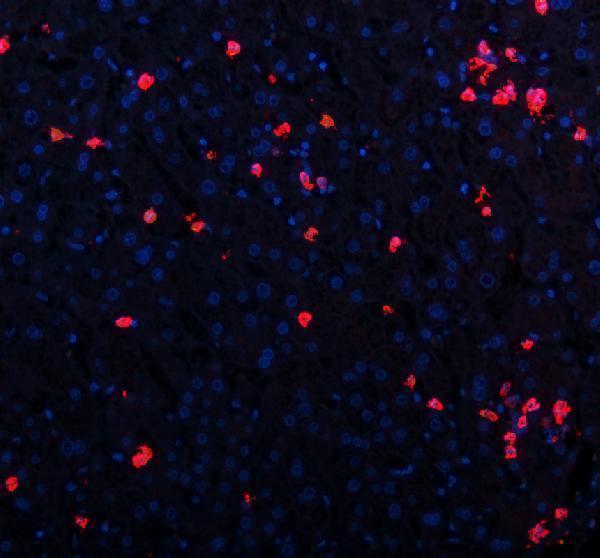
Anti-CXCL12 image data cover paraffin-section IHC in human and rat tissue, tissue IF in human, mouse and rat samples, and ICC in HeLa cells (catalog image captions).

Real IHC data IHC analysis of CXCL12 using anti-CXCL12 antibody (A00053-2). CXCL12 was detected in paraffin-embedded section of human endometrial carcinoma tissue. Heat mediated antigen retrieval was performed in citrate buffer (pH6, epitope retrieval solution) for 20 mins. The tissue section was blocked with 10% goat serum. The tissue section was then incubated with 1μg/ml rabbit anti-CXCL12 Antibody (A00053-2) overnight at 4°C. Biotinylated goat anti-rabbit IgG was used as secondary antibody and incubated for 30 minutes at 37°C. The tissue section was developed using Strepavidin-Biotin-Complex (SABC)(Catalog # SA1022) with DAB as the chromogen.
Anti-CXCL12 Antibody
Cat # A00053-2
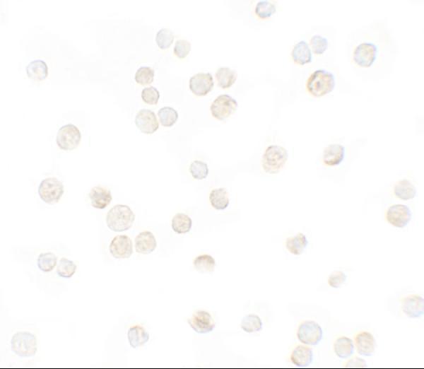
Real IF data Immunocytochemistry of CXCL12 in HeLa cells with CXCL12 antibody at 5 μg/mL.
Anti-Stromal cell-derived factor 1 CXCL12 Antibody
Cat # A00053-3

A00053-2 has paraffin-section IHC images from human endometrial carcinoma, tonsil and liver cancer tissue and rat spleen, plus tissue IF images from human hepatitis and tonsil tissue and mouse and rat spleen (A00053-2 image captions). A00053-3 has an ICC image in HeLa cells at 5 μg/mL (A00053-3 image caption).

Which to pick: For paraffin-section IHC, choose A00053-2: its listed IHC range is 0.5–1 μg/mL, and its IHC captions describe citrate pH 6 or citrate pH 6 retrieval; the fixative is unreported (A00053-2 catalog dilution and IHC image captions). For tissue IF, A00053-2 has section images at 5 μg/mL; for cell ICC, A00053-3 has a HeLa image at 5 μg/mL (A00053-2 IF image captions; A00053-3 image caption). For work across species, the polyclonal A00053-2 lists human, mouse and rat reactivity, with IF images for all three and IHC images for human and rat tissue (A00053-2 catalog dilution_raw, reactivity and image captions).

Each figure is that product's own IHC / IF validation image from its datasheet.

References

  1. UniProt Consortium. UniProt entry P48061 (SDF1_HUMAN, Stromal cell-derived factor 1).
  2. Human Protein Atlas. CXCL12 tissue IHC expression (reliability: Approved).
  3. Human Protein Atlas. CXCL12 subcellular location (ICC-IF): Secreted.
  4. Human Protein Atlas. CXCL12 antibody validation summary (1 antibodies).
  5. CXCL12 is expressed by skeletal muscle cells in tongue oral squamous cell carcinoma. Cancer medicine 2023 — PMC10028106.
  6. Function of chemokine (CXC motif) ligand 12 in periodontal ligament fibroblasts. PloS one 2014 — PMC4012992.
  7. The expression of autophagy-related gene CXCL12 in endometriosis associated ovarian cancer and pan-cancer analysis. Frontiers in endocrinology 2025 — PMC11955448.
  8. The expression of CXCR4, CXCL12 and CXCR7 in malignant pleural mesothelioma. The Journal of pathology 2011 — PMC3085597.
  9. PubMed PMID:7490086 — UniProt-cited evidence.
  10. PubMed PMID:16626895 — UniProt-cited evidence.
  11. PubMed PMID:14702039 — UniProt-cited evidence.