This website uses cookies to ensure you get the best experience on our website.
- Table of Contents
1 Citations 8 Q&As
2 Citations 10 Q&As
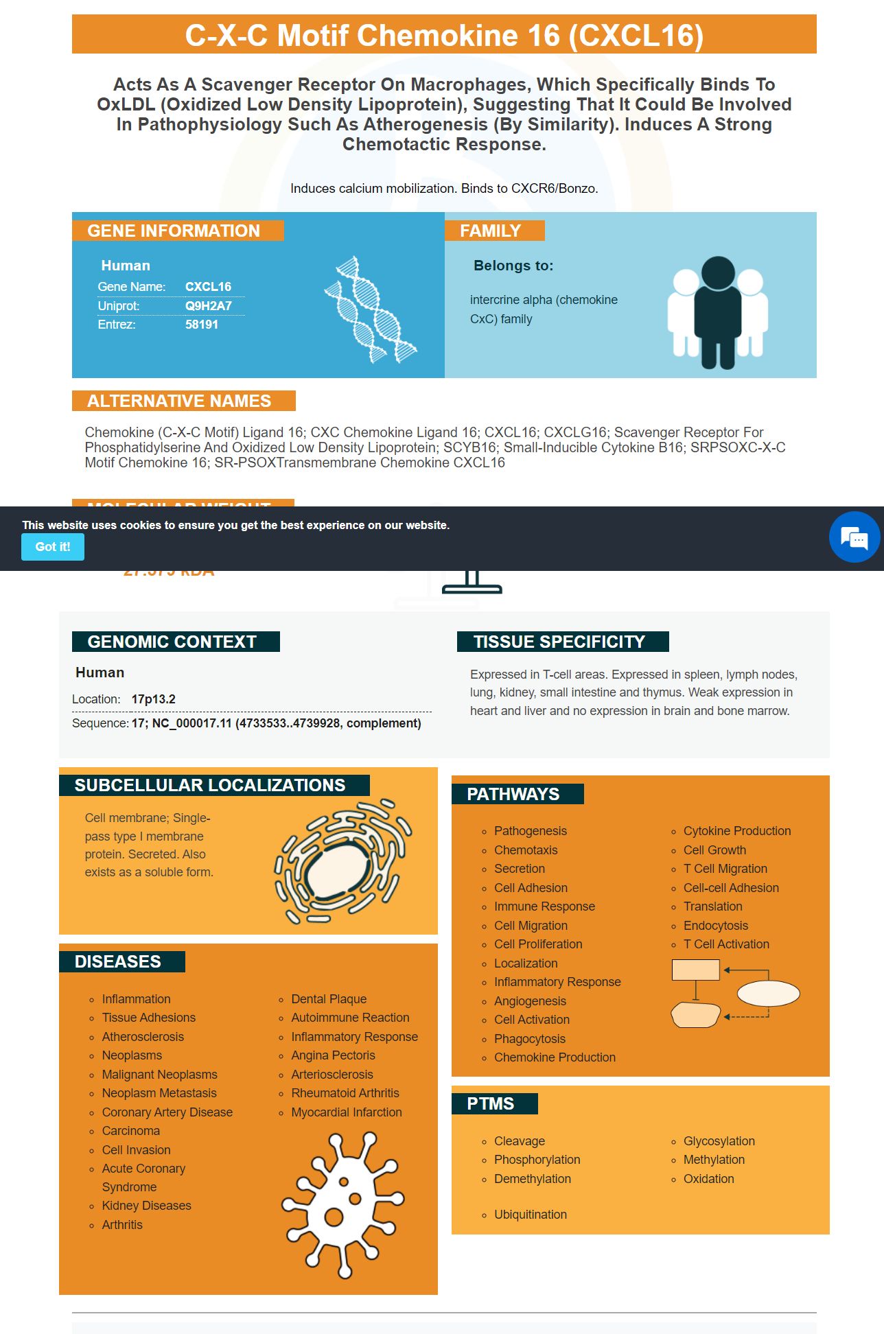
Facts about C-X-C motif chemokine 16.
Induces calcium mobilization. Binds to CXCR6/Bonzo.
Human | |
---|---|
Gene Name: | CXCL16 |
Uniprot: | Q9H2A7 |
Entrez: | 58191 |
Belongs to: |
---|
intercrine alpha (chemokine CxC) family |
chemokine (C-X-C motif) ligand 16; CXC chemokine ligand 16; CXCL16; CXCLG16; Scavenger receptor for phosphatidylserine and oxidized low density lipoprotein; SCYB16; Small-inducible cytokine B16; SRPSOXC-X-C motif chemokine 16; SR-PSOXTransmembrane chemokine CXCL16
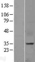
Mass (kDA):
27.579 kDA
Human | |
---|---|
Location: | 17p13.2 |
Sequence: | 17; NC_000017.11 (4733533..4739928, complement) |
Expressed in T-cell areas. Expressed in spleen, lymph nodes, lung, kidney, small intestine and thymus. Weak expression in heart and liver and no expression in brain and bone marrow.
Cell membrane; Single-pass type I membrane protein. Secreted. Also exists as a soluble form.
This article will provide most effective methods to block EAV replication by using a pAb to inhibit the expression of cell surface CXCL16. Boster Pipettes can disperse aqueous solutions in 0.5 milliliters to 1 ml volumes. The Boster Microplate Reader is a useful instrument to measure absorbance at 350nm. It also includes product credits for sharing your results with other researchers.
Researchers have recently reported that reducing the expression of a vital HIV-1 receptor on monocytes may stop the spread of infection. The receptor is vital for HIV-1 replication and may serve as a key determinant of the replication of HIV-1. They also found that siRNA-mediated EqCXCL16 downregulation reduced the rate of replication of viruses. Researchers looked at the reservoirs of viruses inside the body to determine how siRNA might influence the immune system.
In this study, monocytes from humans were transformed into MDMs and transfected with retroviruses that express siRNA. After transfection cells were infected by the NL4-3 Bal-IRES-HSA virus with an MOI of 1.0. The viral titre was determined by X-gal staining, and TZM-bl cells.
The structure of human T-lymphotropic viruses type 1 was published in the journal Nucleic Acids Res. The structure of UPF1-like proteins of the virus (UPF1) was identified. Upf1 is involved in decapsidation of mRNA. It is also involved with the metabolism of mRNA, translation, and other functions.
The study showed that UPF1 and SMG6 decreased their expression at a cellular level by a logarithmic scaling. These proteins play a significant role in the HIV-1's life cycle. These results suggest that the inhibitors may work together. This study suggests that UPF1 could be used to block EAV replication.
Blocking EAV replication with siRNA-mediated EqCXCL16 cell surface protein provides additional advantages. Inhibiting EqCXCL16 could be a promising strategy to stop viral replication and prevent infection of cells infected. This treatment also stops the replication of viruses in stable HEK-EqCXCL16 cell cultures.
EAV replication was inhibited by siRNA-mediated inhibition of the cell surface expression of EqPXCL16. This could be similar to the disease-related state. The knockdown efficiency of cells was determined every 24 hours during a 6-day experiment. Cells were cultured in triplicate for eight months. The knockdown effectiveness of the cells was determined by the qRT-PCR.
The virus was tested in horses by researchers using standardbred horses ST22 and TB10. The genomic DNA was extracted from purified peripheral blood mononuclear cell lines. For cDNA amplification the Qiagen Smart DNA synthesis kit was used. DNA was analysed by using the NanoDrop spectrophotometer.
The study also showed that direct targeting the viral genome of RNA is more effective than indirect targeting of the entry receptor ACE1. The siRNA candidates used in this study, siV1 and siV6, are extremely effective. The siRNAs might prove to be effective therapeutic agents. Researchers have shown that siRNAs targeting these receptors may prevent the replication of viruses and protect human cells from SARS.
The virus's replication was inhibited by siRNA-mediated upregulation EqPXCL16 cells. After 48 hours, the knockdown had reached 90 percent and remained there until the sixth day. The results were confirmed by gene analysis of reporter genes. The authors would like to thank all donors for their cooperation. After all, they are all very thankful for the kindness of their patients and the great care they give to the virus.
This study demonstrated that blocking the cell surface EqCXCL16 by using a Gp Anti-EqCXCL6 Ab inhibits EAV infection in HeK-EqCX16 cells. The antibodies were prediluted with 5 percent nonfat milk powder in TBS-T with 10 mM TrisHCl pH 7.6 150 mM NaCl, and 0.1 percent Tween 20. After incubation for 4°C over an hour, membranes were immunoblotted using an initial Ab (5 milliliters) diluted in bovine se albumin at 5% and the anti GP5 mAb 6D10.
Flow analysis of cytometry proved that the cells used in the production of the pAbs were pure and confirmed the specificity of their reagents. The Gp anti-EqCXCL16 pAb was not reacting with mock-transfected HEK-293T cells. The analysis of flow cytometry showed that the Gp anti-EqCXCL16 polyclonal mAb was able to block the EqCXCL16 protein in HEK cells.
In a subsequent research, we discovered that EqCXCL16 was critical for EAV entry in specific cell populations. Anti-EqCXCL16Gp anti -EqCXCL16 EAV infections in CD14+ monoocytes of the equine. We utilized EAV sVBSmCherry virus as the model for our study.
To determine if Gp anti-EqCXCL6 antigen mAb interferes with mCh expression, we employed siRNAs to suppress EqCXCL16 mRNA. We used a commercially-available siRNA duplex as a negative control. In a subsequent study, we transfected cells HEK-EqCXCL16 with either Gp anti EqCXCL6 mRNA or a scrambled siRNA duplex. The cells were then harvested and Western blot analysis was performed to study the expression of EqCXCL16 and nonstructural viral proteins 1.
Gp anti-EqCXCL6 Abs inhibits the expression nonstructural protein EqCXCL16 which is involved in the development and development of viral infection. We immunized naive HEK-293T as well as stable HEK cells using EqCXCL16 sVBSmCherry , with an MOI of 1.0. We observed that stable cells expressed more EAV and nsp-1, compared to naive cells.
The Gp anti–EqCXCL16.5 mAb blocks Gp–ab, which is believed to be an EAV-binding receptor. Gp anti-EqCXCL16 MAb that targets Gp, which is a potential EAV receptor.
HEK-293T cells were isolated from human fibroblasts. The primary antibodies were dispersed in flow buffer comprising 10% normal goat serum and 0.1 percent sodium azide. After incubating for one hour after which the cells were stained with Alexa Fluor 488. The slides were placed on coverslips using Vectashield mounting medium.
Understanding the role of the antigen is crucial in understanding how to construct antibodies against the EqCXCL16 marker. The immune system of the body produces antibodies, which are proteins. Foreign molecules that enter our bodies can trigger an immune response. The body recognizes these antigens by the immune system and generate antibodies against them. These antibodies circulate through blood and lymph and are able to bind to specific antigens. They can be produced by the body or removed out of circulation.
The procedures for producing antibodies were developed in the 1970s and 80s and remain largely unchanged since the classic work of Harlow and Lane, "Antibodies: A Laboratory Manual," published in 1988. Production of antibodies encompasses the steps that follow making the immunogen, immunizationand creation of hybridomas, collection isotyping, cleaning, and labeling. This procedure is the most crucial step in antibody production.
Protective Ab responses are generated when PCs are activated. In vivo, PCs migrate to the bone marrow where they secrete large amounts of GM-CSF. The PC marker CD138 is produced by innate response activator B cells (IRA B cells) which are the closest to the PCs. PCs are therefore a critical part of the immune system and hematopoiesis.
Cloning a sequence of a horse monoocyte of equine CXCL16 genes is the first step to generate Abs to the EqCXCL16 markers. The sequence of EqCXCL16 was amplified by an PCR procedure using a cDNA sample. Utilizing a Smart CDNA Synthesis kit from Clontech Laboratories Inc., amplifying cDNA was done using a standard laboratory PCR protocol. For this study we used forward primer cx16-15F as well as reverse primer cx16-15R, both from IDT.
It is unclear what the immunological function of antibodies in SARS/CoV-2. However, the antibody response is just one element of a more complex immune response that includes a variety of aspects of innate immunity and T-cell activity that is specific to the virus. Numerous studies have demonstrated positive connections between antibodies and protection, whereas others don't show any connection in any way. It is essential to know how antibodies work.
This method involves staining individual beads at saturation levels. The beads are then combined and analyzed by using flow cytometry. This generates an average curve that shows the geometric average fluorescence intensity against the antibody's binding ability. The antigen density that results is proportional to the surface receptors. Therefore, the antigen density is equal to the total number of surface receptors.
PMID: 11017100 by Matloubian M., et al. A transmembrane CXC chemokine is a ligand for HIV-coreceptor Bonzo.
PMID: 11290797 by Wilbanks A., et al. Expression cloning of the strl33/bonzo/tymstr ligand reveals elements of cc, cxc, and cx3c chemokines.
*More publications can be found for each product on its corresponding product page