This website uses cookies to ensure you get the best experience on our website.


- Table of Contents
Plan DPYSL5 paraffin IHC around cytoplasmic staining in CNS and testis (HPA tissue IHC). Use caudate neurons as a positive reference (HPA tissue IHC), and start the IHC-P antibody M08644-1 at 1:25 (datasheet M08644-1).

Expected localisation, validated protocols, controls and antibodies — the at-a-glance facts below, then the full design guide.
| Expected localisation | Cytoplasmic staining in CNS and testis (HPA tissue IHC) | |
| Staining pattern | Cytoplasmic in caudate neurons and testis spermatids (HPA tissue IHC) | |
| Antigen retrieval | Citrate pH 6 HIER, heat-mediated (datasheet M08644-1) | |
| Positive control | Caudate+4 more · see all | |
| Negative control | Adipose tissue+4 more · see all |
| Fixation | Formaldehyde-fixed paraffin sections were used (selected-SKU IHC image M08644-1) | |
| Caveat | Cerebellar granular cells were undetected (HPA tissue IHC) | |
| Regulation | Expression regulation unreported (UniProt) | |
| Isoform / epitope | No annotated isoforms or processing; epitope impact unknown (UniProt) |
The catalog antibody has an IHC-P protocol (datasheet: M08644-1); the published DPYSL5 IHC methods below provide two additional tissue-staining examples (PMC10796342; PMC5319751).
| Sample | Formaldehyde-fixed, paraffin-embedded tissue sections (datasheet M08644-1; sample unspecified) |
| Fixation | Image formalin-fixed; duration unreported (datasheet M08644-1); verify before use. |
| Sectioning | 4–5 µm sections on charged slides (standard) |
| Deparaffinisation | Xylene, graded ethanol series to water (standard) |
| Antigen retrieval | Heat retrieval: Citrate pH 6 (datasheet M08644-1); 20 min, 95–100 °C (standard) |
| Peroxidase block | 3% H2O2, 10 min, room temperature (standard) |
| Blocking | 10% normal serum of the secondary host, 30 min, room temperature (standard) |
| Primary antibody | Rabbit anti-DPYSL5, 1:25 (datasheet M08644-1) |
| Primary incubation | 1 hours at 37°C (datasheet M08644-1) |
| Detection | HRP-polymer secondary, DAB chromogen 5–10 min (standard) |
| Counterstain | Hematoxylin, blue, dehydrate and mount (standard) |
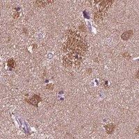
| Expected result | DPYSL5-positive staining in neuronal cells of caudate (HPA tissue IHC: High). HPA tissue profile: Cytoplasmic expression in CNS and testis. No signal in the no-primary control. |
DPYSL5 is annotated as cytoplasmic and has no transmembrane segment (UniProt Q9BPU6). In tissue IHC, expect cytoplasmic staining in caudate neuronal cells, cortical and hippocampal glial cells, and late spermatids; enterocytes also show staining (HPA tissue IHC). HPA rates the tissue pattern Enhanced, citing agreement with RNA data and GTEx support in the CNS (HPA tissue IHC).
| Strong cytoplasmic stain in caudate neuronal cells. | This matches the reported High neuronal staining and cytoplasmic tissue profile (HPA tissue IHC). Judge the pattern in identifiable cells, since a dark area without clear cellular boundaries gives less confidence that the chromogen marks DPYSL5. |
| Moderate cytoplasmic stain in cerebral cortex or hippocampal glial cells, testicular late spermatids, or small-intestinal enterocytes. | Each listed cell population is reported at Medium intensity (HPA tissue IHC). A weaker result than caudate neurons can therefore fit the reference pattern; compare corresponding cell types rather than expecting every positive tissue to stain equally. |
| Predominantly nuclear stain in a paraffin tissue section, with little cytoplasmic signal. | This differs from the reported cytoplasmic tissue IHC profile (HPA tissue IHC; UniProt Q9BPU6). Investigate the tissue pattern and controls before assigning it to DPYSL5. Nuclear staining alone is not proof of artefact: HPA separately approves nucleoplasmic localization in ICC-IF (HPA subcellular). |
| Stain concentrated in an HPA-listed negative cell population, such as adipocytes in adipose tissue or cerebellar granular-layer cells. | Those specific populations are reported Not detected (HPA tissue IHC). Consider cross-reactivity or endogenous detection activity, and review morphology and controls. The HPA result applies to the named cells; it does not declare every cell in the organ negative. |
| Widespread haze across tissue and surrounding section, or no stain in caudate neuronal cells. | A haze obscures the expected cellular pattern and may reflect nonspecific staining or detection background (general IHC practice). No caudate neuronal signal conflicts with the reported High level (HPA tissue IHC); first establish that tissue, staining run, and controls performed adequately. |
| Tissue and cell selection | Caudate neurons offer a High reference; cortical and hippocampal glia, late spermatids, and enterocytes offer Medium references (HPA tissue IHC). Use the named cell population when judging a section; whole-organ labels are too broad for these comparisons. |
| IHC versus ICC-IF localization | Tissue IHC is summarized as cytoplasmic, whereas ICC-IF lists nucleoplasm as the approved main location and cytosol as an approved additional location (HPA tissue IHC; HPA subcellular). An IF image therefore needs its own interpretation; this IHC guide supplies no ICC-IF protocol. |
| Antibody evidence | HPA034544 is rated Enhanced for IHC, while HPA072387 is listed as Approved for ICC with no IHC rating in the supplied record (HPA antibodies). Match the validation to the application; the ICC rating alone does not establish an IHC staining pattern. |
| Topology and processing | DPYSL5 has no transmembrane segment or annotated signal peptide, propeptide, or cleavage into multiple chains; the recorded chain spans residues 1–564 (UniProt Q9BPU6). A cell-surface outline alone would be unexpected from this record and should prompt a control review. |
| Epitope and preparation limits | Five modified residues are annotated, but no antibody epitope, retrieval condition, or target-specific fixation effect is supplied (UniProt Q9BPU6; HPA antibodies). Do not attribute a weak stain to a particular modification or fixation effect without separate evidence. |
| Situation | Likely cause | Next action |
|---|---|---|
| Caudate neuronal cells show no convincing stain. | The result conflicts with their reported High level (HPA tissue IHC); the record does not identify a DPYSL5-specific preparation failure. | Check tissue identity and run controls, then review the IHC-validated antibody's specified paraffin-section procedure and the detection steps (general IHC practice). |
| A brown haze covers positive and negative regions. | Possible nonspecific binding, excess reagent, or detection background (general chromogenic IHC practice); haze cannot be scored as the cytoplasmic pattern (HPA tissue IHC). | Review blocking, washing, antibody concentration, and no-primary control; assess stain only where cell boundaries and compartment remain clear (general IHC practice). |
| Adipose adipocytes or cerebellar granular-layer cells stain prominently. | These named cells are reported Not detected (HPA tissue IHC). Cross-reactivity or endogenous detection activity are possibilities, not established causes. | Confirm cell identity; compare a no-primary control and, where relevant, an endogenous-enzyme block before interpreting the stain as DPYSL5 (general chromogenic IHC practice). |
| Nuclear stain dominates a tissue IHC section. | That differs from the cytoplasmic tissue profile, although nucleoplasm is an approved ICC-IF location (HPA tissue IHC; HPA subcellular). | Check whether cytoplasmic signal is present in known-positive cells, inspect controls, and report the IHC–ICC-IF difference without treating the IF result as an IHC validation. |
| A testis or small-intestine section is scored entirely negative or entirely positive. | HPA reports Medium staining in late spermatids or enterocytes specifically, not a uniform organ-wide signal (HPA tissue IHC). | Locate the named cell population before scoring; compare its cytoplasm with neighboring cells and the run controls (HPA tissue IHC; general IHC practice). |
| Only a sharp cell-edge outline appears, without cytoplasmic stain. | The outline does not match the cytoplasmic tissue profile or the lack of a transmembrane segment (HPA tissue IHC; UniProt Q9BPU6). | Review morphology, no-primary control, and detection background; avoid assigning the edge signal to DPYSL5 unless independent evidence supports it (general IHC practice). |



Comprehensive Human Protein Atlas IHC scoring per tissue (reliability: Enhanced — High consistency between antibody staining and RNA expression data. GTEX supports antibody staining in CNS.). Rows are taken directly from the HPA tissue chart — click any row's HPA link to view the source.
| Tissue | Cell type | Level | Evidence | Source |
|---|---|---|---|---|
| Caudate | Neuronal cells | High | Protein (IHC) | HPA → |
| Cerebral cortex | Glial cells | Medium | Protein (IHC) | HPA → |
| Hippocampus | Glial cells | Medium | Protein (IHC) | HPA → |
| Small intestine | Enterocytes | Medium | Protein (IHC) | HPA → |
| Testis | Elongated or late spermatids | Medium | Protein (IHC) | HPA → |
| Tissue | Cell type | Level | Evidence | Source |
|---|---|---|---|---|
| Adipose tissue | Adipocytes | Not detected | Protein (IHC) | HPA → |
| Adrenal gland | Glandular cells | Not detected | Protein (IHC) | HPA → |
| Appendix | Endocrine cells | Not detected | Protein (IHC) | HPA → |
| Bone marrow | Hematopoietic cells | Not detected | Protein (IHC) | HPA → |
| Breast | Adipocytes | Not detected | Protein (IHC) | HPA → |
Troubleshoot DPYSL5 chromogenic IHC by checking retrieval, tissue controls, cell identity and staining compartment; use the IF guidance for complementary localisation checks.
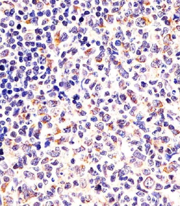
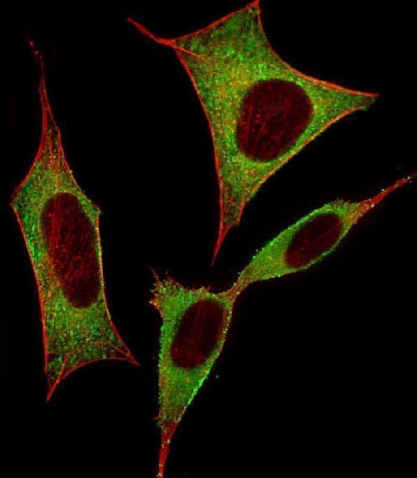
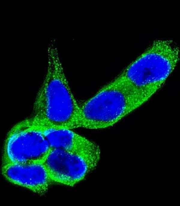
Catalog anti-DPYSL5 antibodies have human tonsil paraffin-section IHC and human SH-SY5Y cell IF images (catalog figure captions); all three list Human, Mouse, and Rat reactivity (catalog reactivity).
M08644-1 has an IHC-P image of human tonsil paraffin sections (M08644-1 figure caption). M08644 and M08644-2 each have an IF image of human SH-SY5Y cells (respective figure captions).
Which to pick: For tissue IHC, choose M08644-1: its own paraffin-section caption reports citrate retrieval at pH 6 and a 1:25 primary dilution (M08644-1 figure caption). For cell IF/ICC, choose rabbit polyclonal M08644 or mouse monoclonal M08644-2; each has a cytoplasmic SH-SY5Y IF image at 1:25 (catalog antibody descriptions and respective figure captions). For Mouse or Rat samples, all three list reactivity, while the supplied images show Human samples only; select by application and validate staining in the intended species (catalog reactivity and figure captions).