This website uses cookies to ensure you get the best experience on our website.
- Table of Contents

Facts about Dentin matrix acidic phosphoprotein 1.

During the osteoblast to osteocyte transition phase it is phosphorylated and exported to the extracellular matrix, where it regulates nucleation of hydroxyapatite. .
| Human | |
|---|---|
| Gene Name: | DMP1 |
| Uniprot: | Q13316 |
| Entrez: | 1758 |

| Belongs to: |
|---|
| No superfamily |

ARHP; ARHR; dentin matrix acidic phosphoprotein 1; dentin matrix acidic phosphoprotein; Dentin matrix protein 1; DMP1; DMP-1
Mass (kDA):
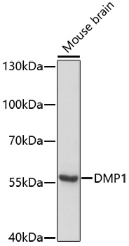
55.782 kDA

| Human | |
|---|---|
| Location: | 4q22.1 |
| Sequence: | 4; NC_000004.12 (87650280..87664357) |
Expressed in tooth particularly in odontoblast, ameloblast and cementoblast.
Nucleus. Cytoplasm. Secreted, extracellular space, extracellular matrix. In proliferating preosteoblasts it is nuclear, during early maturation stage is cytoplasmic and in mature osteoblast localizes in the mineralized matrix. Export from the nucleus of differentiating osteoblast is triggered by the release of calcium from intracellular stores followed by a massive influx of this pool of calcium into the nucleus.


PMID: 9177774 by Hirst K.L., et al. Elucidation of the sequence and the genomic organization of the human dentin matrix acidic phosphoprotein 1 (DMP1) gene: exclusion of the locus from a causative role in the pathogenesis of dentinogenesis imperfecta type II.
PMID: 8586437 by Aplin H.M., et al. Mapping of the human dentin matrix acidic phosphoprotein gene (DMP1) to the dentinogenesis imperfecta type II critical region at chromosome 4q21.