This website uses cookies to ensure you get the best experience on our website.
- Table of Contents
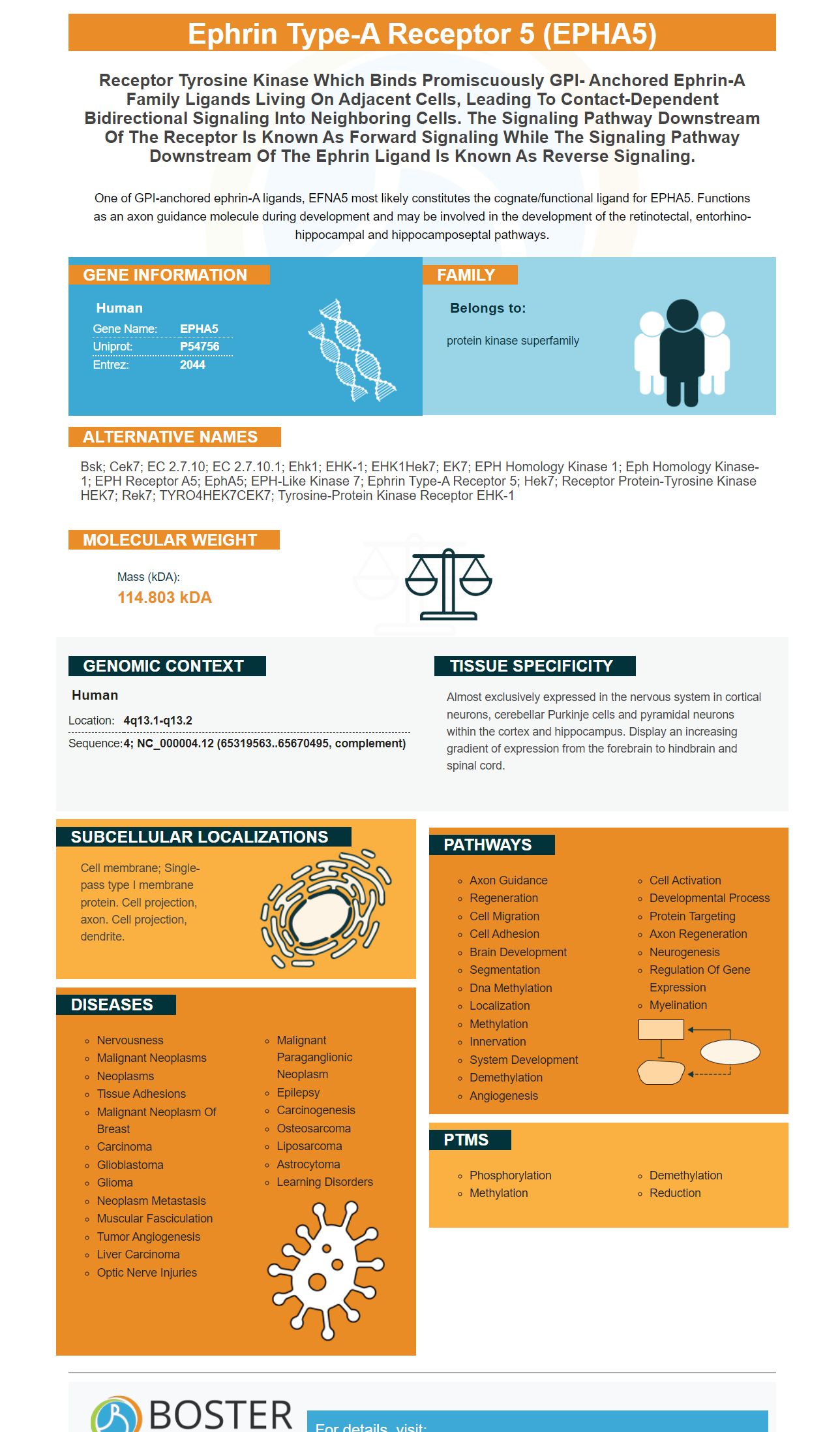
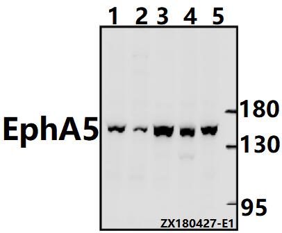
Facts about Ephrin type-A receptor 5.
One of GPI-anchored ephrin-A ligands, EFNA5 most likely constitutes the cognate/functional ligand for EPHA5. Functions as an axon guidance molecule during development and may be involved in the development of the retinotectal, entorhino- hippocampal and hippocamposeptal pathways.
Human | |
---|---|
Gene Name: | EPHA5 |
Uniprot: | P54756 |
Entrez: | 2044 |
Belongs to: |
---|
protein kinase superfamily |
Bsk; Cek7; EC 2.7.10; EC 2.7.10.1; Ehk1; EHK-1; EHK1Hek7; EK7; EPH homology kinase 1; Eph homology kinase-1; EPH receptor A5; EphA5; EPH-like kinase 7; ephrin type-A receptor 5; Hek7; receptor protein-tyrosine kinase HEK7; Rek7; TYRO4HEK7CEK7; tyrosine-protein kinase receptor EHK-1
Mass (kDA):
114.803 kDA
Human | |
---|---|
Location: | 4q13.1-q13.2 |
Sequence: | 4; NC_000004.12 (65319563..65670495, complement) |
Almost exclusively expressed in the nervous system in cortical neurons, cerebellar Purkinje cells and pyramidal neurons within the cortex and hippocampus. Display an increasing gradient of expression from the forebrain to hindbrain and spinal cord.
Cell membrane; Single-pass type I membrane protein. Cell projection, axon. Cell projection, dendrite.
The EPHA5 Cell Based ELISA Kit provides a easy and efficient method to determine the relative amounts of this protein in cells. The EPHA5 Cell Based ELISA Kit can also be used to test for activators and inhibitors that influence EPHA5's expression. However, it is not available in isolation and cannot be used on all samples.
The monoclonal antibody for anti-EphA5 made by BOSTER BIO recognizes the epidermal growth factor receptor (EphA10) that is expressed by MDSC and TAM cells. The antibody recognized EphA10's intact structure and produced a greater intensity of the fluorescence than the normal clone. This antigen was also observed in various tumor regions and immune-suppressive myeloid cells. However, the antibody was not able to show statistical significance.
It is also a marker for the epidermal factor receptor (EGF). These receptors are involved in many developmental processes and have been implicated in neurodegeneration as well as cancer. But, despite the degree of complexity of their role in tumorigenesis, there is still no definitive understanding of how they regulate cell growth. However, there is some connection between the ephrin subfamily and the EGFR (EGFR) which is a family of protein kinases.
The Anti-Ephrin type AB (EphA5) from Boster Bio is a monoclonal antibody made from EPHA5 protein . It is then formulated in PBS with 0.09% sodium azide. The antibody reacts with humans as well as Mice, Rats, and Humans. The antibody should be stored at -20°C when using according to the instructions. Avoid repeated freeze-thaw cycles.
The Anti-Ephrin type A receptor 5 EphA5 monoclonally manufactured by Boster Bio has been clinically examined for its efficacy. The antibody was found to significantly reduce EphA2 expression in HCC cells. EphA2 could also be a target for miR-266b which could enhance the diagnostic imaging.
Boster Bio Cell-Based EPHA5 Marker ELISA Kit allows researchers to quantify the expression of this protein. The primary antibody binds EPHA5 to cells that are targeted. Secondary antibodies conjugate with HRP, an enzyme that causes an achromatic reaction upon the addition of an appropriate substrate. The kit also comes with a variety of methods of normalization, including an Anti-GAPDH Antibody that acts as an internal positive control.
To conduct the test, GCs were transfected using siEfna5 following which they were incubated at 37°C/5%CO2 for 48 hours. After transfection, GC were washed with the FITC, PI and stained with PI. The samples were then incubated for another 48 hours at ambient temperature. The GC cells were then counted using a plate with 96 wells.
EPHA5 is a member of the Eph receptor family, which comprises the largest subset of RTKs. It is most frequently mutated in lung cancers. Patients with EPHA5 mutations demonstrated enhanced infiltration of T cells that are CD8+ with decreased recruitment of immunosuppressive regulatory T cells, and elevated levels of inhibitory immune checkpoints. The mutations were found to cooccur with homologous recombination.
Additionally, patients with cancers expressing the EPHA5 gene showed increased levels of recombination. Recombination is a signaling pathway that prevents DNA damage. The mutations in EPHA5 also boost the rate of DNA replication and repair homologous recombination and the mismatch repair pathway. To determine the function of the marker the marker has to be tested on mouse tumor models.
The EPHA5 mutation is associated with higher levels of TMB, PDL1, CD8+ cell infiltration, as well as the IFN-g signature. These findings suggest that the EPHA5 gene variant could be a prognostic biomarker. Clinical studies have revealed that patients with EPHA5-related mutations have increased survival in immunotherapy cohorts. While there isn't any evidence to suggest that the EPHA5 mutation is responsible for cancer, it can be useful in determining the underlying genetic cause of disease.
Recent studies have proven that the EPHA5 mutation contributes with an increase in the amount of immune infiltration within the TCGA LUAD data. Furthermore, the EPHA5 mutation correlates with a decrease in immune suppressive cells, and an increase in CD8+ T cells and M1 macrophages in the tumor microenvironment. However, the evidence suggests a link between EPHA5 mutations and immune signatures. These genes are linked to an increase in IFN-g levels and Chemokine signatures.
The EPHA5 gene could be used to treat cancer through RNA interference (RNAi). It has been demonstrated to block EPHA5 gene expression. It comes in three molecular weights (nmol), and is available in two vials that contain a single nmol as well as a double nmol. Each siRNA duplex is made up of several different sequences that are designed to knock down EPHA5 genes. Transfecting the duplexes individually or in combination is the way to work. Western Blots or qPCRs are often used to monitor knockdown. In addition, some siRNA oligos are chemically modified (2'-OMe) to ensure greater stability and knockdown in vitro.
PMID: 9191074 by Miescher G.C., et al. Extensive splice variation and localization of the EHK-1 receptor tyrosine kinase in adult human brain and glial tumors.
PMID: 7898931 by Fox G.M., et al. cDNA cloning and tissue distribution of five human EPH-like receptor protein-tyrosine kinases.