ERBB2 / Receptor tyrosine-protein kinase erbB-2 · IHC design guide

Design Immunohistochemistry for ERBB2

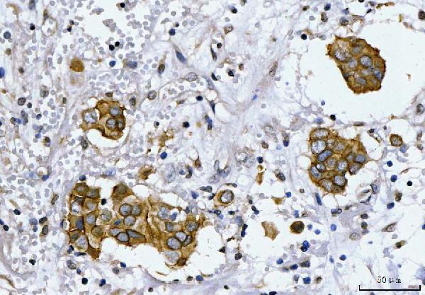
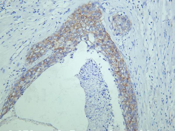
Plan chromogenic ERBB2 IHC in paraffin sections using the catalog antibody’s human breast carcinoma example (datasheet A00010-2). Compare the observed membrane and cytoplasmic staining with the reported tissue pattern (HPA tissue IHC).

Evidence assembled Oct 2026 · For research use; verify linked source records and product datasheet before use
Immunohistochemistry protocol sheet for ERBB2 (IHC for ERBB2): expected localisation Cytoplasmic and membranous staining in several tissues (HPA tissue IHC), antibody A00010-2, validated IHC image, and IHC protocol steps
Printable ERBB2 IHC protocol sheet — expected localisation Cytoplasmic and membranous staining in several tissues (HPA tissue IHC), antibody A00010-2, controls and protocol steps. Open the full ERBB2 IHC guide →

ERBB2 Immunohistochemistry Experimental Design Guide

Expected localisation, validated protocols, controls and antibodies — the at-a-glance facts below, then the full design guide.

Must know before staining
Expected localisation Cytoplasmic and membranous staining in several tissues (HPA tissue IHC)
Staining pattern Glandular cells show cytoplasmic and membranous staining (HPA tissue IHC)
Antigen retrieval EDTA pH 8.0 HIER, heat-mediated (datasheet A00010-2)
Positive control ⓘ Appendix+4 more · see all
Negative control ⓘ Adipose tissue+4 more · see all
Important caveats
Reasons your staining may differ from the expected pattern.
Fixation Keep fixation consistent across sections. (standard IHC practice; not target-specific)
Caveat EGF stimulation can shift ERBB2 from membrane to endosomes (UniProt)
Regulation EGF promotes membrane internalization (UniProt)
Isoform / epitope 6 isoforms; check whether the epitope is extracellular or cytoplasmic (UniProt)
Section 1

Recommended ERBB2 IHC & IF Protocols

The catalog antibody protocol is followed by published ERBB2 IHC methods for lung and breast cancer specimens.

Recommended immunohistochemistry (IHC-P) protocol parameters
SampleParaffin-embedded human breast cancer tissue; fixative not specified (datasheet A00010-2)
FixationImage fixative and duration unreported (datasheet A00010-2); verify before use.
Sectioning4–5 µm sections on charged slides (standard)
DeparaffinisationXylene, graded ethanol series to water (standard)
Antigen retrievalHeat retrieval: EDTA pH 8.0 (datasheet A00010-2); 20 min, 95–100 °C (standard)
Peroxidase block3% H2O2, 10 min, room temperature (standard)
Blocking10% goat serum (datasheet A00010-2)
Primary antibodyRabbit anti-ERBB2, 2-5μg/ml (datasheet A00010-2)
Primary incubationOvernight at 4 °C (datasheet A00010-2)
DetectionHRP-conjugated secondary, DAB chromogen (datasheet A00010-2)
CounterstainHematoxylin, blue, dehydrate and mount (standard)
Expected resultERBB2-positive staining in glandular cells of appendix (HPA tissue IHC: Medium). HPA tissue profile: Cytoplasmic and membranous expression in several tissues. No signal in the no-primary control.
💡Decision noteStart with heat-mediated EDTA retrieval at pH 8.0 (datasheet A00010-2); neither included article specifies ERBB2 retrieval conditions.
Section 2

What Is the Expected ERBB2 Staining Pattern?

ERBB2 is a single-pass cell-surface receptor with an extracellular region at residues 23–652 and a cytoplasmic region at 676–1255 (UniProt P04626 topology). In paraffin-section IHC, expect membranous and sometimes cytoplasmic staining in selected cells, including breast and appendix glandular cells (HPA tissue IHC). HPA rates the tissue profile Enhanced, with medium consistency between staining and RNA expression (HPA tissue IHC).

What am I looking at on my slide?
Membranous staining in breast or appendix glandular cells at roughly medium intensity (HPA tissue IHC).This fits the observed tissue pattern and ERBB2's cell-membrane location (HPA tissue IHC; UniProt P04626 subcellular location). Judge the cell type and compartment together: HPA also reports cytoplasmic staining in several tissues (HPA tissue IHC).
Cytoplasmic signal accompanies membrane staining in an expected cell population (HPA tissue IHC).Cytoplasmic staining can be compatible with the HPA tissue profile; UniProt also lists endosomal and cytoplasmic locations (HPA tissue IHC; UniProt P04626 subcellular location). Record its distribution separately from the membrane signal so a diffuse deposit is not mistaken for cell-associated staining (general IHC practice).
Signal is predominantly in an unexpected compartment, with little convincing membrane staining (UniProt P04626 subcellular location).Review the pattern before calling it positive: HPA's main ICC/IF location is plasma membrane, while nucleoplasm and cytosol are additional supported locations (HPA subcellular). An isolated nuclear or diffuse pattern merits a control check; nuclear signal alone does not prove an artefact (HPA subcellular; general IHC practice).
Strong staining appears in adipocytes or adrenal glandular cells, where HPA reports no detection (HPA tissue IHC).That cell-type mismatch raises possible cross-reactivity or endogenous chromogenic activity, rather than establishing ERBB2 expression (HPA tissue IHC; general IHC practice). Compare with a no-primary control and examine whether the color follows cells or background (general IHC practice).
Breast glandular cells show no signal, although HPA reports medium staining in that cell type (HPA tissue IHC).Treat the result as inconclusive until a positive-control section and detection controls work (general IHC practice). HPA describes an observed profile with medium staining and medium staining–RNA consistency, not a guarantee that every breast section is positive (HPA tissue IHC).
💡Expected ERBB2 appearanceA credible positive is distinct, cell-associated membrane staining, with possible cytoplasmic signal, in expected glandular cells at around the medium HPA level; widespread color outside cells is suspect background (HPA tissue IHC; UniProt P04626 subcellular location; general IHC practice).
How each factor affects the staining
Receptor topology and antibody epitopeERBB2 spans the membrane at residues 653–675, separating extracellular and cytoplasmic regions (UniProt P04626 topology). Check which region the catalog antibody recognizes before interpreting a compartment-specific pattern; its epitope is not supplied here (general IHC practice).
Observed tissue and cell typeHPA reports medium staining in breast and appendix glandular cells, but no detection in adipocytes or adrenal glandular cells (HPA tissue IHC). Select controls by the stated cell population, since a whole tissue can contain several cell types (general IHC practice).
Antibody validationHPA lists IHC validation as Enhanced for HPA001383 and CAB062555, and Supported for CAB000043 and CAB020416 (HPA antibodies). These ratings describe those antibodies; they do not establish equivalent performance for the catalog antibody (HPA antibodies; general IHC practice).
Cellular redistributionUniProt reports ERBB2 internalization from the cell membrane after EGF stimulation and lists early endosomes among its locations (UniProt P04626 subcellular location). This supports considering intracellular signal alongside membrane signal, but does not identify its cause in a particular fixed section (UniProt P04626; general IHC practice).
IF/ICC Q&AQ: Where should ERBB2 appear by IF/ICC? A: Mainly at the plasma membrane, with supported additional nucleoplasmic and cytosolic localization (HPA subcellular). This is an ICC/IF localization observation, not an IHC-P intensity standard or an IF protocol recommendation (HPA subcellular).
Why is my staining missing, weak or wrong?
SituationLikely causeNext action
No staining in breast glandular cells expected to show medium signal (HPA tissue IHC).The assay may have failed, or the sampled cells may differ from HPA's observed sections (HPA tissue IHC; general IHC practice).Run a positive-control section; check the primary-antibody conditions, retrieval and detection steps against the validated IHC-P method (general IHC practice). Do not infer ERBB2-specific fixation sensitivity from this result.
Diffuse chromogen covers cells and intervening tissue.Nonspecific staining or excessive detection background can obscure a cell-associated pattern (general IHC practice).Inspect a no-primary control, check blocking and washes, and reduce primary-antibody or detection strength if the assay controls indicate background (general IHC practice).
Nuclear staining dominates while membrane staining is weak (HPA subcellular).HPA supports additional nucleoplasmic localization, so compartment alone cannot establish either specificity or artefact (HPA subcellular).Compare membrane and nuclear patterns in the expected cell type, then assess no-primary and positive controls before interpreting the nuclear signal (HPA tissue IHC; general IHC practice).
Adipocytes stain strongly despite HPA reporting no detection (HPA tissue IHC).Cell-type discordance can reflect nonspecific binding or endogenous chromogenic activity (HPA tissue IHC; general IHC practice).Check a no-primary control and the distribution of the deposit; confirm that expected glandular-cell staining is present in a positive control (HPA tissue IHC; general IHC practice).
Cytoplasmic staining is present without a clear membrane outline.HPA reports cytoplasmic as well as membranous tissue expression, while ERBB2 also has intracellular locations (HPA tissue IHC; UniProt P04626 subcellular location).Score the cytoplasmic and membrane components separately; compare a positive-control section before treating cytoplasmic-only staining as the expected receptor pattern (general IHC practice).
Staining varies substantially between sections or cell populations.HPA's Enhanced tissue profile still has only medium consistency with RNA expression and reports different staining levels across cell types (HPA tissue IHC).Identify the specific cells being compared and verify controls on each run; avoid treating tissue-wide color intensity as a single ERBB2 result (HPA tissue IHC; general IHC practice).

Sample controls for ERBB2 IHC & IF

🧪Run breast first: its glandular cells should stain (HPA: Medium in breast glandular cells). Use adipose tissue as a negative comparator (HPA: Not detected in adipocytes); on the breast slide, adjacent nonglandular cells should show only background staining, without convincing membrane signal.
Positive control tissue: Appendix (Glandular cells, HPA Medium)
Negative control tissue: Adipose tissue (HPA Not detected)
ICC-IF cell lines (HPA subcellular resource): HPA ICC-IF images show ERBB2 in U-251MG, A-431, U2OS, HaCaT, RT-4, with annotated localisation: Plasma membrane (enhanced) (HPA subcellular).
Technical controls: Include a no-primary (secondary-only) control, a concentration-matched nonimmune rabbit IgG isotype control (caption: rabbit primary antibody), and an ERBB2 knockout biological control if available. Quench endogenous peroxidase and inspect for residual DAB background in breast sections (caption: HRP/DAB detection).
⚠️Feasibility: A target-specific fixation window is unreported, and the selected A00010-2 tissue-IHC caption does not report a fixative. The caption uses heat retrieval in EDTA at pH 8.0, but does not establish that retrieval is required. The supplied evidence does not establish whether frozen sections or IF/ICC are easier; for breast sections, check that endogenous peroxidase does not create apparent staining (caption: HRP/DAB detection).

HPA tissue IHC evidence for ERBB2

Comprehensive Human Protein Atlas IHC scoring per tissue (reliability: Enhanced — Medium consistency between antibody staining and RNA expression data.). Rows are taken directly from the HPA tissue chart — click any row's HPA link to view the source.

Positive expression · recommended positive controls

TissueCell typeLevelEvidenceSource
Appendix Glandular cells Medium Protein (IHC) HPA →
Breast Glandular cells Medium Protein (IHC) HPA →
Cervix Glandular cells Medium Protein (IHC) HPA →
Endometrium Glandular cells Medium Protein (IHC) HPA →
Fallopian tube Ciliated cells (ciliary rootlets) Medium Protein (IHC) HPA →

Undetected expression · recommended negative controls

TissueCell typeLevelEvidenceSource
Adipose tissue Adipocytes Not detected Protein (IHC) HPA →
Adrenal gland Glandular cells Not detected Protein (IHC) HPA →
Caudate Glial cells Not detected Protein (IHC) HPA →
Cerebellum Cells in granular layer Not detected Protein (IHC) HPA →
Cerebral cortex Endothelial cells Not detected Protein (IHC) HPA →
Section 3

Advanced ERBB2 IHC Tips

Troubleshoot ERBB2 staining by checking retrieval, compartment, and controls before comparing signal intensity across paraffin sections.

How should I troubleshoot weak ERBB2 staining after antigen retrieval?
Start with heat-mediated antigen retrieval in EDTA at pH 8.0 (datasheet A00010-2). The selected paraffin-section example used this retrieval before overnight incubation with 2 μg/ml antibody at 4°C (datasheet A00010-2). If staining is weak, check heating uniformity, section adhesion, and whether sections remained covered by buffer throughout retrieval (standard IHC practice). Compare a known positive section in the same run before changing retrieval conditions, because ERBB2 intensity can vary among tissues and cells (HPA: tissue IHC profile). If a fallback retrieval method is tested, change one condition at a time and check whether stronger signal preserves clear cell-membrane staining (HPA: subcellular localisation; standard IHC practice).
Could fixation explain weak or uneven ERBB2 staining?
The selected ERBB2 image documents a paraffin section but does not report its fixative, so target-specific fixation sensitivity is unknown (datasheet A00010-2). Record each specimen’s fixative, fixation interval, section thickness, and storage history before comparing staining across blocks (standard IHC practice). Run a previously satisfactory control block alongside the affected section, using the same EDTA pH 8.0 retrieval and detection sequence (datasheet A00010-2; standard IHC practice). If both sections fail, inspect retrieval and reagents before attributing the result to fixation (standard IHC practice). If only one block fails, compare its processing record and tissue preservation while keeping antibody concentration and detection constant (standard IHC practice).
Is cytoplasmic or nuclear staining compatible with ERBB2?
Prioritise distinct cell-membrane staining when assessing ERBB2, whose main reported location is the plasma membrane (HPA: subcellular localisation). Its extracellular region spans residues 23–652, its transmembrane segment 653–675, and its cytoplasmic region 676–1255 (UniProt P04626 topology). Cytoplasmic staining can be plausible because tissue IHC shows cytoplasmic and membranous expression, while internalisation and perinuclear localisation are reported (HPA: tissue IHC profile; UniProt P04626 localisation). Nuclear signal also has supporting localisation evidence, but assess whether it is reproducible in intact cells and exceeds control-slide background (HPA: subcellular localisation; standard IHC practice). Report membrane, cytoplasmic, and nuclear staining separately rather than combining them into one intensity score (standard IHC practice).
Can an unknown antibody epitope change how I interpret ERBB2 staining?
ERBB2 has 6 listed isoforms, but the supplied antibody caption does not identify the recognised epitope (UniProt P04626 isoforms; datasheet A00010-2). Therefore, do not assume that this stain detects every isoform or distinguishes them in tissue (standard antibody interpretation practice). Check available antibody documentation for an immunogen or mapped epitope before interpreting compartment-specific staining, and describe the epitope as unknown if documentation gives none (standard IHC practice). ERBB2 has extracellular, transmembrane, and cytoplasmic regions, and its extracellular region carries listed glycosylation sites (UniProt P04626 topology and glycosylation). If an orthogonal antibody is available, compare patterns while documenting each antibody’s epitope and retrieval conditions (standard IHC practice).
How can I assess ERBB2 localisation by multiplex IF?
Treat IF as a separate assay requiring its own controls; the selected antibody evidence describes paraffin-section chromogenic IHC (datasheet A00010-2). Pair ERBB2 with a validated marker for the expected cell type in your specimen, and inspect whether the signals occupy the same cells (standard IF practice). Choose a fluorophore channel with low measured tissue autofluorescence, and include single-stain controls to assess spectral bleed-through (standard IF practice). ERBB2 spans extracellular residues 23–652 and cytoplasmic residues 676–1255; an unknown antibody epitope leaves its membrane side unresolved (UniProt P04626 topology; datasheet A00010-2). Select permeabilisation only after establishing epitope accessibility, then compare membrane and intracellular signal under that condition (standard IF practice).
What should I check when ERBB2 DAB staining looks diffuse?
The selected example used 10% goat-serum blocking, a peroxidase-conjugated secondary, and DAB development (datasheet A00010-2). Compare a no-primary control with the stained section to identify detection-system background, and check whether an appropriate peroxidase block reduces enzyme-related signal (standard IHC practice). Inspect tissue edges, folds, and damaged regions separately because local reagent accumulation or poor morphology can mimic stronger staining (standard IHC practice). Optimise washing and detection exposure while retaining the documented 2 μg/ml primary condition as a reference point (datasheet A00010-2; standard IHC practice). Judge improvement by clearer cell borders and lower control-slide signal, consistent with ERBB2’s main plasma-membrane localisation (HPA: subcellular localisation).
How should I quantify ERBB2 staining across paraffin sections? ⚠ ANSWER MARKED FOR VERIFICATION
Define the scored cell population and compartment before reading slides; ERBB2’s main reported location is the plasma membrane (HPA: subcellular localisation). For membrane staining, record the percentage of positive target cells and intensity categories, then calculate an H-score if those categories are prespecified (standard IHC scoring practice). Keep cytoplasmic and nuclear measurements separate because these locations are also reported and answer a different localisation question (HPA: tissue IHC profile; HPA: subcellular localisation). Normalise positive counts to the number of eligible intact target cells, or stained area to analysed tissue area in mm² (standard image-analysis practice). Use the same thresholds, controls, and exclusion rules for every section in a comparison (standard IHC practice).
How do I distinguish true ERBB2 staining from artefact?
Look first for reproducible membrane staining in intact cells, matching ERBB2’s principal plasma-membrane location (HPA: subcellular localisation). Check the stained cell type against the specimen: HPA reports medium signal in breast glandular cells, but its tissue profile also includes cytoplasmic expression (HPA: tissue IHC profile). Review unexpected nuclear or diffuse cytoplasmic signal separately because additional locations are reported, yet localisation alone cannot establish specificity (HPA: subcellular localisation; standard IHC practice). Exclude necrotic areas, torn tissue, folds, and unusually dark section edges from interpretation (standard IHC practice). Compare no-primary and positive controls to identify endogenous enzyme or detection background before calling an isolated DAB deposit positive (standard IHC practice).
Boster reagents

Best ERBB2 / Receptor tyrosine-protein kinase erbB-2 IHC Antibodies

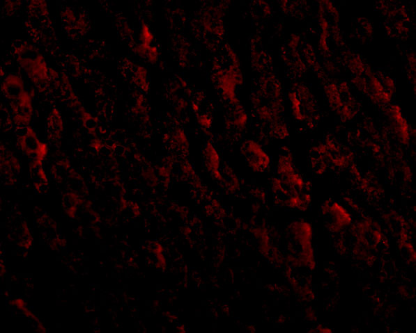
Anti-ERBB2 antibodies have IHC images from human breast cancer, breast carcinoma and liver tissue (catalog IHC captions), plus IF images from human liver tissue and SKBR cells (catalog IF captions).

Real IHC data IHC analysis of ErbB 2 using anti-ErbB 2 antibody (A00010-2). ErbB 2 was detected in a paraffin-embedded section of human breast cancer tissue. Heat mediated antigen retrieval was performed in EDTA buffer (pH 8.0, epitope retrieval solution). The tissue section was blocked with 10% goat serum. The tissue section was then incubated with 2 μg/ml rabbit anti-ErbB 2 Antibody (A00010-2) overnight at 4°C. Peroxidase Conjugated Goat Anti-rabbit IgG was used as secondary antibody and incubated for 30 minutes at 37°C. The tissue section was developed using HRP Conjugated Rabbit IgG Super Vision Assay Kit (Catalog # SV0002) with DAB as the chromogen.
Anti-ErbB 2/ERBB2 Antibody ®
Cat # A00010-2
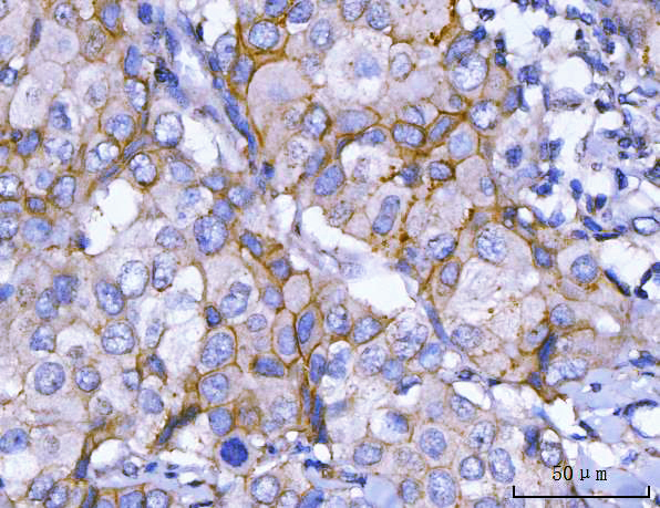
Real IHC data IHC analysis of HER2/ERBB2 using anti-HER2/ERBB2 antibody (A00010-4). HER2/ERBB2 was detected in a paraffin-embedded section of human breast cancer tissue. Heat mediated antigen retrieval was performed in EDTA buffer (pH 8.0, epitope retrieval solution). The tissue section was blocked with 10% goat serum. The tissue section was then incubated with 2 μg/ml rabbit anti-HER2/ERBB2 Antibody (A00010-4) overnight at 4°C. Peroxidase Conjugated Goat Anti-rabbit IgG was used as secondary antibody and incubated for 30 minutes at 37°C. The tissue section was developed using HRP Conjugated Rabbit IgG Super Vision Assay Kit (Catalog # SV0002) with DAB as the chromogen.
Anti-HER2/ERBB2 Antibody ®
Cat # A00010-4
Real IHC data IHC analysis of HER2/ERBB2 using anti-HER2/ERBB2 antibody (A00010-5). HER2/ERBB2 was detected in a paraffin-embedded section of human breast cancer tissue. Heat mediated antigen retrieval was performed in EDTA buffer (pH 8.0, epitope retrieval solution). The tissue section was blocked with 10% goat serum. The tissue section was then incubated with 2 μg/ml rabbit anti-HER2/ERBB2 Antibody (A00010-5) overnight at 4°C. Peroxidase Conjugated Goat Anti-rabbit IgG was used as secondary antibody and incubated for 30 minutes at 37°C. The tissue section was developed using HRP Conjugated Rabbit IgG Super Vision Assay Kit (Catalog # SV0002) with DAB as the chromogen.
Anti-HER2/ERBB2 Antibody ®
Cat # A00010-5
Real IHC data Immunohistochemical analysis of paraffin-embedded Rat stomach, using the Antibody at 1:50 dilution.
Anti-ErbB2 (HER2) Monoclonal Antibody
Cat # M00010-1
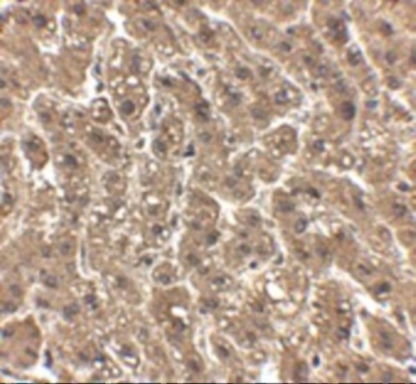
Real IHC data Immunohistochemistry of erbB-2 in human liver tissue with erbB-2 antibody at 2.5 μg/mL.
Anti-erbB-2 Antibody
Cat # A00010
Real IHC data Human breast carcinoma was stained with anti-HER2 rabbit antibody
Anti-HER2 Rabbit Monoclonal Antibody
Cat # M00010-21

A00010-2, A00010-4 and A00010-5 each show paraffin-section human breast cancer IHC (each SKU’s IHC caption), while A00010 shows human liver IHC and IF (A00010 image captions). M00010-1 shows human breast carcinoma IHC and SKBR-cell IF (M00010-1 image captions), and M00010-21 shows human breast carcinoma IHC (M00010-21 IHC caption); M00010-1’s first IHC caption labels rat stomach despite its Human-only listed reactivity (M00010-1 IHC caption; catalog reactivity).

Which to pick: For human paraffin-section IHC, start with A00010-2 at 2–5 μg/mL (A00010-2 datasheet); its own image shows human breast cancer sections with EDTA retrieval at pH 8.0, and the fixative is unreported (A00010-2 IHC caption). For IF/ICC, choose rabbit monoclonal M00010-1 (M00010-1 catalog host, clone and applications), supported by its SKBR-cell IF image (M00010-1 IF caption). For cross-species planning, A00010 lists Human, Mouse and Rat reactivity and IHC-P and IF applications (A00010 catalog), though its IHC and IF images document human liver only (A00010 image captions).

Each figure is that product's own IHC / IF validation image from its datasheet.

References

  1. UniProt Consortium. UniProt entry P04626 (ERBB2_HUMAN, Receptor tyrosine-protein kinase erbB-2).
  2. Human Protein Atlas. ERBB2 tissue IHC expression (reliability: Enhanced).
  3. Human Protein Atlas. ERBB2 subcellular location (ICC-IF): Mainly localized to the plasma membrane. In addition localized to the nucleoplasm and cytosol..
  4. Human Protein Atlas. ERBB2 antibody validation summary (7 antibodies).
  5. Sequencing of 279 cancer genes in ampullary carcinoma reveals trends relating to histologic subtypes and frequent amplification and overexpression of ERBB2 (HER2). Modern pathology : an official journal of the United States and Canadian Academy of Pathology, Inc 2015 — PMC4977532.
  6. Exploration of HER2 (ERBB2) immunohistochemistry in non-small cell lung cancer: correlation with ERBB2 mutational status: experimental research. Annals of medicine and surgery (2012) 2023 — PMC10289698.
  7. RNAi knockdown of C-erbB2 expression inhibits salivary gland adenoid cystic carcinoma SACC-83 cell growth in vitro. Journal of biomedical research 2010 — PMC3596557.
  8. Strong EGFR signaling in cell line models of ERBB2-amplified breast cancer attenuates response towards ERBB2-targeting drugs. Oncogenesis 2012 — PMC3412653.
  9. PubMed PMID:3003577 — UniProt-cited evidence.
  10. PubMed PMID:2999974 — UniProt-cited evidence.
  11. PubMed PMID:15489334 — UniProt-cited evidence.