EZH2 / Histone-lysine N-methyltransferase EZH2 · IHC design guide

Design Immunohistochemistry for EZH2

Plan EZH2 IHC in paraffin sections around nuclear staining in testis and lymphoid tissues (HPA tissue IHC). This guide covers fixation, controls, detection, and scoring of cell-specific nuclear signal.

Evidence assembled Oct 2026 · For research use; verify linked source records and product datasheet before use
Immunohistochemistry protocol sheet for EZH2 (IHC for EZH2): expected localisation Nuclear in several tissues (HPA tissue IHC), antibody M00050-1, validated IHC image, and IHC protocol steps
Printable EZH2 IHC protocol sheet — expected localisation Nuclear in several tissues (HPA tissue IHC), antibody M00050-1, controls and protocol steps. Open the full EZH2 IHC guide →

EZH2 Immunohistochemistry Experimental Design Guide

Expected localisation, validated protocols, controls and antibodies — the at-a-glance facts below, then the full design guide.

Must know before staining
Expected localisation Nuclear in several tissues (HPA tissue IHC)
Staining pattern Nuclear in pachytene spermatocytes and germinal center cells (HPA tissue IHC)
Antigen retrieval Tris-EDTA pH 9.0 HIER, 95–98 °C, 20 min (rule: nuclear antigen)
Positive control ⓘ Bone marrow+4 more · see all
Negative control ⓘ Adipose tissue+4 more · see all
Important caveats
Reasons your staining may differ from the expected pattern.
Fixation Matched tissue-IHC evidence does not establish the fixation claim. Validate the specimen-specific method before use. (selected-SKU IHC image M00050-1)
Caveat Staining varies by cell type within tissues (HPA tissue IHC)
Regulation Overexpressed in several tumor types (UniProt)
Isoform / epitope 5 isoforms; check antibody epitope coverage (UniProt)
Section 1

Recommended EZH2 IHC & IF Protocols

Compare the catalog antibody’s IHC-P protocol with published EZH2 protocols for cervical carcinoma (PMC10148987), bone marrow (PMC13105204), and lung carcinoma (PMC3890101).

Recommended immunohistochemistry (IHC-P) protocol parameters
SampleParaffin-embedded human tonsil tissue; fixative not specified (datasheet M00050-1)
FixationImage fixative and duration unreported (datasheet M00050-1); verify before use.
Sectioning4–5 µm sections on charged slides (standard)
DeparaffinisationXylene, graded ethanol series to water (standard)
Antigen retrievalHeat-induced epitope retrieval in Tris-EDTA buffer, pH 9.0, 20 min at 95–98 °C (standard rule: nuclear antigen)
Peroxidase block3% H2O2, 10 min, room temperature (standard)
Blocking10% normal serum of the secondary host, 30 min, room temperature (standard)
Primary antibodyRabbit monoclonal (clone IDF-5) anti-EZH2, 1:50-1:200 (datasheet M00050-1)
Primary incubationOvernight at 4 °C (standard)
DetectionHRP-polymer secondary, DAB chromogen 5–10 min (standard)
CounterstainHematoxylin, blue, dehydrate and mount (standard)
Expected resultEZH2-positive staining in hematopoietic cells of bone marrow (HPA tissue IHC: High). HPA tissue profile: Nuclear expression in several tissues, most abundant in testis and lymphoid tissues. No signal in the no-primary control.
💡Decision noteStart with Tris-EDTA pH 9.0 at 95–98 °C for 20 min (page retrieval rule: nuclear antigen); test citrate pH 6.0 if needed (PMC13105204; PMC3890101).
Section 2

What Is the Expected EZH2 Staining Pattern?

EZH2 is nuclear (UniProt Q15910: no transmembrane segment) and stains germinal center cells (HPA: High; IHC reliability Enhanced).

What am I looking at on my slide?
Strong nuclear staining in tonsil germinal center cells (HPA: High).Expected EZH2 pattern (HPA: nuclear tissue expression; UniProt Q15910: nucleus).
Predominantly cytoplasmic staining.Suspect artefact; EZH2 is nuclear (UniProt Q15910; HPA: nuclear tissue expression).
Strong staining of adipocytes (HPA: Not detected).Consider cross-reactivity or endogenous detection activity (HPA: adipocytes Not detected).
Diffuse staining across nuclei and surrounding tissue.Background may obscure the nuclear pattern (HPA: nuclear tissue expression; general IHC practice).
No signal in tonsil germinal center cells (HPA: High).The expected positive control has failed (HPA: High in germinal center cells).
💡Expected EZH2 appearanceHigh nuclear staining in germinal center cells (HPA); diffuse cytoplasm is suspect (UniProt Q15910).
How each factor affects the staining
Cell compartmentEZH2 is nuclear and has no transmembrane segment (UniProt Q15910).
Tissue and cell choiceTonsil germinal center cells are High; adipocytes are Not detected (HPA tissue IHC).
Antibody validationCAB009589 is Enhanced for IHC; HPA029131 has no IHC rating (HPA antibodies).
IsoformsFive isoforms are listed; antibody epitope coverage is unspecified (UniProt Q15910).
Why is my staining missing, weak or wrong?
SituationLikely causeNext action
No nuclear signal in tonsil germinal centers (HPA: High).A failed stain or detection step is possible (general IHC practice).Check positive control, retrieval and detection reagents (general IHC practice).
Cytoplasmic signal dominates.This conflicts with nuclear EZH2 localisation (UniProt Q15910).Check antibody specificity and detection controls (general IHC practice).
Adipocytes stain strongly (HPA: Not detected).Cross-reactivity or endogenous activity is possible (general IHC practice).Run a primary-omission control and review blocking (general IHC practice).
Diffuse background obscures nuclei.Excess antibody or incomplete washing is possible (general IHC practice).Optimise dilution and washing using controls (general IHC practice).
Nuclear signal is weak in an expected positive cell.Retrieval or detection may be suboptimal (general IHC practice).Review retrieval and detection against a positive control (general IHC practice).
What should IF/ICC show?EZH2 localises to the nucleoplasm (HPA: enhanced ICC-IF).Look for nucleoplasmic signal relative to a nuclear counterstain (HPA: nucleoplasm).

Sample controls for EZH2 IHC & IF

🧪Run tonsil first: germinal center cells should show nuclear EZH2 staining (HPA: High in tonsil germinal center cells; UniProt Q15910: nucleus). Use adipose tissue as the negative tissue: adipocytes are listed as not detected (HPA: adipose tissue adipocytes); on the tonsil slide, internal negative cells should have counterstained nuclei without specific chromogen, but their identity must be confirmed rather than inferred from location alone (standard IHC interpretation).
Positive control tissue: Bone marrow (Hematopoietic cells, HPA High)
Negative control tissue: Adipose tissue (HPA Not detected)
ICC-IF cell lines (HPA subcellular resource): HPA ICC-IF images show EZH2 in REH, SiHa, U2OS, A-431, U-251MG, RPTEC/TERT1, hTERT-RPE1 (serum starved), KOLF2.1J, with annotated localisation: Nucleoplasm (enhanced) (HPA subcellular).
Technical controls: Run a no-primary, secondary-only control; use host-species- and isotype-matched IgG for a monoclonal antibody, or host-matched normal IgG for a polyclonal antibody; and include EZH2 knockout material as a biological negative, with immunizing-peptide competition as an option if the peptide is available (standard IHC controls). Block endogenous peroxidase in tonsil for chromogenic detection (standard IHC practice).
⚠️Feasibility: Paraffin-section tonsil IHC is documented for the catalog antibody, but the selected SKU M00050-1 caption does not report a fixative; no target-specific fixation window or fixation effect is reported in the supplied evidence (selected SKU tissue-IHC caption). Antigen-retrieval dependency is unreported, so retrieval conditions need empirical optimization (supplied target/application evidence; standard IHC practice). The evidence does not establish whether frozen sections or IF/ICC are easier; HPA reports nucleoplasmic ICC-IF localization, while densely packed tonsil germinal center nuclei warrant careful nuclear scoring (HPA: nucleoplasm enhanced; HPA: High in tonsil germinal center cells; standard IHC interpretation).

HPA tissue IHC evidence for EZH2

Comprehensive Human Protein Atlas IHC scoring per tissue (reliability: Enhanced — Medium consistency between antibody staining and RNA expression data.). Rows are taken directly from the HPA tissue chart — click any row's HPA link to view the source.

Positive expression · recommended positive controls

TissueCell typeLevelEvidenceSource
Bone marrow Hematopoietic cells High Protein (IHC) HPA →
Placenta Cytotrophoblasts High Protein (IHC) HPA →
Skin Cells in granular layer High Protein (IHC) HPA →
Testis Pachytene spermatocytes High Protein (IHC) HPA →
Tonsil Germinal center cells High Protein (IHC) HPA →

Undetected expression · recommended negative controls

TissueCell typeLevelEvidenceSource
Adipose tissue Adipocytes Not detected Protein (IHC) HPA →
Breast Adipocytes Not detected Protein (IHC) HPA →
Caudate Glial cells Not detected Protein (IHC) HPA →
Cerebellum Cells in granular layer Not detected Protein (IHC) HPA →
Cerebral cortex Endothelial cells Not detected Protein (IHC) HPA →
Section 3

Advanced EZH2 IHC Tips

Troubleshoot EZH2 chromogenic IHC by checking nuclear staining, tissue controls, processing conditions and cell specific scoring (UniProt: nucleus; HPA: tissue IHC profile).

What retrieval should I try when EZH2 nuclear staining is weak?
Start with Tris-EDTA, pH 9.0, heat-induced retrieval at 95–98 °C for 20 min (page retrieval setting). Use the same section thickness, heating vessel and cooling procedure across control and test slides so a change in nuclear signal can be attributed to retrieval (standard IHC practice). Compare germinal center cells in tonsil, which show high EZH2 staining, with adjacent cells on the same section (HPA: high in tonsil germinal center cells). If staining remains weak, trial another retrieval condition on serial sections while keeping antibody and detection settings fixed; assess both nuclear signal and tissue damage (standard IHC practice).
Could fixation explain variable EZH2 staining between paraffin blocks?
The selected antibody image documents paraffin-embedded human tonsil but does not state its fixative (M00050-1 tissue-IHC caption). EZH2-specific fixation sensitivity is therefore unknown from the supplied evidence; differences between blocks should be investigated rather than assigned to a particular fixative effect (M00050-1 tissue-IHC caption). Record each block’s documented fixation and processing history, then stain comparable sections together with one known positive tonsil control (standard IHC practice; HPA: high in tonsil germinal center cells). If a block gives weak staining, review preservation, retrieval consistency and detection controls before drawing a biological conclusion from its EZH2 score (standard IHC practice).
How should I assess EZH2 staining outside the nucleus?
Expect predominantly nuclear EZH2 staining in tissue sections (UniProt: nucleus; HPA: nuclear expression in several tissues). Examine whether chromogen aligns with intact nuclei under the counterstain, particularly in germinal center cells of tonsil, where high expression is reported (HPA: high in tonsil germinal center cells; standard IHC practice). Diffuse cytoplasmic or extracellular color without convincing nuclear signal warrants checks of section integrity, antibody concentration, detection background and the negative control (standard IHC practice). EZH2 has no transmembrane segment, so a crisp cell membrane pattern alone would not match its annotated topology or localisation (UniProt: no transmembrane segment; UniProt: nucleus).
Can this IHC result distinguish EZH2 isoforms or epitope changes?
EZH2 has 5 annotated isoforms, but the supplied product caption does not specify this antibody’s epitope or isoform coverage (UniProt: isoforms 1–5; M00050-1 tissue-IHC caption). Its CXC and SET domains occupy residues 503–605 and 612–727, respectively; those coordinates alone cannot establish where this antibody binds (UniProt: domains). Annotated phosphorylation sites include residues 21, 76, 339, 345 and 363, without supplied evidence that any alters this assay (UniProt: modified residues). Interpret nuclear staining as antibody-detected EZH2 immunoreactivity, and seek epitope documentation or independently validated reagents before making isoform or modification-specific claims (standard IHC practice).
How can I assess EZH2 alongside a cell marker by IF?
For a separate IF assessment, pair EZH2 with a validated marker identifying the cell population under study, then assess whether EZH2 signal falls within those cells’ nuclei (UniProt: nucleus; standard IF practice). Choose spectrally separated fluorophores after inspecting tissue autofluorescence in an unstained section, and include single-stain controls to assess channel bleed-through (standard IF practice). Because EZH2 is nucleoplasmic and lacks a transmembrane segment, use a permeabilisation condition that permits antibody access to the nucleus while preserving cell boundaries (HPA: nucleoplasm; UniProt: no transmembrane segment; standard IF practice). HPA reports nucleoplasmic localisation in its subcellular dataset; verify that finding in the specimen and antibody used for IF (HPA: nucleoplasm; standard IF practice).
What should I check when chromogen obscures EZH2-positive nuclei?
First compare a no-primary control with the stained section to determine whether color arises from the chromogenic detection system (standard IHC practice). Check the peroxidase block, antibody concentration, wash steps and chromogen development time; excessive development can obscure the nuclear boundaries needed for EZH2 assessment (standard IHC practice; UniProt: nucleus). Examine sections for edge-concentrated color, folds and damaged areas, and avoid using those fields to judge specific staining (standard IHC practice). Reassess signal in intact tonsil germinal center cells alongside the control, since high EZH2 expression is reported there (HPA: high in tonsil germinal center cells).
How should I score EZH2 across tissue regions? ⚠ ANSWER MARKED FOR VERIFICATION
Define the cell population and tissue region before counting, then score nuclei rather than total chromogen area because EZH2 is a nuclear protein (UniProt: nucleus; standard IHC practice). Report the percentage of positive nuclei and, when staining intensity is reproducible, an H-score using the same intensity categories and positivity threshold across slides (standard IHC practice). For discrete infiltrates, positive nuclei per mm² can supplement those measures if the sampled tissue area is documented (standard IHC practice). Normalise counts to eligible, intact nuclei in the defined compartment, exclude necrotic and damaged areas, and document how fields were selected (standard IHC practice).
How do I distinguish genuine EZH2 staining from an artefact?
A persuasive positive result places chromogen in intact nuclei of the assessed cells, consistent with EZH2 localisation (UniProt: nucleus; HPA: nuclear tissue expression). Tonsil germinal center cells provide a high-expression comparison, while adipocytes are reported as not detected in HPA tissue IHC; compare like cell populations when interpreting either observation (HPA: high in tonsil germinal center cells; HPA: not detected in adipocytes; standard IHC practice). Treat isolated cytoplasmic color, section-edge staining and necrotic fields cautiously because they do not establish an intact nuclear pattern (UniProt: nucleus; standard IHC practice). If a no-primary slide retains color, investigate endogenous enzyme activity or detection background before calling the EZH2 stain positive (standard IHC practice).
Boster reagents

Best EZH2 / Histone-lysine N-methyltransferase EZH2 IHC Antibodies

Anti-EZH2 antibodies have IHC images from human tonsil and IF/ICC images from human cells (catalog image captions); listed reactivity spans human, mouse, and rat (catalog reactivity).

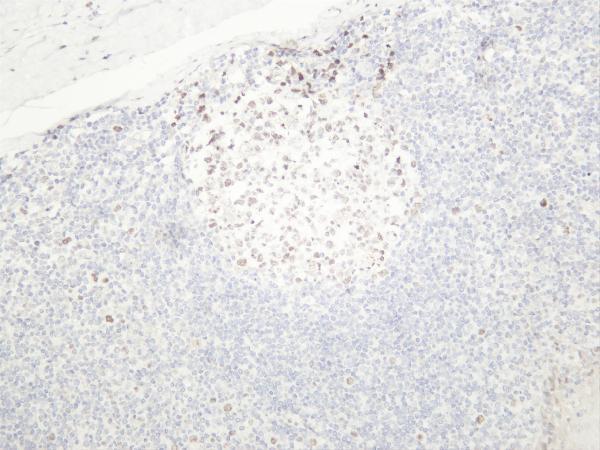
Real IHC data Immunohistochemical analysis of paraffin-embedded human tonsil, using KMT6 / EZH2 Antibody.
Anti-KMT6 / EZH2 Rabbit Monoclonal Antibody
Cat # M00050-1
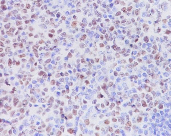
Real IHC data Humantonsil was stained with anti-EZH2 rabbit antibody
Anti-EZH2 Rabbit Monoclonal Antibody
Cat # M00050-2
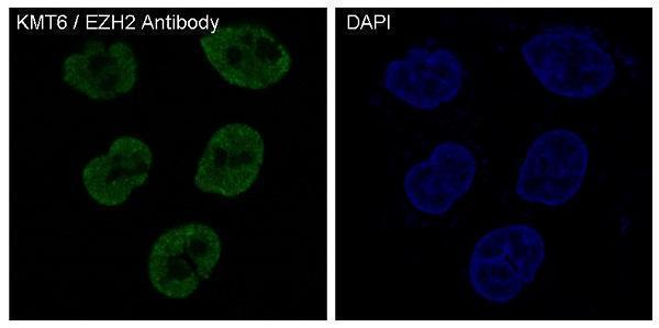
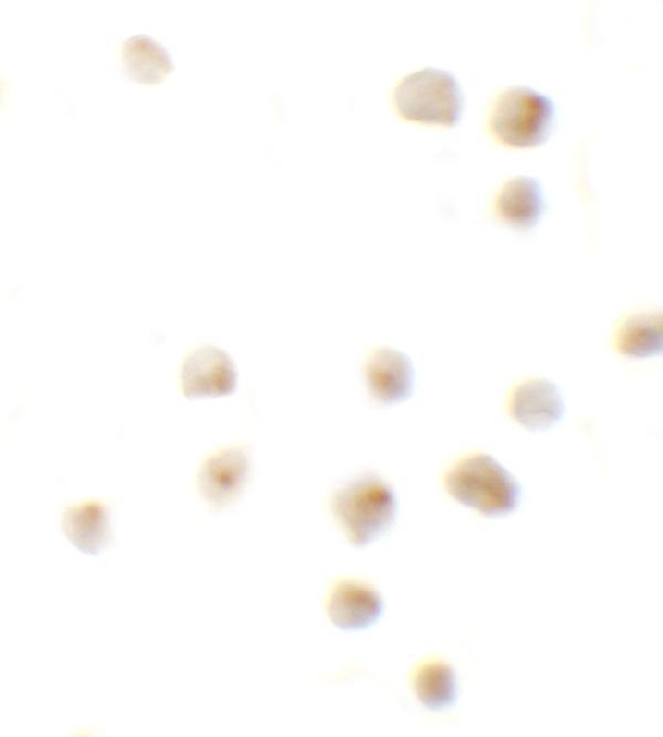
Real IF data Immunocytochemistry of EZH2 in 293 cells with EZH2 antibody at 10 μg/mL.
Anti-EZH2 Antibody
Cat # A00050
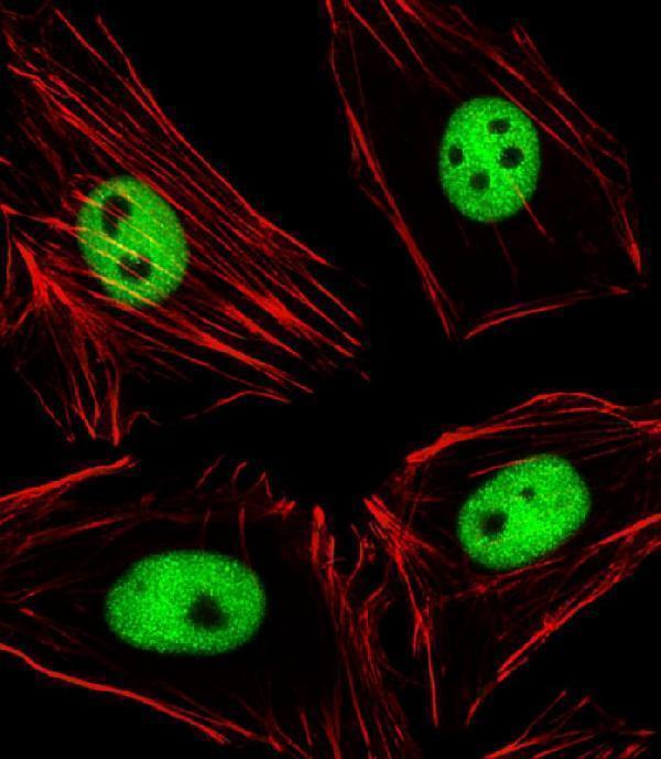
Real IF data Immunofluorescent analysis of 4% paraformaldehyde-fixed, 0. 1% Triton X-100 permeabilized Hela (Human cervical epithelial adenocarcinoma cell line) cells labeling EZH2 with A00050-3 at 1/25 dilution, followed by Alexa Fluor 488-conjugated goat anti-rabbit IgG secondary antibody at 1/400 dilution (green). Confocal image showing nuclear staining on Hela cell line. Cytoplasmic actin is detected with Alexa Fluor® 555 conjugated with Phalloidin at 1/100 dilution (red).
Anti-EZH2 Antibody
Cat # A00050-3

M00050-1 has a paraffin-embedded human tonsil IHC image, and M00050-2 has a human tonsil IHC image with processing unspecified (catalog image captions). A00050 has 293-cell ICC/IF images, while A00050-3 has a paraformaldehyde-fixed HeLa-cell IF image and lists IHC-P; both list human and mouse reactivity (catalog image captions; catalog applications and reactivity).

Which to pick: For paraffin-section IHC, start with rabbit monoclonal M00050-1: its own IHC image shows paraffin-embedded human tonsil; the fixative is unreported (catalog host, description, and IHC image caption). For cell IF/ICC, choose A00050 for its 293-cell images and listed ICC/IF applications, or A00050-3 for its HeLa IF image and listed IF application (catalog image captions and applications). For work across human, mouse, and rat, M00050-1 and M00050-2 both list all three species and IHC/IF applications; M00050-2’s tonsil caption does not report section processing or fixative (catalog reactivity, applications, and IHC image caption).

Each figure is that product's own IHC / IF validation image from its datasheet.

References

  1. UniProt Consortium. UniProt entry Q15910 (EZH2_HUMAN, Histone-lysine N-methyltransferase EZH2).
  2. Human Protein Atlas. EZH2 tissue IHC expression (reliability: Enhanced).
  3. Human Protein Atlas. EZH2 subcellular location (ICC-IF): Localized to the nucleoplasm..
  4. Human Protein Atlas. EZH2 antibody validation summary (2 antibodies).
  5. Evaluation of Immunohistochemical Expression of Enhancer of Zeste Homolog 2 (EZH2) and Its Association With Clinicopathological Variables in Carcinoma Cervix. Cureus 2023 — PMC10148987.
  6. Immunohistochemical loss of enhancer of Zeste Homolog 2 (EZH2) protein expression correlates with EZH2 alterations and portends a worse outcome in myelodysplastic syndromes. Modern pathology : an official journal of the United States and Canadian Academy of Pathology, Inc 2022 — PMC13105204.
  7. EZH2 protein expression associates with the early pathogenesis, tumor progression, and prognosis of non-small cell lung carcinoma. Clinical cancer research : an official journal of the American Association for Cancer Research 2013 — PMC3890101.
  8. Prognostic impact of c-MYC and EZH2 expression in small cell and non-small cell lung carcinoma: a single-center retrospective study. Discover oncology 2026 — PMC13172210.
  9. PubMed PMID:8954776 — UniProt-cited evidence.
  10. PubMed PMID:9214638 — UniProt-cited evidence.
  11. PubMed PMID:14702039 — UniProt-cited evidence.