This website uses cookies to ensure you get the best experience on our website.


- Table of Contents
Plan FMR1 chromogenic IHC in paraffin sections around its cytoplasmic tissue pattern, with distinct staining in neuronal cells and spermatogonia (HPA tissue IHC). Use the guide to choose controls, assess cell types and interpret staining with the antibody’s epitope coverage in mind (UniProt).

Expected localisation, validated protocols, controls and antibodies — the at-a-glance facts below, then the full design guide.
| Expected localisation | Cytoplasmic tissue staining (HPA tissue IHC) | |
| Staining pattern | Cytoplasmic staining, distinct in neurons and spermatogonia (HPA tissue IHC) | |
| Antigen retrieval | EDTA pH 8.0 HIER, heat-mediated (datasheet PB9703) | |
| Positive control | Cerebellum+4 more · see all | |
| Negative control | Adipose tissue |
| Fixation | Keep fixation consistent across sections. (standard IHC practice; not target-specific) | |
| Caveat | Cortical glia can also stain strongly (HPA tissue IHC) | |
| Regulation | Staining varies by cell type (HPA tissue IHC) | |
| Isoform / epitope | 11 isoforms; verify epitope coverage (UniProt) |
The catalog antibody protocol uses EDTA pH 8.0 retrieval (datasheet: PB9703). The published IHC protocols below describe FMR1 staining in gastric cancer tissue (PMC13006672; PMC12581516).
| Sample | Paraffin-embedded human seminoma testis tissue; fixative not specified (datasheet PB9703) |
| Fixation | Image fixative and duration unreported (datasheet PB9703); verify before use. |
| Sectioning | 4–5 µm sections on charged slides (standard) |
| Deparaffinisation | Xylene, graded ethanol series to water (standard) |
| Antigen retrieval | Heat retrieval: EDTA pH 8.0 (datasheet PB9703); 20 min, 95–100 °C (standard) |
| Peroxidase block | 3% H2O2, 10 min, room temperature (standard) |
| Blocking | 10% goat serum (datasheet PB9703) |
| Primary antibody | Rabbit anti-FMR1, 2-5 μg/ml (datasheet PB9703) |
| Primary incubation | Overnight at 4 °C (datasheet PB9703) |
| Detection | HRP-conjugated secondary, DAB chromogen (datasheet PB9703) |
| Counterstain | Hematoxylin, blue, dehydrate and mount (standard) |
| Expected result | FMR1-positive staining in cells in molecular layer of cerebellum (HPA tissue IHC: High). HPA tissue profile: Cytoplasmic expression in all tissues. Distinct expression in neuronal cells and in spermatogonia in seminiferous ducts. No signal in the no-primary control. |
FMR1 staining should be predominantly cytoplasmic in paraffin sections (HPA tissue IHC: cytoplasmic expression across tissues; HPA: Approved, with medium consistency between antibody staining and RNA expression). Look for distinct staining in hippocampal neuronal cells and testicular spermatogonia (HPA tissue IHC). FMR1 has no transmembrane segment, although UniProt also describes nuclear and neuronal process localisation (UniProt Q06787: subcellular location and topology).
| Cytoplasmic staining is clear in hippocampal neurons; nearby cells vary in intensity (HPA: High in hippocampal neuronal cells). | This fits the reported positive pattern (HPA tissue IHC). Judge the signal in identified cells, since HPA reports cytoplasmic expression across tissues and different staining levels among cell types (HPA tissue IHC). |
| The strongest signal is nuclear, with little cytoplasmic staining. | Recheck localisation against the predominant cytoplasmic tissue pattern (HPA tissue IHC). Nuclear FMR1 is described, so nuclear signal alone cannot prove an artefact; an isolated strong nuclear pattern warrants review of controls and detection background (UniProt Q06787: subcellular location; general IHC practice). |
| Adipocytes stain strongly while an expected positive population is weak. | Treat this as a specificity warning, since adipocytes were not detected in the HPA tissue IHC record (HPA: Not detected in adipocytes). Cross-reactivity or endogenous detection activity are possible explanations to investigate, rather than a diagnosis from one slide (general IHC practice). |
| Colour spreads evenly across tissue, empty spaces, or several unrelated cell types. | A diffuse deposit does not establish cellular FMR1 localisation (general IHC practice). Check background with an appropriate primary-omission control and inspect blocking, washes, chromogen development, and tissue-associated endogenous activity (general IHC practice). |
| No stain appears in hippocampal neuronal cells or another documented high-staining population (HPA tissue IHC). | An uninformative negative result can reflect failed staining or detection, so first verify an on-slide or same-run positive control (general IHC practice). HPA's Approved tissue profile has medium RNA–staining consistency; its reported level is a guide, not a guarantee for every specimen (HPA tissue IHC). |
| Which cells anchor interpretation? | HPA reports High staining in hippocampal neuronal cells, cerebral cortex glia, kidney tubule cells, and lung macrophages, among other populations; adipocytes are Not detected (HPA tissue IHC). Compare like cell types within the specimen rather than assigning one expected intensity to an entire organ (general IHC practice). |
| What compartment should be scored? | The tissue profile is cytoplasmic, while UniProt also lists perikaryon, neurites, synaptic sites, nucleus, and nucleolus (HPA tissue IHC; UniProt Q06787: subcellular location). Tissue IHC does not by itself resolve every listed fine structure, so score the visible cellular compartment at the section's resolution (general IHC practice). |
| How firm is the tissue evidence? | The HPA tissue IHC reliability is Approved, with medium consistency between staining and RNA expression (HPA tissue IHC). Listed antibodies HPA050118, HPA056084, and CAB012444 are individually IHC Approved; the supplied record does not assign IHC Enhanced status to this target (HPA antibodies). |
| Do isoforms or processing predict a different stain? | UniProt lists 11 isoforms, one chain spanning residues 1–632, and no signal peptide, propeptide, or transmembrane segment (UniProt Q06787: isoforms, processing, topology). The supplied data give no antibody epitope, so they cannot predict which isoforms a given antibody detects or a different pattern after antigen retrieval. |
| Can IF/ICC help check localisation? | Yes, as a separate imaging comparison: HPA ICC-IF reports cytosol as the enhanced main location and lists A-431, U-251MG, U2OS, and NIH 3T3 images (HPA subcellular ICC-IF). That cell-image result supports a cytosolic expectation but does not establish the staining intensity of a paraffin tissue section (HPA subcellular ICC-IF; general IHC practice). |
| Situation | Likely cause | Next action |
|---|---|---|
| Expected cytoplasmic cells are negative. | The staining run, detection, or control may have failed; absence of signal alone does not establish absent FMR1 (general IHC practice). | Check a same-run positive population, antibody and detection reagents, and the recorded antigen-retrieval conditions (general IHC practice). HPA reports High staining in hippocampal neuronal cells (HPA tissue IHC). |
| The slide is dominated by nuclear colour. | Background or misassigned compartment is possible, while nuclear localisation remains biologically described (UniProt Q06787: subcellular location; general IHC practice). | Compare the primary-omission control and inspect cytoplasm in identifiable positive cells before calling the pattern specific (general IHC practice; HPA tissue IHC: cytoplasmic expression). |
| Adipocytes or empty spaces carry conspicuous colour. | The distribution conflicts with the reported undetected adipocytes and cellular cytoplasmic pattern; nonspecific deposit is possible (HPA tissue IHC; general IHC practice). | Review the primary-omission control, blocking and wash steps, and chromogen development; score signal only where it belongs to identifiable cells (general IHC practice). |
| Lung macrophages appear intensely coloured alongside broad tissue background. | Macrophages are a documented High population, but surrounding diffuse colour can obscure whether their staining is specific (HPA: High in lung macrophages; general IHC practice). | Assess macrophage cytoplasm separately from extracellular colour and compare the background control on the same run (general IHC practice). |
| A low-staining population is used as the sole negative control. | Low is not equivalent to Not detected: HPA lists Low hepatocytes, cardiomyocytes, and several other populations (HPA tissue IHC). | Use the reported undetected adipocyte population as a pattern reference and include technical controls when assessing background (HPA: Not detected in adipocytes; general IHC practice). |
| Different antibody runs give different cell or compartment patterns. | The listed antibodies have individual IHC Approved status, and the supplied evidence does not establish identical epitopes or isoform coverage (HPA antibodies; UniProt Q06787: isoforms). | Compare matched sections and controls, record the antibody used, and avoid treating one divergent pattern as proof of a new FMR1 location (general IHC practice). |


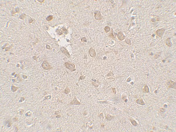
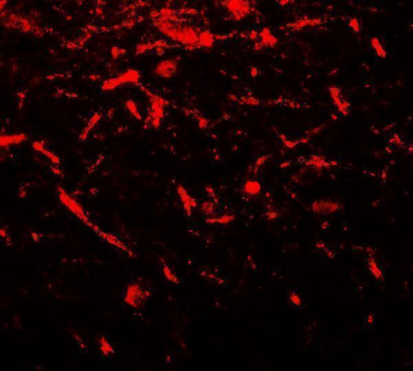


Comprehensive Human Protein Atlas IHC scoring per tissue (reliability: Approved — Medium consistency between antibody staining and RNA expression data.). Rows are taken directly from the HPA tissue chart — click any row's HPA link to view the source.
| Tissue | Cell type | Level | Evidence | Source |
|---|---|---|---|---|
| Cerebellum | Cells in molecular layer | High | Protein (IHC) | HPA → |
| Cerebral cortex | Glial cells | High | Protein (IHC) | HPA → |
| Epididymis | Glandular cells | High | Protein (IHC) | HPA → |
| Fallopian tube | Glandular cells | High | Protein (IHC) | HPA → |
| Hippocampus | Neuronal cells | High | Protein (IHC) | HPA → |
| Tissue | Cell type | Level | Evidence | Source |
|---|---|---|---|---|
| Adipose tissue | Adipocytes | Not detected | Protein (IHC) | HPA → |
Troubleshoot FMR1 staining in paraffin section IHC by checking retrieval, cell identity and subcellular pattern before comparing signal intensity (datasheet PB9703; HPA tissue IHC; UniProt Q06787).
Anti-FMR1 antibodies have IHC images from human seminoma testis paraffin sections and human brain tissue, plus IF images from human brain tissue and HeLa cells (catalog image captions).
PB9703 has an IHC image from a human seminoma testis paraffin section and an IF/ICC image from HeLa cells (PB9703 image captions). A00192 has IHC and IF images from human brain tissue (A00192 image captions).
Which to pick: Choose PB9703 for paraffin section IHC: its caption documents EDTA retrieval and DAB detection, but does not report the fixative (PB9703 IHC caption). For IF, A00192 has a human brain tissue image; for ICC/IF, PB9703 has a HeLa cell image (A00192 and PB9703 IF captions). Both list Human, Mouse, and Rat reactivity, but their IHC images document human samples only (catalog reactivity; IHC image captions).