This website uses cookies to ensure you get the best experience on our website.
- Table of Contents
4 Citations
1 Citations 4 Q&As


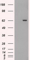
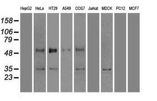
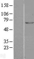
Facts about Glutamate decarboxylase 1.

Catalyzes the production of GABA.
.| Human | |
|---|---|
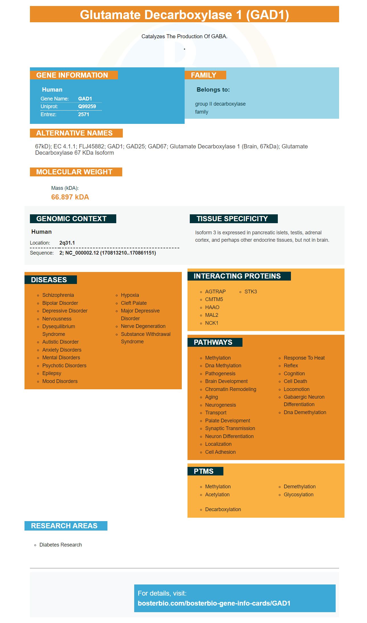
| Gene Name: | GAD1 |
| Uniprot: | Q99259 |
| Entrez: | 2571 |

| Belongs to: |
|---|
| group II decarboxylase family |

67kD); EC 4.1.1; FLJ45882; GAD1; GAD25; GAD67; glutamate decarboxylase 1 (brain, 67kDa); Glutamate decarboxylase 67 kDa isoform
Mass (kDA):
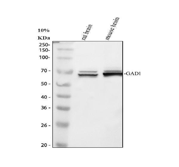
66.897 kDA

| Human | |
|---|---|
| Location: | 2q31.1 |
| Sequence: | 2; NC_000002.12 (170813210..170861151) |
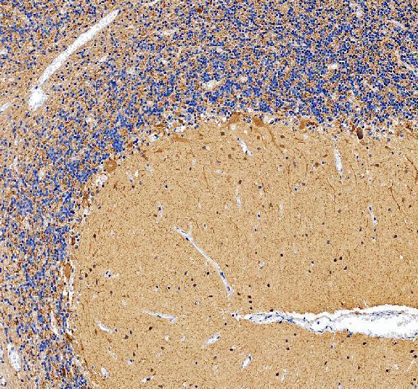
Isoform 3 is expressed in pancreatic islets, testis, adrenal cortex, and perhaps other endocrine tissues, but not in brain.


PMID: 1549570 by Bu D.-F., et al. Two human glutamate decarboxylases, 65-kDa GAD and 67-kDa GAD, are each encoded by a single gene.
PMID: 8088791 by Bu D.-F., et al. The exon-intron organization of the genes (GAD1 and GAD2) encoding two human glutamate decarboxylases (GAD67 and GAD65) suggests that they derive from a common ancestral GAD.
*More publications can be found for each product on its corresponding product page