Click image to see more details
-

-
-
-
-
+9
Product Info Summary
| SKU: | M02002-1 |
|---|---|
| Size: | 100 µl |
| Reactive Species: | Human, Monkey, Mouse, Rat |
| Host: | Mouse |
| Application: | IP, IF, IHC, WB |
Customers Who Bought This Also Bought

Product info
Product Name
Anti-GAD67 (GAD1) Mouse Monoclonal Antibody [Clone ID: OTI3G9]
SKU/Catalog Number
M02002-1
Size
100 µl
Description
Boster Bio GAD1 (GAD67) mouse monoclonal antibody, clone OTI3G9 (formerly 3G9). Catalog# M02002-1. Tested in IF, IHC, IP, WB. This antibody reacts with Human, Monkey, Mouse, Rat.
Storage & Handling
Store at -20°C as received.
Cite This Product
Anti-GAD67 (GAD1) Mouse Monoclonal Antibody [Clone ID: OTI3G9] (Boster Biological Technology, Pleasanton CA, USA, Catalog # M02002-1)
Host
Mouse
Contents
PBS (pH 7.3) containing 1% stabilizing protein, 50% glycerol and 0.02% sodium azide.
This antibody is supplied in a stabilized formulation.
Compatibility with conjugation reactions depends on the chemistry of the conjugation method used.
For conjugation methods that are not compatible with the stabilizing components present in this formulation, a carrier-free antibody format is required.
Clonality
Monoclonal
Clone Number
OTI3G9
Isotype
IgG2b
Immunogen
Full length human recombinant protein of human GAD1 (NP_000808) produced in HEK293T cell.
Reactive Species
M02002-1 is reactive to GAD1 in Human, Monkey, Mouse, Rat
Calculated molecular weight
66.9 kDa
Antibody Validation
Boster validates all antibodies on WB, IHC, ICC, Immunofluorescence, and ELISA with known positive control and negative samples to ensure specificity and high affinity, including thorough antibody incubations.
Application & Images
Applications
M02002-1 is guaranteed for IP, IF, IHC, WB Boster Guarantee
Recommend Dilution
WB 1:1000-1:2000
IHC 1:50
IF 1:50
IP: 4ug/mL
Validation Images & Assay Conditions
Click image to see more details
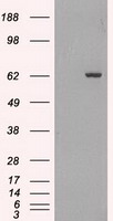
HEK293T cells were transfected with the pCMV6-ENTRY control (Left lane) or pCMV6-ENTRY GAD1 (Right lane) cDNA for 48 hrs and lysed. Equivalent amounts of cell lysates (5 ug per lane) were separated by SDS-PAGE and immunoblotted with anti-GAD1.

Click image to see more details
Western blot analysis of extracts (35ug) from 9 different cell lines by using anti-GAD1 monoclonal antibody.

Click image to see more details
Immunohistochemical staining of paraffin-embedded Human Ovary tissue within the normal limits using anti-GAD1 mouse monoclonal antibody. (Heat-induced epitope retrieval by 10mM citric buffer

Click image to see more details
Immunohistochemical staining of paraffin-embedded Adenocarcinoma of Human ovary tissue using anti-GAD1 mouse monoclonal antibody. (Heat-induced epitope retrieval by 10mM citric buffer

Click image to see more details
Immunohistochemical staining of paraffin-embedded Human pancreas tissue within the normal limits using anti-GAD1 mouse monoclonal antibody. (Heat-induced epitope retrieval by 10mM citric buffer

Click image to see more details
Immunohistochemical staining of paraffin-embedded Human thyroid tissue within the normal limits using anti-GAD1 mouse monoclonal antibody. (Heat-induced epitope retrieval by 10mM citric buffer

Click image to see more details
Immunohistochemical staining of paraffin-embedded Human endometrium tissue within the normal limits using anti-GAD1 mouse monoclonal antibody. (Heat-induced epitope retrieval by 10mM citric buffer

Click image to see more details
Immunohistochemical staining of paraffin-embedded Carcinoma of Human bladder tissue using anti-GAD1 mouse monoclonal antibody. (Heat-induced epitope retrieval by 10mM citric buffer

Click image to see more details
Immunohistochemical staining of paraffin-embedded Adenocarcinoma of Human breast tissue using anti-GAD1 mouse monoclonal antibody. (Heat-induced epitope retrieval by 10mM citric buffer

Click image to see more details
Immunohistochemical staining of paraffin-embedded Human colon tissue within the normal limits using anti-GAD1 mouse monoclonal antibody. (Heat-induced epitope retrieval by 10mM citric buffer

Click image to see more details
Immunohistochemical staining of paraffin-embedded Carcinoma of Human kidney tissue using anti-GAD1 mouse monoclonal antibody. (Heat-induced epitope retrieval by 10mM citric buffer

Click image to see more details
Immunohistochemical staining of paraffin-embedded Human lung tissue within the normal limits using anti-GAD1 mouse monoclonal antibody. (Heat-induced epitope retrieval by 10mM citric buffer

Click image to see more details
Immunofluorescent staining of A549 cells using anti-GAD1 mouse monoclonal antibody (M02002-1).
Specific Publications For Anti-GAD67 (GAD1) Mouse Monoclonal Antibody [Clone ID: OTI3G9] (M02002-1)
Loading publications
Recommended Resources
Here are featured tools and databases that you might find useful.
- Boster's Pathways Library
- Protein Databases
- Bioscience Research Protocol Resources
- Data Processing & Analysis Software
- Photo Editing Software
- Scientific Literature Resources
- Research Paper Management Tools
- Molecular Biology Software
- Primer Design Tools
- Bioinformatics Tools
- Phylogenetic Tree Analysis
Customer Reviews
Have you used Anti-GAD67 (GAD1) Mouse Monoclonal Antibody [Clone ID: OTI3G9]?
Share your experimental results or join a short interview to earn up to $1,000 in product credits or other rewards.
0 Reviews For Anti-GAD67 (GAD1) Mouse Monoclonal Antibody [Clone ID: OTI3G9]
Customer Q&As
Have a question?
Find answers in Q&As, reviews.
Can't find your answer?
Submit your question
