GFRA1 / GDNF family receptor alpha-1 · IHC design guide

Design Immunohistochemistry for GFRA1

Plan chromogenic GFRA1 IHC in paraffin sections using the reported glandular-cell cytoplasmic pattern (HPA tissue IHC). Interpret positive staining cautiously because its agreement with RNA expression is very low (HPA tissue IHC).

Evidence assembled Oct 2026 · For research use; verify linked source records and product datasheet before use
Immunohistochemistry protocol sheet for GFRA1 (IHC for GFRA1): expected localisation Cytoplasm in tissue IHC (HPA tissue IHC); mainly plasma membrane molecularly (UniProt), antibody PB9202, validated IHC image, and IHC protocol steps
Printable GFRA1 IHC protocol sheet — expected localisation Cytoplasm in tissue IHC (HPA tissue IHC); mainly plasma membrane molecularly (UniProt), antibody PB9202, controls and protocol steps. Open the full GFRA1 IHC guide →

GFRA1 Immunohistochemistry Experimental Design Guide

Expected localisation, validated protocols, controls and antibodies — the at-a-glance facts below, then the full design guide.

Must know before staining
Expected localisation Cytoplasm in tissue IHC (HPA tissue IHC); mainly plasma membrane molecularly (UniProt)
Staining pattern Glandular cells show cytoplasmic staining (HPA tissue IHC)
Antigen retrieval EDTA pH 8.0 HIER, heat-mediated (datasheet PB9202)
Positive control ⓘ Appendix+4 more · see all
Negative control ⓘ Adipose tissue+4 more · see all
Important caveats
Reasons your staining may differ from the expected pattern.
Fixation Keep fixation consistent across paraffin sections (standard IHC practice; not target-specific)
Caveat Staining has very low concordance with RNA expression (HPA tissue IHC)
Regulation Expression regulation is unreported (UniProt)
Isoform / epitope 2 isoforms; mature chain spans residues 25–429; verify epitope (UniProt)
Section 1

Recommended GFRA1 IHC & IF Protocols

The catalog antibody protocol uses EDTA pH 8.0 retrieval (PB9202 datasheet). The published protocols below describe chromogenic GFRA1 staining in rat testis and human breast tumors (PMC12670479; PMC11937365).

Recommended immunohistochemistry (IHC-P) protocol parameters
SampleParaffin-embedded human lung cancer tissue; fixative not specified (datasheet PB9202)
FixationImage fixative and duration unreported (datasheet PB9202); verify before use.
Sectioning4–5 µm sections on charged slides (standard)
DeparaffinisationXylene, graded ethanol series to water (standard)
Antigen retrievalHeat retrieval: EDTA pH 8.0 (datasheet PB9202); 20 min, 95–100 °C (standard)
Peroxidase block3% H2O2, 10 min, room temperature (standard)
Blocking10% goat serum (datasheet PB9202)
Primary antibodyRabbit anti-GFRA1, 0.5-1μg/ml (datasheet PB9202)
Primary incubationOvernight at 4 °C (datasheet PB9202)
DetectionStreptavidin-biotin complex (SABC), DAB chromogen (datasheet PB9202)
CounterstainHematoxylin, blue, dehydrate and mount (standard)
Expected resultGFRA1-positive staining in glandular cells of appendix (HPA tissue IHC: High). HPA tissue profile: Cytoplasmic expression in several tissues. No signal in the no-primary control.
💡Decision noteStart with EDTA pH 8.0 for the catalog antibody (PB9202 datasheet); use citrate pH 6 when reproducing either published protocol (PMC12670479; PMC11937365).
Section 2

What Is the Expected GFRA1 Staining Pattern?

GFRA1 is mainly at the plasma membrane, with vesicular localisation under some conditions; it has no transmembrane segment (UniProt P56159 topology and subcellular annotation). In tissue IHC, HPA reports cytoplasmic staining in several tissues, including high staining in appendix, duodenum and rectum glandular cells (HPA tissue IHC). Treat that pattern as provisional: HPA rates the tissue staining Uncertain because antibody staining and RNA expression show very low consistency (HPA tissue IHC).

What am I looking at on my slide?
Glandular cells stain strongly in appendix, duodenum or rectum, with a discernible cell-associated signal (HPA: High in those cells).This matches the reported tissue IHC pattern, which HPA describes as cytoplasmic (HPA tissue IHC). Assess the stained cells against adjacent tissue and controls before calling them GFRA1-positive: HPA rates the IHC pattern Uncertain, so a matching location and intensity alone do not establish antibody specificity (HPA tissue IHC).
A cell-associated membrane outline or intracellular puncta appear in a stained cell.These compartments are biologically plausible: GFRA1 localises mainly to the plasma membrane and can shift into trans-Golgi and endosomal vesicles in the presence of SORL1 (UniProt P56159 subcellular annotation). HPA's tissue IHC summary instead reports cytoplasmic staining, so compare the pattern with the tissue control; do not require crisp membrane staining in every paraffin section (HPA tissue IHC; UniProt P56159).
Staining is predominantly nuclear, or appears in a compartment that cannot be distinguished from background.Reassess it as a possible artefact in tissue IHC: HPA's tissue summary is cytoplasmic, while UniProt places GFRA1 mainly at the membrane and also in vesicular compartments (HPA tissue IHC; UniProt P56159). A nuclear IF observation cannot by itself validate nuclear IHC: HPA separately reports approved nucleoplasmic staining in ICC-IF (HPA subcellular ICC-IF).
Strong staining appears in an HPA-listed undetected cell population, such as adipocytes in adipose tissue or respiratory epithelium in bronchus (HPA tissue IHC).Check for antibody cross-reactivity or endogenous detection activity before assigning a GFRA1-positive result (general IHC practice). The HPA comparison is cell-specific: its 'Not detected' entries refer to the listed cells and sampled staining, not a guarantee that every cell in that organ lacks GFRA1 (HPA tissue IHC).
Diffuse colour covers cells and surrounding tissue, or no signal appears in appendix, duodenum or rectum glandular cells.Diffuse signal weakens localisation and may reflect background from blocking, detection or overdevelopment; a blank reported-positive control may reflect assay failure or variable tissue staining (general IHC practice; HPA: High in the listed glandular cells). Neither outcome alone proves GFRA1 absence or presence, especially given HPA's Uncertain tissue IHC rating (HPA tissue IHC).
💡Expected GFRA1 appearanceCall a result provisionally positive when glandular cells in appendix, duodenum or rectum show strong, cell-associated cytoplasmic staining (HPA: High; HPA tissue IHC); a uniform haze or strong signal in an HPA-listed undetected cell population warrants background or specificity checks (HPA tissue IHC; general IHC practice).
How each factor affects the staining
Tissue and cell choiceHPA reports High glandular-cell staining in appendix, duodenum and rectum, Medium staining in colon, endometrium and gallbladder glandular cells, and Not detected staining in selected cell populations (HPA tissue IHC). Select controls by the named cell population, and interpret them provisionally because HPA rates the tissue pattern Uncertain (HPA tissue IHC).
Topology and traffickingGFRA1 has no transmembrane segment and carries a GPI-anchor keyword; UniProt places it mainly at the plasma membrane but describes SORL1-associated internalisation into trans-Golgi, endosomal and multivesicular compartments (UniProt P56159). This supports assessing both cell-surface and intracellular patterns without claiming that either must dominate a given IHC section.
Processing and antibody epitopeUniProt annotates a signal peptide at residues 1–24, a mature chain at 25–429 and a propeptide at 430–465 (UniProt P56159 processing). The supplied record gives no antibody epitope, so these annotations cannot predict whether the IHC antibody recognises a processed form; check its documented immunogen or epitope before drawing that conclusion.
IF/ICC pattern?HPA reports Golgi as the main approved ICC-IF location and nucleoplasm as an additional approved location, with images in MCF-7, U-251MG and U2OS (HPA subcellular ICC-IF). This is context for interpreting an IF image, not an IF protocol or proof that the same compartments should dominate paraffin-section IHC (HPA subcellular ICC-IF; HPA tissue IHC).
Why is my staining missing, weak or wrong?
SituationLikely causeNext action
No signal in appendix, duodenum or rectum glandular cells (HPA: High).The IHC run may have failed, or the selected section may differ from the reported HPA pattern (general IHC practice; HPA tissue IHC, Uncertain).Review tissue identity, cell type, antibody exposure, retrieval, detection and a working run control; adjust one general IHC step at a time. Do not infer a GFRA1-specific retrieval requirement from the supplied sources.
Weak signal in colon, endometrium, gallbladder, heart or kidney.Medium is the reported HPA level in the specified glandular cells, cardiomyocytes or tubular cells, respectively (HPA tissue IHC).Score the named cell population against the slide's background and a stronger reported-positive control. Avoid treating a Medium HPA entry as an expected High-intensity result (HPA tissue IHC).
Strong colour appears across most structures, including spaces between cells.Nonspecific detection, inadequate blocking or excessive chromogen development can obscure cell-associated staining (general IHC practice).Compare a negative reagent control and the reported-positive tissue; then review blocking, detection and development settings (general IHC practice). Retain only a cell-associated pattern as interpretable.
Strong staining appears in an HPA-listed Not detected cell type.Possible cross-reactivity or endogenous detection activity; HPA's tissue IHC assessment is Uncertain (general IHC practice; HPA tissue IHC).Check the listed cell identity, negative reagent control and detection chemistry. Seek independent validation before calling unexpected staining GFRA1-specific (general IHC practice; HPA tissue IHC, Uncertain).
An apparently nuclear-only IHC pattern conflicts with the expected tissue pattern.HPA reports cytoplasmic tissue IHC, although its separate ICC-IF dataset also reports approved nucleoplasmic localisation (HPA tissue IHC; HPA subcellular ICC-IF).Compare cell-associated cytoplasmic signal and negative controls on the IHC slide. Report nuclear staining as unresolved unless independently validated in that tissue and method; do not transfer the ICC-IF call directly to IHC.
A section shows puncta rather than a crisp cell-surface outline.Intracellular localisation is plausible when SORL1 promotes GFRA1 internalisation (UniProt P56159 subcellular annotation), but puncta alone do not establish that mechanism.Assess whether puncta are confined to the expected cells and exceed background; compare the reported-positive tissue and controls (HPA tissue IHC; general IHC practice). Describe the observed compartment without claiming SORL1 activity.

Sample controls for GFRA1 IHC & IF

🧪Run appendix first and expect staining in its glandular cells (HPA: High in appendix glandular cells); use adipose tissue adipocytes as the negative tissue (HPA: Not detected in adipocytes). On the appendix slide, internal negative cells should remain unstained; identify them by morphology and verify them against the control slides rather than assuming a specific cell type is negative.
Positive control tissue: Appendix (Glandular cells, HPA High)
Negative control tissue: Adipose tissue (HPA Not detected)
ICC-IF cell lines (HPA subcellular resource): HPA ICC-IF images show GFRA1 in MCF-7, U-251MG, U2OS, with annotated localisation: Golgi apparatus (approved) (HPA subcellular).
Technical controls: Include a no-primary (secondary-only) slide, a concentration-matched nonimmune rabbit IgG isotype control (caption: rabbit primary antibody), and, if available, a GFRA1 knockout specimen or immunizing-peptide competition control. For appendix, check endogenous peroxidase and biotin background before interpreting DAB staining because the selected IHC caption uses biotin-based SABC–DAB detection (caption: SABC–DAB).
⚠️Feasibility: A target-specific fixation window or fixation effect is unreported, and the selected PB9202 paraffin-section caption does not state its fixative (caption: fixative not stated). That caption uses heat retrieval in EDTA at pH 8.0 for lung cancer tissue, but does not establish that retrieval is required for appendix (caption: EDTA retrieval in paraffin-embedded lung cancer tissue). No supplied evidence shows that frozen sections or IF are easier; assess glandular background in appendix with the controls above (HPA: High in appendix glandular cells; caption: SABC–DAB).

HPA tissue IHC evidence for GFRA1

Comprehensive Human Protein Atlas IHC scoring per tissue (reliability: Uncertain — Very low consistency between antibody staining and RNA expression data. Pending external verification.). Rows are taken directly from the HPA tissue chart — click any row's HPA link to view the source.

Positive expression · recommended positive controls

TissueCell typeLevelEvidenceSource
Appendix Glandular cells High Protein (IHC) HPA →
Duodenum Glandular cells High Protein (IHC) HPA →
Rectum Glandular cells High Protein (IHC) HPA →
Colon Glandular cells Medium Protein (IHC) HPA →
Endometrium Glandular cells Medium Protein (IHC) HPA →

Undetected expression · recommended negative controls

TissueCell typeLevelEvidenceSource
Adipose tissue Adipocytes Not detected Protein (IHC) HPA →
Adrenal gland Glandular cells Not detected Protein (IHC) HPA →
Bronchus Respiratory epithelial cells Not detected Protein (IHC) HPA →
Caudate Glial cells Not detected Protein (IHC) HPA →
Cerebellum Cells in granular layer Not detected Protein (IHC) HPA →
Section 3

Advanced GFRA1 IHC Tips

Troubleshoot GFRA1 chromogenic IHC in paraffin sections using the selected antibody’s tissue image, protein annotations, and cautious interpretation of tissue staining.

Which retrieval conditions should I try first when GFRA1 staining is weak?
Start with heat-mediated EDTA at pH 8.0 for paraffin sections (datasheet PB9202). The selected tissue image used this retrieval before incubating human lung cancer sections with 1 μg/ml primary antibody overnight at 4°C (PB9202 caption). If signal is weak, verify deparaffinisation and reagent performance, then compare retrieval times while keeping the buffer and pH fixed (standard IHC practice). Inspect section morphology and matched controls after each adjustment, since excessive heating can damage tissue and increase diffuse staining (standard IHC practice). Record membrane and intracellular staining separately because GFRA1 has both reported locations (UniProt P56159 localisation).
How should I assess whether fixation is affecting GFRA1 IHC?
The selected image identifies a paraffin section but does not report its fixative, so target-specific fixation sensitivity is unknown (PB9202 caption). Do not infer a preferred fixative or fixation duration from the tissue staining profile or GFRA1 protein annotations (PB9202 caption; HPA tissue IHC; UniProt P56159). For a controlled comparison, process matched specimens with documented fixation conditions, then apply the same retrieval, antibody concentration, and detection workflow to every section (standard IHC practice). Include a matched tissue control and assess signal alongside morphology, because poor preservation can complicate cell-level scoring (standard IHC practice). Report fixation details with the results so the comparison can be reproduced (standard IHC practice).
Should GFRA1 staining be membranous or cytoplasmic in paraffin sections?
Assess membranous staining because GFRA1 localizes mainly to the plasma membrane (UniProt P56159 localisation). Intracellular puncta can also be plausible: GFRA1 is reported in the trans-Golgi network, endosomes, and multivesicular bodies, with SORL1 associated with redistribution to these structures (UniProt P56159 localisation). HPA describes cytoplasmic tissue staining, but rates its tissue IHC reliability as uncertain because staining and RNA expression have very low consistency (HPA tissue IHC). Record membrane and cytoplasmic patterns separately, including the stained cell type and distribution across intact tissue (standard IHC practice). Treat isolated nuclear staining cautiously even though HPA reports nucleoplasmic localisation in subcellular imaging (HPA subcellular; standard IHC practice).
Can this IHC stain distinguish GFRA1 isoforms or processing states?
GFRA1 has 2 annotated isoforms, but the supplied antibody evidence does not map its epitope or establish isoform selectivity (UniProt P56159 isoforms; PB9202 caption). The precursor has a signal peptide at residues 1–24, a reported chain at 25–429, and a propeptide at 430–465 (UniProt P56159 processing). Glycosylation is annotated at residues 59, 347, and 406; these annotations alone do not establish whether this antibody detects every processed form (UniProt P56159 glycosylation; standard assay interpretation). Score the observed staining as GFRA1 immunoreactivity unless epitope mapping or independent validation supports a narrower claim (standard IHC practice). Use identically processed controls when comparing staining patterns (standard IHC practice).
How can I check a GFRA1 IHC pattern with multiplex immunofluorescence?
Use IF as a complementary localisation check and retain the paraffin IHC result as the primary readout; the selected antibody evidence describes chromogenic IHC, not an IF protocol (PB9202 caption). Pair GFRA1 with a validated marker for the expected cell type in the specimen, and include single-label controls to check channel overlap (standard IF practice). Choose fluorophores after inspecting tissue autofluorescence, favouring a channel with clear separation from that background (standard IF practice). Set permeabilisation according to the antibody’s mapped epitope and whether intracellular access is required; GFRA1 has no transmembrane segment and is annotated as GPI-anchored (UniProt P56159 topology and keywords; standard IF practice). Compare membrane and vesicular signals without treating fluorescence alone as proof of specificity (UniProt P56159 localisation; standard IF practice).
What should I change if GFRA1 staining is diffuse or patchy?
Begin by comparing stained sections with a no-primary control and checking whether background follows tissue edges, damaged areas, or the detection reagents (standard IHC practice). The selected workflow used 10% goat serum blocking, a biotinylated secondary antibody, a streptavidin–biotin complex, and DAB (PB9202 caption). Check endogenous peroxidase blocking and assess nonspecific signal from the biotin-based detection system with appropriate controls (standard IHC practice). If background persists, titrate the primary below the documented 1 μg/ml condition while monitoring a matched positive section (PB9202 caption; standard IHC practice). Interpret diffuse cytoplasmic colour cautiously because HPA tissue IHC has uncertain reliability (HPA tissue IHC).
How should I score GFRA1 IHC across heterogeneous tissue sections? ⚠ ANSWER MARKED FOR VERIFICATION
Define the tissue compartment and eligible intact cells before scoring, then report the percentage of positive cells and staining intensity separately (standard IHC practice). An H-score can combine intensity with the percentage of cells at each intensity; specify the scoring scale and keep thresholds fixed across sections (standard IHC practice). For spatially restricted staining, positive-cell density per mm² is another option when the analysed tissue area is recorded (standard IHC practice). Normalise counts to the number of eligible cells or the measured tissue area, and exclude necrosis and section folds consistently (standard IHC practice). Report membrane and cytoplasmic scores separately because GFRA1 has both reported locations (UniProt P56159 localisation).
When does a GFRA1-positive pattern warrant scepticism?
Give more weight to staining in intact cells with coherent membrane or vesicular localisation than to colour confined to section edges or necrosis (UniProt P56159 localisation; standard IHC practice). Check the stained cell type against specimen morphology and matched controls; HPA reports high glandular-cell staining in appendix, duodenum, and rectum, but rates its tissue IHC evidence uncertain (HPA tissue IHC). A no-primary control helps reveal detection-related colour, including endogenous enzyme activity that can mimic weak DAB signal (standard IHC practice). Treat isolated nuclear staining cautiously: HPA subcellular imaging reports nucleoplasmic localisation, while UniProt describes predominantly plasma-membrane localisation (HPA subcellular; UniProt P56159 localisation). Confirm disputed patterns with an independent specificity control before making a biological claim (standard IHC practice).
Boster reagents

Best GFRA1 / GDNF family receptor alpha-1 IHC Antibodies

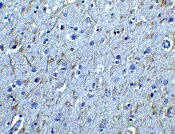
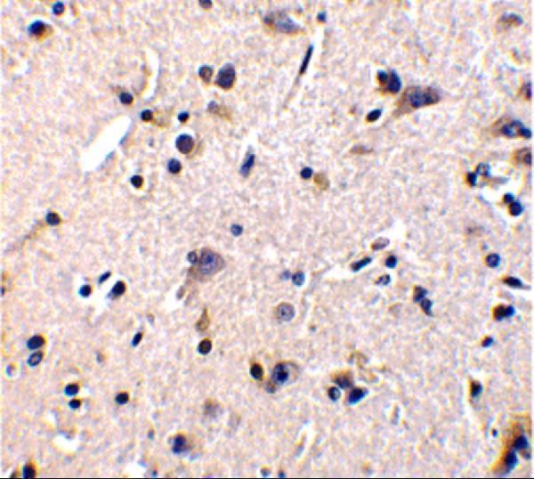
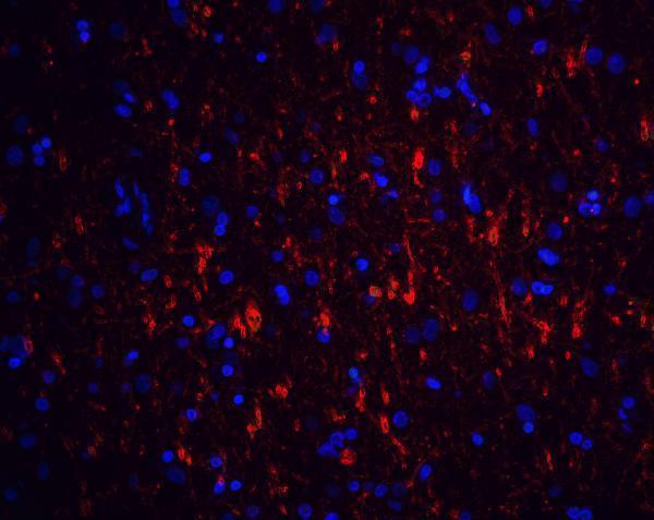
Validated anti-GFRA1 antibodies have IHC images from human lung cancer and brain tissue, plus a human brain IF image (PB9202 IHC caption; A02897 IHC/IF captions).

Real IHC data IHC analysis of GFRA1 using anti-GFRA1 antibody (PB9202). GFRA1 was detected in a paraffin-embedded section of human lung cancer tissue. Heat mediated antigen retrieval was performed in EDTA buffer (pH 8.0, epitope retrieval solution). The tissue section was blocked with 10% goat serum. The tissue section was then incubated with 1 μg/ml rabbit anti-GFRA1 Antibody (PB9202) overnight at 4°C. Biotinylated goat anti-rabbit IgG was used as secondary antibody and incubated for 30 minutes at 37°C. The tissue section was developed using Strepavidin-Biotin-Complex (SABC) (Catalog # SA1022) with DAB as the chromogen.
Anti-GDNF Receptor alpha 1/GFRA1 Antibody ®
Cat # PB9202
Real IHC data Immunohistochemistry of GFR alpha 1 in human brain tissue with GFR alpha 1 antibody at 2.5 μg/ml.
Anti-GFR alpha 1 GFRA1 Antibody
Cat # A02897

PB9202 is listed for human IHC and has a paraffin-section image from human lung cancer tissue (PB9202 applications/reactivity; PB9202 IHC caption). A02897 is listed for human, mouse and rat IHC-P and IF, with IHC and IF images from human brain tissue (A02897 applications/reactivity; A02897 IHC/IF captions).

Which to pick: Choose PB9202 for human tissue IHC when its documented paraffin-section workflow is useful: EDTA retrieval at pH 8.0 and 1 μg/ml primary antibody overnight at 4°C (PB9202 IHC caption). Choose A02897 for IF or work involving mouse or rat, based on its listed applications and reactivity; its IF image is from human brain tissue, while ICC validation and clonality are unreported (A02897 applications/reactivity; A02897 IF caption; catalog clone field). The fixative is unreported for both IHC images (PB9202 IHC caption; A02897 IHC captions).

Each figure is that product's own IHC / IF validation image from its datasheet.

References

  1. UniProt Consortium. UniProt entry P56159 (GFRA1_HUMAN, GDNF family receptor alpha-1).
  2. Human Protein Atlas. GFRA1 tissue IHC expression (reliability: Uncertain).
  3. Human Protein Atlas. GFRA1 subcellular location (ICC-IF): Mainly localized to the Golgi apparatus. In addition localized to the nucleoplasm..
  4. Human Protein Atlas. GFRA1 antibody validation summary (1 antibodies).
  5. Effects of neonatal hypothyroidism on testicular development and undifferentiated spermatogonia in prepubertal rats. Andrology 2026 — PMC12670479.
  6. Characterization, isolation, and culture of spermatogonial stem cells in Macaca fascicularis. Asian journal of andrology 2021 — PMC8152426.
  7. Molecular classification of hormone receptor-positive /HER2-positive breast cancer reveals potential neoadjuvant therapeutic strategies. Signal transduction and targeted therapy 2025 — PMC11937365.
  8. In Vitro and In Vivo Determinations of The Anti-GDNF Family Receptor Alpha 1 Antibody in Mice by Immunochemistry and RT-PCR. International journal of fertility & sterility 2020 — PMC7604702.
  9. PubMed PMID:8674117 — UniProt-cited evidence.
  10. PubMed PMID:9177201 — UniProt-cited evidence.
  11. PubMed PMID:9545641 — UniProt-cited evidence.