GLG1 / Golgi apparatus protein 1 · IHC design guide

Design Immunohistochemistry for GLG1

Plan GLG1 staining in paraffin sections using the granular cytoplasmic tissue profile (HPA tissue IHC). Interpret the signal alongside GLG1’s Golgi membrane localisation and check which region of the protein the antibody recognizes (UniProt).

Evidence assembled Oct 2026 · For research use; verify linked source records and product datasheet before use
Immunohistochemistry protocol sheet for GLG1 (IHC for GLG1): expected localisation Granular cytoplasm (HPA tissue IHC); Golgi membrane (UniProt), antibody A07510-1, validated IHC image, and IHC protocol steps
Printable GLG1 IHC protocol sheet — expected localisation Granular cytoplasm (HPA tissue IHC); Golgi membrane (UniProt), antibody A07510-1, controls and protocol steps. Open the full GLG1 IHC guide →

GLG1 Immunohistochemistry Experimental Design Guide

Expected localisation, validated protocols, controls and antibodies — the at-a-glance facts below, then the full design guide.

Must know before staining
Expected localisation Granular cytoplasm (HPA tissue IHC); Golgi membrane (UniProt)
Staining pattern Widespread granular cytoplasmic staining (HPA tissue IHC)
Antigen retrieval EDTA pH 8.0 HIER, heat-mediated (datasheet A07510-1)
Positive control ⓘ Adrenal gland+4 more · see all
Negative control ⓘ None in HPA (detected in all 45 tissues); use no-primary + isotype controls
Important caveats
Reasons your staining may differ from the expected pattern.
Fixation Keep fixation conditions consistent across sections. (standard IHC practice; not target-specific)
Caveat Lung type I alveolar cells show low staining (HPA tissue IHC)
Regulation No expression regulator specified (UniProt)
Isoform / epitope 3 isoforms; map epitopes against the membrane topology (UniProt)
Section 1

Recommended GLG1 IHC & IF Protocols

The catalog IHC-P protocol (datasheet A07510-1) is accompanied by three published GLG1 IHC protocols (PMC7139395; PMC5788584; PMC4224387).

Recommended immunohistochemistry (IHC-P) protocol parameters
SampleParaffin-embedded mouse kidney tissue; fixative not specified (datasheet A07510-1)
FixationImage fixative and duration unreported (datasheet A07510-1); verify before use.
Sectioning4–5 µm sections on charged slides (standard)
DeparaffinisationXylene, graded ethanol series to water (standard)
Antigen retrievalHeat retrieval: EDTA pH 8.0 (datasheet A07510-1); 20 min, 95–100 °C (standard)
Peroxidase block3% H2O2, 10 min, room temperature (standard)
Blocking10% goat serum (datasheet A07510-1)
Primary antibodyRabbit anti-GLG1, 2-5 μg/ml (datasheet A07510-1)
Primary incubationOvernight at 4 °C (datasheet A07510-1)
DetectionHRP-conjugated secondary, DAB chromogen (datasheet A07510-1)
CounterstainHematoxylin, blue, dehydrate and mount (standard)
Expected resultGLG1-positive staining in glandular cells of adrenal gland (HPA tissue IHC: High). HPA tissue profile: Ubiquitous cytoplasmic expression in a granular pattern. No signal in the no-primary control.
💡Decision noteStart with heat-mediated EDTA pH 8.0 retrieval for the catalog antibody (datasheet A07510-1). Use each article’s stated retrieval when reproducing its protocol (PMC7139395; PMC5788584; PMC4224387).
Section 2

What Is the Expected GLG1 Staining Pattern?

GLG1 is a Golgi membrane protein, with its large luminal domain spanning residues 30–1145 (UniProt Q92896 topology). In paraffin section IHC, expect granular cytoplasmic staining in many cell types, including glandular, respiratory epithelial, and neuronal cells (HPA tissue IHC: ubiquitous granular cytoplasmic expression; High in these listed cells). HPA rates the tissue pattern Supported, with medium consistency between antibody staining and RNA expression (HPA tissue IHC).

What am I looking at on my slide?
Granular cytoplasmic chromogen in glandular cells of colon or duodenum.This fits the reported pattern and cell distribution (HPA tissue IHC: ubiquitous granular cytoplasmic expression; High in colon and duodenal glandular cells). Assess the granules within intact cells, since pooled chromogen can obscure their location (general IHC practice).
Predominantly nuclear staining, or a uniform cell-surface rim without cytoplasmic granules.These compartments do not match the principal Golgi localization (UniProt Q92896 subcellular location; HPA subcellular: Golgi apparatus, supported). Check counterstain, precipitate, and detection controls before interpreting the signal as GLG1 (general IHC practice).
Strong staining in an unexpected cell population while a listed high-staining population is faint.For example, alveolar type I cells are listed Low, whereas bronchial respiratory epithelial cells are High (HPA tissue IHC). Confirm cell identity and distribution; cross-reactivity or endogenous detection activity is possible, but this contrast alone does not establish either cause (general IHC practice).
Diffuse haze across tissue, extracellular spaces, or a largely cell-free area.This differs from the reported intracellular granular pattern (HPA tissue IHC). Uneven reagent coverage, inadequate washes, or chromogen deposition can produce background; compare a control lacking primary antibody and inspect tissue edges (general IHC practice).
No visible staining in a section containing colon or duodenal glandular cells.Both are listed High and provide useful positive-reference cell populations (HPA tissue IHC). First check tissue integrity, detection reagents, and the catalog antibody's IHC-P instructions; a negative slide alone does not prove GLG1 absence (general IHC practice).
💡Expected GLG1 appearanceA convincing positive result is intracellular granular cytoplasmic chromogen in a listed High cell population, such as colon glandular cells (HPA tissue IHC), consistent with Golgi localization (UniProt Q92896; HPA subcellular), whereas dominant nuclear staining or diffuse extracellular color is suspect (general IHC practice).
How each factor affects the staining
Membrane topology and compartmentGLG1 has one transmembrane segment at 1146–1166 and a large luminal domain (UniProt Q92896 topology). The observed IHC pattern is granular cytoplasmic (HPA tissue IHC); topology alone does not identify the catalog antibody's epitope.
Tissue and cell selectionHPA lists glandular cells in colon and duodenum as High and alveolar type I cells as Low (HPA tissue IHC). Use the listed cell types when comparing slides; Low is not a validated negative control (HPA tissue IHC).
Antibody evidenceThe listed antibody HPA010815 has Supported IHC status, while the tissue profile has medium antibody–RNA consistency (HPA antibody validation; HPA tissue IHC). Treat a matching pattern as supporting evidence, especially when evaluating weak or unexpected staining.
Antigen retrievalUse the catalog antibody's IHC-P retrieval instructions as the starting condition (general IHC practice). The supplied UniProt and HPA records do not establish GLG1-specific retrieval requirements or fixation sensitivity.
Isoforms and processingThree isoforms are listed, and the signal peptide occupies residues 1–29 (UniProt Q92896 isoforms; processing). Without an antibody epitope or isoform-validation record, do not assign a staining difference to a particular isoform or to shedding.
IF/ICC Q&A: where should GLG1 appear?Mainly at the Golgi apparatus (HPA subcellular: supported), consistent with the Golgi membrane annotation (UniProt Q92896). IF/ICC has its own guide; this localization is an interpretation reference, not an IF protocol.
Why is my staining missing, weak or wrong?
SituationLikely causeNext action
Listed High cells show no chromogen.A failed detection step or an unsuitable assay condition is possible; colon and duodenal glandular cells are listed High (HPA tissue IHC; general IHC practice).Check a known-working IHC control and the catalog antibody's IHC-P instructions, then verify primary antibody, detection reagents, and chromogen were applied (general IHC practice).
Signal is mainly nuclear.Nuclear-dominant signal conflicts with the Golgi annotation and reported granular cytoplasmic IHC profile (UniProt Q92896; HPA tissue IHC).Compare with a primary-omission control, inspect counterstain and precipitate, and reassess localization in intact cells (general IHC practice).
Tissue-wide brown haze hides individual cells.Nonspecific reagent binding or incomplete washing may obscure the reported granular pattern (HPA tissue IHC; general IHC practice).Review blocking, reagent concentration, wash coverage, and chromogen development; compare a control lacking primary antibody (general IHC practice).
Color persists in the primary-omission control.Endogenous detection activity or reagent deposition can generate color without the primary antibody (general chromogenic IHC practice).Inspect the detection chemistry and its appropriate endogenous-activity block; check for deposits before scoring GLG1 staining (general chromogenic IHC practice).
Alveolar type I cells appear stronger than bronchial respiratory epithelial cells.This reverses their listed Low and High levels, respectively (HPA tissue IHC); cell identification or background may explain the discrepancy (general IHC practice).Confirm the compared cell types, inspect a primary-omission control, and repeat the comparison in well-preserved regions (general IHC practice).
Granules are difficult to distinguish from dark deposits.Excess chromogen or precipitate can obscure subcellular detail (general chromogenic IHC practice); the reference pattern is granular cytoplasmic (HPA tissue IHC).Inspect deposits outside cells, review chromogen development, and score only clearly localized intracellular signal (general chromogenic IHC practice).

Sample controls for GLG1 IHC & IF

🧪Run bronchus first and expect staining in respiratory epithelial cells (HPA: High in bronchus respiratory epithelial cells). HPA detects GLG1 in all 45 scored tissues, so use no-primary and isotype controls in place of a negative tissue; no cell type on the positive slide is established as GLG1-negative, and unstained cells should be treated only as a background reference (HPA: no negative tissue rows).
Positive control tissue: Adrenal gland (Glandular cells, HPA High)
Negative control tissue: None in HPA: GLG1 is detected in all 45 scored tissues. Use a no-primary (secondary-only) and an isotype control instead.
ICC-IF cell lines (HPA subcellular resource): HPA ICC-IF images show GLG1 in U-251MG, U2OS, Sperm, NIH 3T3, with annotated localisation: Golgi apparatus (supported) (HPA subcellular).
Technical controls: Include a no-primary, secondary-only slide and an isotype control matched to the primary antibody’s rabbit host and verified clonality (selected-SKU tissue-IHC caption: rabbit primary; clonality unreported). Use a matched GLG1-knockout specimen as a biological negative if available, and quench endogenous peroxidase before HRP/DAB detection in bronchus (standard IHC practice; selected-SKU tissue-IHC caption: HRP/DAB).
⚠️Feasibility: No target-specific fixation window or fixation effect is reported, and the A07510-1 paraffin-section caption does not state a fixative (selected-SKU tissue-IHC caption). That caption uses heat retrieval in EDTA at pH 8.0, but provides no comparison establishing that retrieval is required (selected-SKU tissue-IHC caption). Frozen-section performance and whether IF is easier are unreported; for bronchus, assess intracellular Golgi-pattern staining against luminal mucus and background, while HPA supports Golgi localization in ICC-IF (UniProt Q92896 localization; HPA: Golgi apparatus supported; standard IHC practice).

HPA tissue IHC evidence for GLG1

Comprehensive Human Protein Atlas IHC scoring per tissue (reliability: Supported — Medium consistency between antibody staining and RNA expression data.). Rows are taken directly from the HPA tissue chart — click any row's HPA link to view the source.

Positive expression · recommended positive controls

TissueCell typeLevelEvidenceSource
Adrenal gland Glandular cells High Protein (IHC) HPA →
Appendix Glandular cells High Protein (IHC) HPA →
Bronchus Respiratory epithelial cells High Protein (IHC) HPA →
Caudate Neuronal cells High Protein (IHC) HPA →
Cerebellum Cells in molecular layer High Protein (IHC) HPA →

Undetected expression · recommended negative controls

TissueCell typeLevelEvidenceSource
None in HPA: GLG1 is detected in all 45 scored tissues. Use a no-primary (secondary-only) and an isotype control instead.
Section 3

Advanced GLG1 IHC Tips

Troubleshoot GLG1 staining by checking retrieval, Golgi-pattern specificity, controls, and cell-specific scoring in paraffin-section IHC (UniProt Q92896 localisation; HPA: granular cytoplasmic profile).

What should I adjust when GLG1 staining is weak after antigen retrieval?
Start with heat-mediated EDTA at pH 8.0 for paraffin-section IHC with A07510-1 (datasheet A07510-1). The documented mouse kidney condition used 2 μg/ml primary overnight at 4°C after a 10% goat-serum block (datasheet A07510-1). If signal remains weak, compare retrieval times on adjacent sections while keeping primary concentration and DAB development constant, then assess tissue morphology and no-primary controls (standard IHC practice). Score recovery as a granular cytoplasmic pattern consistent with Golgi localisation, since diffuse brown staining alone does not establish GLG1 detection (HPA: granular cytoplasmic profile; UniProt Q92896 localisation).
Can fixation explain weak or uneven GLG1 staining in paraffin sections?
GLG1-specific fixation sensitivity is unknown, and the A07510-1 mouse kidney paraffin-section caption does not state a fixative (datasheet A07510-1). Record fixative, fixation duration, processing history, and section age before attributing unequal staining to the antibody (standard IHC practice). Compare sections processed under matched conditions, holding EDTA retrieval at pH 8.0 and the documented 2 μg/ml primary condition constant (datasheet A07510-1; standard IHC practice). If morphology or staining differs across batches, repeat with matched controls; neither the tissue expression profile nor GLG1 topology establishes a target-specific fixation effect (standard IHC practice; HPA: tissue IHC profile; UniProt Q92896 topology).
How should I judge a GLG1-positive compartment in chromogenic IHC?
Look for granular cytoplasmic staining that fits GLG1's principal Golgi localisation, including its annotation in medial cisternae (HPA: granular cytoplasmic profile; UniProt Q92896 localisation). Interpret apparent nuclear-only or uniformly diffuse staining cautiously and compare it with adjacent intact cells and a no-primary control (UniProt Q92896 localisation; standard IHC practice). GLG1 is also annotated at postsynaptic Golgi outposts and microtubule-organizing regions, but a brown punctum alone cannot identify one of those structures (UniProt Q92896 localisation; standard IHC practice). Compare cell types within the section: neuronal cells in cerebral cortex have high reported staining, whereas staining intensity can vary by cell type (HPA: High in cerebral-cortex neuronal cells; HPA: tissue IHC profile).
How do epitope position and GLG1 isoforms affect IHC interpretation?
GLG1 has 3 annotated isoforms, but the supplied A07510-1 caption does not locate its epitope or establish isoform coverage (UniProt Q92896 isoforms; datasheet A07510-1). The canonical topology places residues 30–1145 outside the cytoplasm, a membrane span at 1146–1166, and a cytoplasmic tail at 1167–1179 (UniProt Q92896 topology). In a Golgi membrane, accessibility can therefore depend on which side contains the epitope and how the section is processed (UniProt Q92896 topology; standard IHC practice). Five annotated glycosylation sites lie in the large noncytoplasmic region, so epitope-dependent staining differences require direct validation rather than assignment to an isoform or modification from IHC appearance alone (UniProt Q92896 glycosylation; standard IHC practice).
How can IF help check a questionable GLG1 IHC pattern?
Use complementary IF to ask whether a Golgi-localised signal appears in the cell population scored by IHC; the supported subcellular location is Golgi (HPA: Golgi apparatus supported). Multiplex with a marker identifying the expected cell type, such as a respiratory epithelial-cell marker when evaluating bronchus, and include single-channel controls (HPA: High in bronchus respiratory epithelial cells; standard IF practice). Choose a fluorophore channel after checking tissue autofluorescence and spectral overlap, with red or far-red detection when background is lower there (standard IF practice). Because the A07510-1 epitope is unspecified, assess permeabilisation against whether the recognised site is luminal or cytoplasmic; GLG1 spans residues 1146–1166 (datasheet A07510-1; UniProt Q92896 topology; standard IF practice).
What controls distinguish GLG1 staining from chromogenic background?
Begin with the documented 10% goat-serum block and 2 μg/ml primary condition, then compare a no-primary section processed through the same detection steps (datasheet A07510-1; standard IHC practice). The A07510-1 image used a peroxidase-conjugated secondary and DAB; blocking endogenous peroxidase is a general chromogenic IHC precaution, not evidence of GLG1 expression (datasheet A07510-1; standard IHC practice). If both test and no-primary sections brown in the same region, inspect endogenous enzyme activity, pigment, and reagent carryover before adjusting antibody concentration (standard IHC practice). Retain only reproducible granular cytoplasmic staining in intact cells as a candidate target pattern (HPA: granular cytoplasmic profile; standard IHC practice).
How should I score GLG1 IHC across heterogeneous tissue regions? ⚠ ANSWER MARKED FOR VERIFICATION
Define the cell population and intact tissue area before scoring, because GLG1 staining can differ among cell types despite its broad expression (HPA: tissue IHC profile; UniProt Q92896 tissue specificity; standard IHC practice). For graded staining, calculate an H-score by summing each intensity grade 0–3 times its percentage of cells, yielding 0–300 (standard IHC practice). Alternatively, report the percentage of positive target cells or positive-cell density per mm² when segmentation is reliable (standard IHC practice). Normalize cell counts to all evaluable cells of the same type, or density to viable tissue area, and keep retrieval and DAB development matched across samples (standard IHC practice).
When is a GLG1-positive IHC result convincing rather than artefactual?
A convincing result shows reproducible granular cytoplasmic staining in intact cells, consistent with the reported Golgi pattern (HPA: granular cytoplasmic profile; HPA: Golgi apparatus supported). Check cell identity against the tissue context: bronchial respiratory epithelial cells have high reported staining, while lung alveolar type I cells are reported low (HPA: High in bronchus respiratory epithelial cells; HPA: Low in lung alveolar type I cells). Treat nuclear-only signal, section-edge accentuation, and staining confined to necrotic areas as suspect until controls resolve them (UniProt Q92896 localisation; standard IHC practice). Brown signal persisting without primary antibody suggests detection-system background, including endogenous peroxidase, rather than evidence for GLG1 (standard IHC practice).
Boster reagents

Best GLG1 / Golgi apparatus protein 1 IHC Antibodies

Two anti-GLG1 antibodies have IHC images from paraffin sections: skeletal muscle for A07510 and mouse and rat kidney for A07510-1 (catalog image captions). Neither has IF/ICC data (catalog applications and image lists).

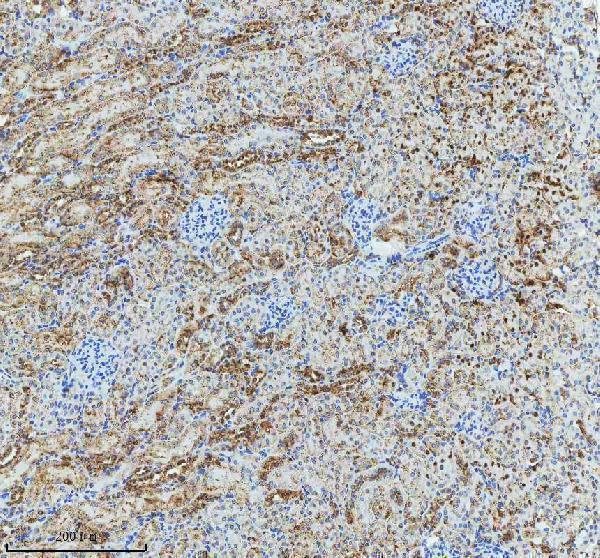
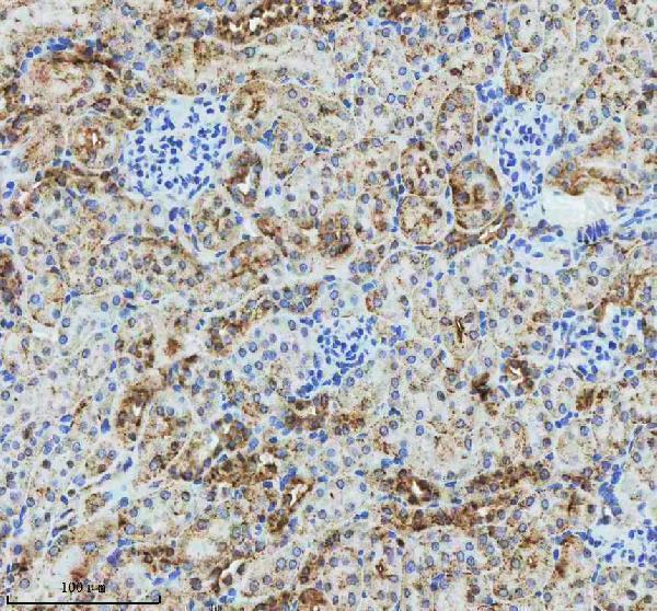
Real IHC data IHC analysis of GLG1 using anti-GLG1 antibody (A07510-1). GLG1 was detected in a paraffin-embedded section of mouse kidney tissue. Heat mediated antigen retrieval was performed in EDTA buffer (pH 8.0, epitope retrieval solution). The tissue section was blocked with 10% goat serum. The tissue section was then incubated with 2 μg/ml rabbit anti-GLG1 Antibody (A07510-1) overnight at 4°C. Peroxidase Conjugated Goat Anti-rabbit IgG was used as secondary antibody and incubated for 30 minutes at 37°C. The tissue section was developed using HRP Conjugated Rabbit IgG Super Vision Assay Kit (Catalog # SV0002) with DAB as the chromogen.
Anti-GLG1 Antibody ®
Cat # A07510-1
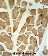
Real IHC data GLG1 Antibody (C-term) IHC analysis in formalin fixed and paraffin embedded skeletal muscle followed by peroxidase conjugation of the secondary antibody and DAB staining. This data demonstrates the use of the GLG1 Antibody (C-term) for immunohistochemistry. Clinical relevance has not been evaluated.
Anti-GLG1 Antibody (C-term)
Cat # A07510

A07510 has an IHC-P image from formalin-fixed, paraffin-embedded skeletal muscle; the caption does not identify the species (A07510 image caption). A07510-1 has IHC images from mouse and rat paraffin-embedded kidney; the fixative is unreported (A07510-1 image captions).

Which to pick: For tissue IHC, choose A07510 for paraffin-section, paraffin-embedded skeletal muscle or A07510-1 for mouse or rat paraffin-embedded kidney (respective image captions). Neither SKU lists IF/ICC validation, so there is no supported IF/ICC pick (catalog applications and image lists). For broader species reactivity, A07510-1 lists human, mouse and rat, but its IHC conditions specify mouse and rat; A07510 is rabbit polyclonal and lists human and mouse reactivity (catalog reactivity, dilution and clonality fields). The selected A07510-1 tissue-IHC caption documents paraffin sections, but does not specify the fixative (selected-SKU IHC image A07510-1).

Each figure is that product's own IHC / IF validation image from its datasheet.

References

  1. UniProt Consortium. UniProt entry Q92896 (GSLG1_HUMAN, Golgi apparatus protein 1).
  2. Human Protein Atlas. GLG1 tissue IHC expression (reliability: Supported).
  3. Human Protein Atlas. GLG1 subcellular location (ICC-IF): Mainly localized to the Golgi apparatus. In addition localized to the mid piece, principal piece and end piece..
  4. Human Protein Atlas. GLG1 antibody validation summary (1 antibodies).
  5. High Specificity of BCL11B and GLG1 for EWSR1-FLI1 and EWSR1-ERG Positive Ewing Sarcoma. Cancers 2020 — PMC7139395.
  6. GLG1 in cancer-associated fibroblasts promotes bladder cancer growth while limiting tumor apoptosis. Apoptosis : an international journal on programmed cell death 2026 — PMC13601173.
  7. Robust diagnosis of Ewing sarcoma by immunohistochemical detection of super-enhancer-driven EWSR1-ETS targets. Oncotarget 2018 — PMC5788584.
  8. Comparative proteomics analysis of gastric cancer stem cells. PloS one 2014 — PMC4224387.
  9. PubMed PMID:8985126 — UniProt-cited evidence.
  10. PubMed PMID:14702039 — UniProt-cited evidence.
  11. PubMed PMID:15616553 — UniProt-cited evidence.