HAVCR1 / Hepatitis A virus cellular receptor 1 · IHC design guide

Design Immunohistochemistry for HAVCR1

Plan chromogenic HAVCR1 IHC-P using kidney tubular cells and glandular epithelia as staining references (HPA tissue IHC). Interpret staining cautiously because tissue IHC reliability is uncertain (HPA tissue IHC).

Evidence assembled Oct 2026 · For research use; verify linked source records and product datasheet before use
Immunohistochemistry protocol sheet for HAVCR1 (IHC for HAVCR1): expected localisation Membranous and cytoplasmic staining in glandular epithelia (HPA tissue IHC), antibody PA1624, validated IHC image, and IHC protocol steps
Printable HAVCR1 IHC protocol sheet — expected localisation Membranous and cytoplasmic staining in glandular epithelia (HPA tissue IHC), antibody PA1624, controls and protocol steps. Open the full HAVCR1 IHC guide →

HAVCR1 Immunohistochemistry Experimental Design Guide

Expected localisation, validated protocols, controls and antibodies — the at-a-glance facts below, then the full design guide.

Must know before staining
Expected localisation Membranous and cytoplasmic staining in glandular epithelia (HPA tissue IHC)
Staining pattern Most glandular epithelia show cytoplasmic and membranous staining (HPA tissue IHC)
Antigen retrieval Citrate pH 6.0 HIER, 95–98 °C, 20 min (rule: cytoplasmic / membrane antigen)
Positive control ⓘ Appendix+4 more · see all
Negative control ⓘ Adipose tissue+4 more · see all
Important caveats
Reasons your staining may differ from the expected pattern.
Fixation Matched tissue-IHC evidence does not establish the fixation claim. Validate the specimen-specific method before use. (selected-SKU IHC image PA1624)
Caveat Low staining–RNA consistency limits confidence in tissue patterns (HPA tissue IHC)
Regulation Expressed by injured kidney tubules (UniProt)
Isoform / epitope No isoforms listed; extracellular versus cytoplasmic epitope matters (UniProt)
Section 1

Recommended HAVCR1 IHC & IF Protocols

The catalog antibody’s IHC-P protocol is accompanied by four published HAVCR1 IHC protocols (PMC12094995; PMC12419044; PMC11702950; PMC11585238).

Recommended immunohistochemistry (IHC-P) protocol parameters
SampleTissue sections; selected-image fixative not specified (standard IHC workflow)
FixationImage fixative and duration unreported (datasheet PA1624); verify before use.
Sectioning4–5 µm sections on charged slides (standard)
DeparaffinisationXylene, graded ethanol series to water (standard)
Antigen retrievalHeat-induced epitope retrieval in citrate buffer, pH 6.0, 20 min at 95–98 °C (standard rule: cytoplasmic / membrane antigen)
Peroxidase block3% H2O2, 10 min, room temperature (standard)
Blocking10% normal serum of the secondary host, 30 min, room temperature (standard)
Primary antibodyRabbit anti-HAVCR1, 0.5-1μg/ml (datasheet PA1624)
Primary incubationOvernight at 4 °C (standard)
DetectionHRP-polymer secondary, DAB chromogen 5–10 min (standard)
CounterstainHematoxylin, blue, dehydrate and mount (standard)
Expected resultHAVCR1-positive staining in glandular cells of appendix (HPA tissue IHC: Medium). HPA tissue profile: Cytoplasmic and membranous expression in most glandular epithelia. No signal in the no-primary control.
💡Decision noteStart with citrate pH 6.0 HIER at 95–98 °C for 20 min (page antigen-retrieval rule); compare the published retrieval methods below.
Section 2

What Is the Expected HAVCR1 Staining Pattern?

HAVCR1 stains tubular and glandular cells at the membrane and in the cytoplasm (HPA tissue IHC: uncertain; UniProt Q96D42 topology).

What am I looking at on my slide?
Membrane and cytoplasmic staining in tubules or glandsFits the reported pattern; staining is medium in listed tissues (HPA tissue IHC).
Nuclear-only stainingUnexpected for HAVCR1; assess nonspecific signal (UniProt Q96D42 subcellular location).
Staining in adipocytes or cardiomyocytesNeither is detected by HPA; assess cross-reactivity or endogenous activity (HPA tissue IHC).
Diffuse staining across the sectionMay be background; review blocking and washes (standard IHC practice).
No staining in kidney tubulesHPA reports medium staining; check the positive control and detection (HPA tissue IHC).
💡Expected HAVCR1 appearancePositive: medium tubular/glandular membrane and cytoplasm; nuclear-only signal is suspect (HPA tissue IHC).
How each factor affects the staining
Topology (UniProt Q96D42)Extracellular aa 21–295; cytoplasmic aa 317–364 (UniProt Q96D42).
Subcellular pools (UniProt Q96D42)Membrane, phagocytic cup and phagosome are annotated (UniProt Q96D42).
IHC confidence (HPA antibodies)CAB075697 IHC validation is uncertain; tissue staining has low RNA agreement (HPA antibodies; HPA tissue IHC).
IF/ICC: where is signal expected?Vesicles are reported with uncertain localisation; see the separate IF/ICC guide (HPA ICC-IF).
Why is my staining missing, weak or wrong?
SituationLikely causeNext action
Nuclear-only signalThis conflicts with annotated HAVCR1 locations (UniProt Q96D42).Check a no-primary control and antibody dilution (standard IHC practice).
Adipocytes stainHPA reports adipocytes as not detected (HPA tissue IHC).Check secondary-only control for endogenous activity (standard IHC practice).
Diffuse backgroundInsufficient blocking or washing is possible (standard IHC practice).Optimize blocking and washes (standard IHC practice).
Kidney tubules lack signalHPA reports medium tubular staining (HPA tissue IHC).Check retrieval, positive control and detection reagents (standard IHC practice).
Chromogen obscures cell boundariesOverdevelopment can hide localisation (standard IHC practice).Shorten chromogen development and compare controls (standard IHC practice).

Sample controls for HAVCR1 IHC & IF

🧪Run kidney first and look for staining in tubular cells (HPA: Medium in kidney cells in tubules). Run adipose tissue as the negative tissue, where adipocytes are not detected; on the kidney slide, non-tubular cells without target staining should show only counterstain and background, but their biological negativity is unverified (HPA: Not detected in adipocytes; HPA: no kidney internal-negative cell class supplied).
Positive control tissue: Appendix (Glandular cells, HPA Medium)
Negative control tissue: Adipose tissue (HPA Not detected)
ICC-IF cell lines (HPA subcellular resource): HPA ICC-IF images show HAVCR1 in A-549, CACO-2, U2OS, RPTEC/TERT1, hTERT-RPE1 (serum starved), with annotated localisation: Vesicles (uncertain) (HPA subcellular).
Technical controls: Include no-primary (secondary-only), an isotype control matched to the primary antibody’s host species and class, and a HAVCR1 knockout biological negative (standard IHC control practice). Quench endogenous peroxidase and check for endogenous biotin in kidney if using avidin–biotin detection (standard kidney IHC practice).
⚠️Feasibility: A target-specific fixation window or fixation effect is unreported, and the selected PA1624 IHC(P) tonsil caption does not report its fixative (supplied caption). Retrieval dependence is unreported, so optimize antigen retrieval with the controls; the supplied evidence does not establish that frozen sections or IF are easier (supplied target/application evidence; standard IHC practice). Endogenous biotin in kidney can produce background with avidin–biotin detection (standard kidney IHC practice).

HPA tissue IHC evidence for HAVCR1

Comprehensive Human Protein Atlas IHC scoring per tissue (reliability: Uncertain — Low consistency between antibody staining and RNA expression data.). Rows are taken directly from the HPA tissue chart — click any row's HPA link to view the source.

Positive expression · recommended positive controls

TissueCell typeLevelEvidenceSource
Appendix Glandular cells Medium Protein (IHC) HPA →
Colon Glandular cells Medium Protein (IHC) HPA →
Gallbladder Glandular cells Medium Protein (IHC) HPA →
Kidney Cells in tubules Medium Protein (IHC) HPA →
Liver Cholangiocytes Medium Protein (IHC) HPA →

Undetected expression · recommended negative controls

TissueCell typeLevelEvidenceSource
Adipose tissue Adipocytes Not detected Protein (IHC) HPA →
Bone marrow Hematopoietic cells Not detected Protein (IHC) HPA →
Caudate Glial cells Not detected Protein (IHC) HPA →
Cerebellum Cells in granular layer Not detected Protein (IHC) HPA →
Esophagus Squamous epithelial cells Not detected Protein (IHC) HPA →
Section 3

Advanced HAVCR1 IHC Tips

Troubleshoot HAVCR1 staining in paraffin sections by checking retrieval, cell type, compartment, controls and scoring before interpreting signal.

How should I adjust retrieval when HAVCR1 staining is weak or diffuse?
Use citrate pH 6.0 heat induced epitope retrieval at 95–98 °C for 20 min in paraffin sections (page retrieval setting). Run a known positive section alongside the test section, and keep cooling time and antibody incubation identical so retrieval is the variable under study (standard IHC practice). If staining remains weak, compare a second retrieval condition on adjacent sections as a fallback, while checking whether stronger treatment also increases background or damages morphology (standard IHC practice). Judge improvement in the expected cell membrane and cytoplasmic compartments, particularly tubular cells in kidney, rather than by overall darkness (UniProt Q96D42 subcellular location; HPA tissue IHC: kidney cells in tubules, Medium).
Could fixation explain weak HAVCR1 staining in paraffin sections?
Target specific fixation sensitivity is unknown from the supplied evidence; the PA1624 tissue caption identifies paraffin IHC in human tonsil but gives no fixative (PA1624 tissue IHC caption). Compare sections with documented fixation histories and identical retrieval, staining and detection conditions before assigning weak signal to fixation (standard IHC practice). Record fixative, time in fixative and processing history for each section; use matched positive controls to distinguish a specimen wide failure from a local staining pattern (standard IHC practice). Do not infer a HAVCR1 fixation preference from its membrane topology, glycosylation sites or HPA tissue pattern (UniProt Q96D42 topology and glycosylation; HPA tissue IHC: reliability Uncertain).
Where should genuine HAVCR1 staining appear within a positive cell?
Assess membranous and cytoplasmic staining in the relevant cell type: UniProt places HAVCR1 at the cell membrane, phagocytic cup and phagosome, while HPA describes cytoplasmic and membranous epithelial staining (UniProt Q96D42 subcellular location; HPA tissue IHC profile). Its extracellular region spans residues 21–295, the transmembrane segment 296–316, and the cytoplasmic tail 317–364 (UniProt Q96D42 topology). Examine intact cell boundaries at high magnification and compare adjacent sections with and without primary antibody when a diffuse deposit obscures the compartment (standard IHC practice). Treat vesicle like staining cautiously because HPA's vesicular IF assignment is uncertain and its tissue IHC reliability is Uncertain (HPA subcellular; HPA tissue IHC reliability).
How can antibody epitope location change the HAVCR1 pattern?
Check the catalog antibody's stated immunogen or mapped epitope before interpreting a negative section; the supplied record does not specify that antibody's epitope (PA1624 tissue IHC caption; UniProt Q96D42 topology). HAVCR1 has an extracellular region at residues 21–295, a membrane span at 296–316, and a cytoplasmic tail at 317–364 (UniProt Q96D42 topology). The record lists glycosylation sites at 65, 263, 277 and 291 and 0 annotated isoforms, so do not assign differing staining patterns to a named isoform without further evidence (UniProt Q96D42 glycosylation and isoforms). Compare an independent, documented epitope with the same tissue controls if staining and expected cell distribution disagree (standard IHC practice).
How should I investigate a conflicting HAVCR1 IF result?
Keep the paraffin chromogenic IHC result as the reference for this page, and evaluate IF with its own fixation and staining controls (PA1624 tissue IHC caption; standard IF practice). Multiplex HAVCR1 with a marker for the expected cell type, such as a tubular epithelial marker in kidney, because HPA reports medium staining in tubular cells (HPA tissue IHC: kidney cells in tubules, Medium). Choose fluorophores after inspecting unstained tissue for autofluorescence, and include single color controls when separating overlapping signals (standard IF practice). Match permeabilisation to the documented epitope: access to the cytoplasmic tail at residues 317–364 generally requires permeabilisation, whereas an extracellular epitope may be assessed without it (UniProt Q96D42 topology; standard IF practice).
What should I check when HAVCR1 staining covers the whole section?
Inspect a section without primary antibody to separate antibody dependent staining from chromogen deposits or endogenous enzyme activity (standard chromogenic IHC practice). For peroxidase detection, verify the peroxidase block and examine whether background tracks tissue edges, folds or damaged areas rather than intact cells (standard chromogenic IHC practice). Titrate the primary antibody and detection reagent on adjacent sections, changing 1 condition at a time while retaining the same citrate pH 6.0 retrieval (page retrieval setting; standard IHC practice). Compare the residual pattern with HPA's reported epithelial membrane and cytoplasmic staining, but keep its Uncertain reliability in view when deciding whether broad signal is specific (HPA tissue IHC profile and reliability).
How should I score HAVCR1 staining across specimens? ⚠ ANSWER MARKED FOR VERIFICATION
Define the cell population and compartment before scoring: HPA reports medium staining in kidney tubular cells and describes epithelial cytoplasmic and membranous expression (HPA tissue IHC profile). For comparable regions, record the percentage of positive target cells and an H-score using intensity categories 0–3; keep membrane and cytoplasm scores separate when the distinction matters (standard IHC scoring practice). Normalise cell based measures to the number of evaluable cells of that type, or report positive cell density per mm² of viable tissue when cell counts are impractical (standard IHC quantification practice). Use the same threshold, region selection and control sections across specimens, and report HPA's Uncertain tissue IHC reliability alongside biological conclusions (standard IHC practice; HPA tissue IHC reliability).
How can I distinguish true HAVCR1 signal from artefact?
Give most weight to staining in intact cells with plausible membrane or cytoplasmic localisation, particularly kidney tubular cells, where HPA reports medium signal (UniProt Q96D42 subcellular location; HPA tissue IHC: kidney cells in tubules, Medium). Recheck staining confined to section edges, folds or necrotic areas against a section without primary antibody and an appropriately blocked peroxidase run (standard chromogenic IHC practice). Diffuse nuclear only staining conflicts with the supplied HAVCR1 localisation, while a positive tonsil section alone establishes neither its expressing cell type nor its fixative (UniProt Q96D42 subcellular location; PA1624 tissue IHC caption). Resolve uncertain cases with cell type markers and matched controls, and qualify conclusions because HPA rates its tissue staining reliability Uncertain owing to low agreement with RNA expression (standard IHC practice; HPA tissue IHC reliability).
Boster reagents

Best HAVCR1 / Hepatitis A virus cellular receptor 1 IHC Antibodies

Anti-HAVCR1 antibodies have IHC data from human tonsil (PA1624 IHC image caption) and IF data from human testis and mouse kidney (A01306-1 IF image captions).

Real IHC data Anti-TIM 1 antibody, PA1624, IHC(P) IHC(P): Human Tonsil Tissue
Anti-TIM 1/HAVCR1 Antibody ®
Cat # PA1624
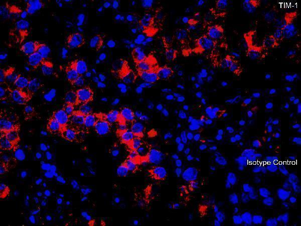
Real IF data Immunofluorescence Validation of TIM-1 in Human Testis Immunofluorescent analysis of 4% paraformaldehyde-fixed human testis tissue labeling TIM-1 with A01306-1 at 10 μg/mL, followed by goat anti-rabbit IgG secondary antibody at 1/500 dilution (red) and DAPI staining (blue).
Anti-TIM-1 HAVCR1 Antibody
Cat # A01306-1

PA1624 is listed for IHC, IF and ICC in human samples (catalog: applications and reactivity), with an IHC image of a human tonsil paraffin section (PA1624 IHC image caption). A01306-1 is listed for IF in human and mouse samples (catalog: applications and reactivity), with IF images of human testis and mouse kidney (A01306-1 IF image captions).

Which to pick: For tissue IHC, choose PA1624: its image documents a human tonsil paraffin section (PA1624 IHC image caption); the fixative is unreported (PA1624 IHC image caption). For IF or ICC in human samples, PA1624 lists both applications (catalog: applications and reactivity); for IF across human and mouse samples, choose the polyclonal A01306-1, which lists both species and has IF images from each (catalog: dilution_raw and reactivity; A01306-1 IF image captions).

Each figure is that product's own IHC / IF validation image from its datasheet.

References

  1. UniProt Consortium. UniProt entry Q96D42 (HAVR1_HUMAN, Hepatitis A virus cellular receptor 1).
  2. Human Protein Atlas. HAVCR1 tissue IHC expression (reliability: Uncertain).
  3. Human Protein Atlas. HAVCR1 subcellular location (ICC-IF): Localized to vesicles..
  4. Human Protein Atlas. HAVCR1 antibody validation summary (2 antibodies).
  5. Combining biomarkers to construct a novel predictive model for predicting preoperative lymph node metastasis in early gastric cancer. Frontiers in oncology 2025 — PMC12094995.
  6. Sinapic acid alleviates renal ischemia-reperfusion injury by regulating oxidative stress, apoptosis, and inflammation. Turkish journal of medical sciences 2025 — PMC12419044.
  7. Macrophage autophagy protects against acute kidney injury by inhibiting renal inflammation through the degradation of TARM1. Autophagy 2025 — PMC11702950.
  8. Integration of the bulk transcriptome and single-cell transcriptome reveals efferocytosis features in lung adenocarcinoma prognosis and immunotherapy by combining deep learning. Cancer cell international 2024 — PMC11585238.
  9. PubMed PMID:9658108 — UniProt-cited evidence.
  10. PubMed PMID:15372022 — UniProt-cited evidence.
  11. PubMed PMID:15489334 — UniProt-cited evidence.