Click image to see more details
-

-
-
-
-
+2
Product Info Summary
| SKU: | A01306-1 |
|---|---|
| Size: | 0.1 mg |
| Reactive Species: | Human, Mouse |
| Host: | Rabbit |
| Application: | ELISA, IF, WB |
Customers Who Bought This Also Bought

Product info
Product Name
Anti-TIM-1 HAVCR1 Antibody
SKU/Catalog Number
A01306-1
Size
0.1 mg
Form
Liquid
Description
Boster Bio Anti-TIM-1 HAVCR1 Antibody (Catalog # A01306-1). Tested in ELISA, WB, IF applications. This antibody reacts with Human, Mouse.
Storage & Handling
TIM-1 antibody can be stored at 4°C up to one year. Antibodies should not be exposed to prolonged high temperatures.
Cite This Product
Anti-TIM-1 HAVCR1 Antibody (Boster Biological Technology, Pleasanton CA, USA, Catalog # A01306-1)
Host
Rabbit
Contents
TIM-1 Antibody is supplied in PBS containing 0.02% sodium azide.
Clonality
Polyclonal
Isotype
IgG
Immunogen
Anti-TIM-1 antibody was raised against a peptide corresponding to 16 amino acids near the amino terminus of human TIM-1. The immunogen is located within amino acids 50 - 100 of TIM-1.
Reactive Species
A01306-1 is reactive to HAVCR1 in Human, Mouse
Observed Molecular Weight
68 kDa
Calculated molecular weight
39.3 kDa
Background of HAVCR1
The human form of TIM-1 was initially discovered as a membrane glycoprotein through which the hepatitis A virus can gain entry into a cell. It was also identified as kidney injury molecule 1 (Kim-1), a predicted adhesion molecule that is upregulated on the surfaces of kidney epithelia. It is also expressed on T helper 2 (Th2) cells of the immune system, and following the binding of its natural ligand TIM-4, stimulates T cell expansion and cytokine production in response to viral challenge. It has been suggested that hyperactivation of TIM-1 leads to an increased level of Th2 responsiveness and asthma susceptibility, and antibodies to TIM-1 may therefore be a novel approach to treating asthma.
Antibody Validation
Boster validates all antibodies on WB, IHC, ICC, Immunofluorescence, and ELISA with known positive control and negative samples to ensure specificity and high affinity, including thorough antibody incubations.
Application & Images
Applications
A01306-1 is guaranteed for ELISA, IF, WB Boster Guarantee
Recommend Dilution
WB: 1 - 8 μg/mL (overnight incubation at 4˚ C)
IHC-P/IF: 10-20 μg/mL.
Antibody validated: Western Blot in human and mouse samples; Immunofluorescence in human and mouse samples. All other applications and species not yet tested.
Validation Images & Assay Conditions
Click image to see more details
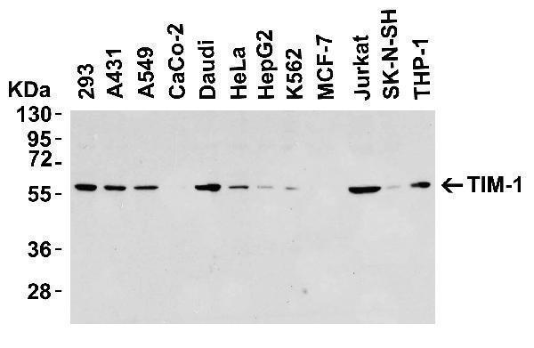
Western Blot Validation in Human Cell Lines
Loading: 15 μg of lysates per lane.
Antibodies: TIM-1 A01306-1 (8 μg/mL), overnight incubation at 4˚ C in 5% NFDM/TBST.
Secondary: Goat anti-rabbit IgG HRP conjugate at 1:10000 dilution.

Click image to see more details
Independent Antibody Validation (IAV) via Protein Expression Profile in Cell Lines
Loading: 15 μg of lysates per lane.
Antibodies: TIM-1 A01306-1 (8 μg/mL), TIM-1 3811 (1 μg/mL), beta-actin (1 μg/mL), and GAPDH (0.02 μg/mL), overnight incubation at 4˚ C (A01306-1) or 1h incubation at RT in 5% NFDM/TBST.
Secondary: Goat anti-rabbit IgG HRP conjugate at 1:10000 dilution.

Click image to see more details
Validation with TIM-1 siRNA Knockdown in Hela Cells
HeLa cells were transfected with control siRNAs (lane 1) or TIM-1 siRNAs (lane 2)
Loading: 15 μg of HeLa whole cell lysates per lane.
Antibodies: A01306-1 (8 μg/mL), 1 h incubation at RT in 5% NFDM/TBST.
Secondary: Goat anti-rabbit IgG HRP conjugate at 1:10000 dilution.

Click image to see more details
Western Blot Validation in Mouse Tissues
Loading: 15 μg of lysates.
Antibodies: TIM-1 A01306-1, 2 μ g/mL, 1h incubation at RT in 5% NFDM/TBST.
Secondary: Goat anti-rabbit IgG HRP conjugate at 1:10000 dilution.

Click image to see more details
Immunofluorescence Validation of TIM-1 in Human Testis
Immunofluorescent analysis of 4% paraformaldehyde-fixed human testis tissue labeling TIM-1 with A01306-1 at 10 μg/mL, followed by goat anti-rabbit IgG secondary antibody at 1/500 dilution (red) and DAPI staining (blue).

Click image to see more details
Immunofluorescence Validation of TIM-1 in Mouse Kidney
Immunofluorescent analysis of 4% paraformaldehyde-fixed mouse kidney tissue labeling TIM-1 with A01306-1 at 10 μg/mL, followed by goat anti-rabbit IgG secondary antibody at 1/500 dilution (red) and DAPI staining (blue).
Specific Publications For Anti-TIM-1 HAVCR1 Antibody (A01306-1)
Loading publications
Recommended Resources
Here are featured tools and databases that you might find useful.
- Boster's Pathways Library
- Protein Databases
- Bioscience Research Protocol Resources
- Data Processing & Analysis Software
- Photo Editing Software
- Scientific Literature Resources
- Research Paper Management Tools
- Molecular Biology Software
- Primer Design Tools
- Bioinformatics Tools
- Phylogenetic Tree Analysis
Customer Reviews
Have you used Anti-TIM-1 HAVCR1 Antibody?
Share your experimental results or join a short interview to earn up to $1,000 in product credits or other rewards.
0 Reviews For Anti-TIM-1 HAVCR1 Antibody
Customer Q&As
Have a question?
Find answers in Q&As, reviews.
Can't find your answer?
Submit your question
