This website uses cookies to ensure you get the best experience on our website.
- Table of Contents
1 Citations 15 Q&As
1 Citations 4 Q&As
Facts about HLA class I histocompatibility antigen, A alpha chain.
Please shorten your article and try again.
Human | |
---|---|
Gene Name: | HLA-A |
Uniprot: | P01891 |
Entrez: | 3105 |
Belongs to: |
---|
MHC class I family |
A-10 alpha chain; FLJ26655; HLA class I histocompatibility antigen, A-1 alpha chain; HLA class I histocompatibility antigen, A-28 alpha chain; HLA class I histocompatibility antigen, A-9 alpha chain; HLAA; major histocompatibility complex, class I, A; MHC class I antigen A*1; MHC class I antigen A*11; MHC class I antigen A*80
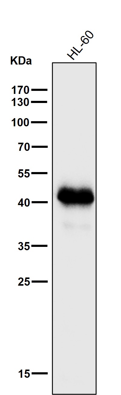
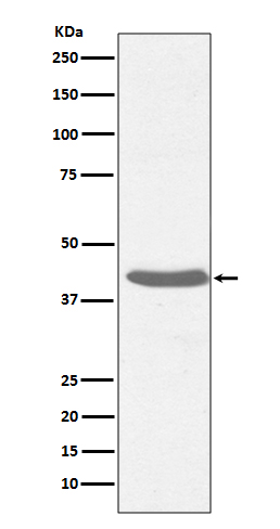
Mass (kDA):
40.841 kDA
Human | |
---|---|
Location: | 6p22.1 |
Sequence: | 6; NC_000006.12 (29942532..29945870) |
Medical researchers may have heard about the HLA-B27 locus. This article will explore this gene, the mixed-HLA antibody method as well as flow cytometry. Boster Bio's HLA platform will be discussed. Be sure to remember the following guidelines.
The surface proteins of white blood cells, known as human leukocyte antigens (or HLAs, help the immune system recognize non-self and self. Everyone inherits a distinct combination of HLA genes each one of which codes for one of several antigens that are found on cell surfaces. HLA-B27 is found in around 6% of the U.S. population, and is associated with an increased risk of developing autoimmune diseases and inflammatory bowel disease.
Positive HLA-B27 tests can be used to confirm the diagnosis of reactive arthritis or ankylosing Spondylitis. They can also be used for excluding certain autoimmune diseases. Although the HLA-B27 test does not provide an indication of a diagnosis, it does provide valuable information. It is often combined with other tests such as C-reactive proteins and erythrocyte segregation rate to give a precise diagnosis.
Although HLA B27 is linked to autoimmune diseases, it is unclear if this gene causes them. Recent research has revealed that HLA B27 was detected in two families, putting the risk of developing the HLA–B27 related diseases. More research is required in order to determine the role played by HLA-227 as a factor in autoimmune diseases like spondyloarthritis.
Researchers are working on developing new indicators to determine the susceptibility of individuals to HLA-B27 in the population. A new method could aid in identifying patients at a high risk of developing uveitis. Although the incidence of uveitis is low, about one percent HLA-B27 positive individuals are likely to develop it. This will allow doctors to improve their treatment and preventive treatment. What are the most effective uses for the HLA-B27 locus?
The HLA system is the largest gene that is distributed in the human population. It encodes serologically recognized antigens, also known as antigens. DNA sequencing of these alleles can identify them by their standard designations. The gene's name is followed by an asterisk (or colon) and the designations are their names. They also carry numbers that represent the specific allele. Additional numbers in the DNA sequence identify similar proteins or polymorphisms in 5' and 3 regions that are not translated.
This mixed-HLA antibody test is one of two types of HLA tests. One type identifies non-HLA antibodies and the other one identifies HLA antigens that the body has a high affinity for. Both tests employ well-defined lymphocytes or purified HLA antigens, which are then paired with specific microparticles to determine whether an individual is sensitized to these antigens. Additionally, the Luminex HLA Single Antigen Class I and II Luminex bead test assesses reactivity with specific donor antigens.
To find out which method is more effective to detect the difference, the FCM mixed HLA antibody method and the Luminex mixed HLA antibody method were compared. The FCM method uses an anti-human IgG FITC antibody, whereas the Luminex method employs PE-conjugated goat anti-human IgG. Both methods use the exact amount of antigens to target and the concentrations are similar. The Luminex method also included MICA*008 along with MICA*027 in its test panel.
The Luminex mixed-HLA antibody method uses the polystyrene microsphere and fluorochromes of different intensities to provide a unique signal. These beads are connected to a microsphere array at three different levels. The first level is comprised of a large number of class 1 and class 2 molecules. The second level is composed of two molecules for each allele, so the mixtures of two alleles can be identified.
The Luminex mixed-HLA antibody method was also examined for HLA-DR antibodies but only a fraction of samples had positive results. This method requires an anti-Human IgG antibody which is directed towards HLA class I and HLA class II antigens. Furthermore the method's sensitivity, as well as specificity have improved. However, there are a few important issues to be considered when selecting the most reliable anti-HLA assay.
The Luminex Labscreen mixed-HLA antigen antibody test was developed by One Lambda Inc. in 2003. This test detects binding antibodies of HLA class I or II. The results are available as the form of a batch electronic file output. The researcher can customize the results to suit his/her needs. A unique modification of the HLA Single Antigen Luminex Bead Test is the HLA Single Antigen-C1q-C1qScreen. C1q is an important component of the classic complement cascade. Luminex has developed a method of detecting C1q binding antibodies.
Different cells can be identified by the use of fluorescence. The fluorescent probe targets specific epitopes and gives quantitative and qualitative data. Fluorescence measurements in flowcytometry employ distinct fluorescence channels. Fluorescence probes are made from fluorescent dyes. The measurements of fluorescence can be used for a variety purposes, including the identification of distinct cell populations, measurement of nucleic acids content and apoptosis. Fluorescent probes are ideal for flow cytometry since they can measure multiple parameters at once.
It's very similar to shopping at a supermarket. A clerk sorts your purchases when you go to the supermarket. One by one, she scans the items with a laser , then sorts them by the type. Similar items are put in a shopping cart. Similar cell sorting happens in the same way. The histogram for F4/80 illustrates the results for R2 and R3.
Flow cytometry is another test to determine the HLA-A marker. These results can be used to assist doctors in identifying different kinds of cancer. HLA-A-positive patients are more likely to be diagnosed with B-cell lineage (also known as lymphoma) Additional antibodies detected in blood can aid in the diagnosis of the disease.
Flow cytometry can be used to determine the HLA A marker in various ways. Flow cytometry is among the most reliable and common ways to determine the HLA A marker. This method uses HLA-A antigens to identify immune cells, which is an important part of immunohistochemistry. In this method an antibody will react with a collection of different HLA antigens.
Another method to study the immune system is by using flow cytometry. It makes use of a mixture of monoclonal antibodies as well as CD markers to determine the levels of immunoglobulins found in B-lymphocytes. The sample is typically stained with three to ten tubes. The sample should be isotype-matched, because blood serum immunoglobulins block the detection of immunoglobulins.
Boster Bio HLA A ELSA was specifically developed to detect antibodies against the human HLA A polymorphism. The test is performed with a cell line that has the chimeric genes C8166–CCR5. The antibody responses are high in the 3 and 1 groups however, the results were lower in group 4. The CCR5 antibody was anti-C8166 and was associated with cumulative responses to anti-A*01 peptides , as well as anti-DRB1*.
The ELISA kit was tested on the lymphoblast cell line T2. This type of cell is TAP-deficient and expresses low levels HLA-A*0201 molecules in normal culture conditions. T2 cells were used to determine the binding affinity of this cell to the HLA A molecule. The results showed that T2 cells are capable of binding to HLA molecules in the lab.
To determine the sensitivity of Boster Bio HLA A ELSA, T2 cells were incubated for a period of time with the HLA-A peptides (50 umol/L) 1 umol/L human b2-microglobulin (HLA-A*0201) and serum-free RPMI 1640 medium. The analysis of the flow cytometry of T2 cells followed by the use of PE-labeled anti–HLA A*0201m. The FI is the average fluorescence of HLA A*0201 molecules inside the cells.
Boster Bio HLA A ELSA is used to test the immune system’s ability to recognize and block certain antigens. HLA ELISA HLA A is compatible with HLA A*0201 and HLA B*01 which are two of the most commonly used forms of HFRS. HLA-B*0201 mice did not have the antigens that are corresponding to HLA-A in PBMCs.
Utilizing the Boster Bio HLA-A ELSA to test for this is a great option for a wide range of applications. People who are positive for HLA-A may see more positive results than those who are in the negative group. This is especially true when HIV is a concern. As a preventative allovaccine the vaccine must contain sufficient HLA lines to trigger an immune response in over 90% of the population.
The Boster Bio HLA-A ELSA also detects anti-HLA B cells in blood. People with HLA-A are more likely to have higher antibody titers than people who aren't. The serum was also tested against the SHIV-SF162P4/C virus , in the absence and presence of complement. The positive results were in line with those obtained using HLA-A-ELISA in human subjects.
PMID: 2431040 by Wan A.M., et al. The primary structure of HLA-A32 suggests a region involved in formation of the Bw4/Bw6 epitopes.
PMID: 3496393 by Holmes N., et al. Multiple genetic mechanisms have contributed to the generation of the HLA- A2/A28 family of class I MHC molecules.
*More publications can be found for each product on its corresponding product page