HSPA5 / Endoplasmic reticulum chaperone BiP · IHC design guide

Design Immunohistochemistry for HSPA5

Plan chromogenic IHC for HSPA5 in paraffin sections using its cytoplasmic tissue staining pattern (HPA tissue IHC). Choose positive reference cells and interpret widespread staining using the reported tissue profile (HPA tissue IHC).

Evidence assembled Oct 2026 · For research use; verify linked source records and product datasheet before use
Immunohistochemistry protocol sheet for HSPA5 (IHC for HSPA5): expected localisation Cytoplasmic tissue staining (HPA tissue IHC); ER lumen localization (UniProt), antibody M00955, validated IHC image, and IHC protocol steps
Printable HSPA5 IHC protocol sheet — expected localisation Cytoplasmic tissue staining (HPA tissue IHC); ER lumen localization (UniProt), antibody M00955, controls and protocol steps. Open the full HSPA5 IHC guide →

HSPA5 Immunohistochemistry Experimental Design Guide

Expected localisation, validated protocols, controls and antibodies — the at-a-glance facts below, then the full design guide.

Must know before staining
Expected localisation Cytoplasmic tissue staining (HPA tissue IHC); ER lumen localization (UniProt)
Staining pattern Abundant cytoplasmic staining in immune, neuronal and thyroid follicular cells (HPA tissue IHC)
Antigen retrieval Citrate pH 6.0 HIER, 95–98 °C, 20 min (rule: cytoplasmic / membrane antigen)
Positive control ⓘ Caudate+4 more · see all
Negative control ⓘ None in HPA (detected in all 45 tissues); use no-primary + isotype controls
Important caveats
Reasons your staining may differ from the expected pattern.
Fixation Matched tissue-IHC evidence does not establish the fixation claim. Validate the specimen-specific method before use. (selected-SKU IHC image M00955)
Caveat Ubiquitous staining limits negative tissue controls (HPA tissue IHC)
Regulation Staining intensity regulation is unreported (UniProt)
Isoform / epitope No annotated isoforms; signal peptide spans residues 1–18 (UniProt)
Section 1

Recommended HSPA5 IHC & IF Protocols

Compare the catalog antibody’s IHC-P protocol with published HSPA5 staining methods for placenta and glioma tissue (PMC9926443; PMC12325653; PMC8121073).

Recommended immunohistochemistry (IHC-P) protocol parameters
SampleParaffin-embedded tissue sections; fixative not specified (datasheet M00955; sample unspecified)
FixationImage fixative and duration unreported (datasheet M00955); verify before use.
Sectioning4–5 µm sections on charged slides (standard)
DeparaffinisationXylene, graded ethanol series to water (standard)
Antigen retrievalHeat-induced epitope retrieval in citrate buffer, pH 6.0, 20 min at 95–98 °C (standard rule: cytoplasmic / membrane antigen)
Peroxidase block3% H2O2, 10 min, room temperature (standard)
Blocking10% normal serum of the secondary host, 30 min, room temperature (standard)
Primary antibodyRabbit monoclonal (clone BOO-8) anti-HSPA5, 1:50-1:200 (datasheet M00955)
Primary incubationOvernight at 4 °C (standard)
DetectionHRP-polymer secondary, DAB chromogen 5–10 min (standard)
CounterstainHematoxylin, blue, dehydrate and mount (standard)
Expected resultHSPA5-positive staining in neuronal cells of caudate (HPA tissue IHC: High). HPA tissue profile: Ubiquitous cytoplasmic expression, highly abundant in immune, neuronal cells and thyroid follicular cells. No signal in the no-primary control.
💡Decision noteStart with citrate pH 6.0 retrieval at 95–98 °C for 20 min (page antigen-retrieval setting); adjust using the reported glioma method if needed (PMC12325653).
Section 2

What Is the Expected HSPA5 Staining Pattern?

HSPA5/BiP is an ER-lumen protein without a transmembrane segment (UniProt P11021 topology). Expect cytoplasmic staining across tissues, with high abundance in immune, neuronal and thyroid follicular cells (HPA tissue IHC). HPA rates tissue IHC reliability Enhanced, with medium antibody–RNA consistency (HPA tissue IHC).

What am I looking at on my slide?
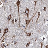
Strong cytoplasmic stain in hippocampal neuronsExpected: neuronal cells are High (HPA tissue IHC); BiP is ER-luminal (UniProt P11021).
Strong nuclear-only stainAtypical for ER-lumen BiP (UniProt P11021); investigate staining artefact.
Strong stain in oral squamous cellsThese cells are Low (HPA tissue IHC); check cross-reactivity or endogenous detection activity.
Uniform haze across tissue and blank areasSuggests background; review blocking and detection controls (general IHC practice).
No stain in hippocampal neuronsUnexpected: neuronal cells are High (HPA tissue IHC); check assay performance.
💡Expected HSPA5 appearanceA positive result shows high cytoplasmic staining in hippocampal neurons (HPA tissue IHC); nuclear-only or blank-area stain suggests artefact (UniProt P11021; general IHC practice).
How each factor affects the staining
Target locationBiP is ER-luminal and has no transmembrane segment (UniProt P11021 topology).
Antibody epitopeCheck epitope position: residues 1–18 are signal sequence; the mature chain is 19–654 (UniProt P11021).
IHC validationHPA038845, HPA038846 and CAB005221 each have Enhanced IHC validation (HPA antibodies).
IF/ICC location?Annulus is approved; cytosol is uncertain (HPA subcellular). Interpret IF/ICC in its own guide.
Why is my staining missing, weak or wrong?
SituationLikely causeNext action
No hippocampal neuronal signalNeuronal cells are High (HPA tissue IHC); assay failure is possible.Check a positive control, retrieval and dilution (general IHC practice).
Nuclear-only signalBiP is ER-luminal (UniProt P11021); nonspecific signal is possible.Review the cytoplasmic pattern and an omit-primary control (general IHC practice).
Strong oral squamous signalOral squamous cells are Low (HPA tissue IHC); nonspecific detection is possible.Check omit-primary and detection controls (general IHC practice).
Diffuse section-wide hazeBackground or endogenous detection activity is possible (general IHC practice).Review blocking, washes and chromogen development (general IHC practice).
Patchy weak staining in positive tissueUneven retrieval or reagent coverage is possible (general IHC practice).Check retrieval and reagent coverage against a positive control (general IHC practice).

Sample controls for HSPA5 IHC & IF

🧪Run duodenum first and look for staining in enterocytes (HPA: High in duodenal enterocytes). HPA detects HSPA5 in all 45 scored tissues, so use no-primary and isotype controls for the negative comparison; no cell population on the positive slide is established as an internal negative, and any unstained cells should be recorded as observed rather than presumed HSPA5-negative (HPA: no negative tissue rows).
Positive control tissue: Caudate (Neuronal cells, HPA High)
Negative control tissue: None in HPA: HSPA5 is detected in all 45 scored tissues. Use a no-primary (secondary-only) and an isotype control instead.
ICC-IF cell lines (HPA subcellular resource): HPA ICC-IF images show HSPA5 in A-431, U-251MG, U2OS, Sperm, with annotated localisation: Cytosol (uncertain), Annulus (approved) (HPA subcellular).
Technical controls: Include a no-primary, secondary-only control; an isotype control matched to the primary antibody’s host and immunoglobulin class, with subclass matching for a monoclonal; and an HSPA5 knockout specimen or immunogen peptide-block control if suitable material is available (standard IHC practice). For duodenal chromogenic IHC, quench endogenous peroxidase and, if detection uses biotin, block endogenous biotin (standard IHC practice).
⚠️Feasibility: A target-specific fixation window or fixation effect is unreported, and the selected M00955 paraffin-section caption does not state a fixative (caption: fixative unreported). Retrieval dependency is unreported; optimize antigen retrieval with a matched untreated section, and do not assume frozen sections or IF/ICC are easier from the supplied evidence (standard IHC practice; HPA: ICC-IF images available). In duodenum, distinguish cellular staining from luminal debris when scoring enterocytes (standard IHC practice).

HPA tissue IHC evidence for HSPA5

Comprehensive Human Protein Atlas IHC scoring per tissue (reliability: Enhanced — Medium consistency between antibody staining and RNA expression data.). Rows are taken directly from the HPA tissue chart — click any row's HPA link to view the source.

Positive expression · recommended positive controls

TissueCell typeLevelEvidenceSource
Caudate Neuronal cells High Protein (IHC) HPA →
Cerebellum Cells in molecular layer High Protein (IHC) HPA →
Cerebral cortex Neuronal cells High Protein (IHC) HPA →
Colon Endocrine cells High Protein (IHC) HPA →
Duodenum Enterocytes High Protein (IHC) HPA →

Undetected expression · recommended negative controls

TissueCell typeLevelEvidenceSource
None in HPA: HSPA5 is detected in all 45 scored tissues. Use a no-primary (secondary-only) and an isotype control instead.
Section 3

Advanced HSPA5 IHC Tips

Troubleshooting HSPA5/BiP staining in paraffin section IHC, with a focused note on IF multiplexing.

How should I retrieve HSPA5 in paraffin sections when staining is weak?
Start with citrate buffer at pH 6.0, heated to 95–98 °C for 20 min (page retrieval rule). Allow sections to cool consistently, then compare a dilution series around the caption's 1:100 antibody dilution before increasing retrieval intensity (selected M00955 tissue-IHC caption; standard IHC practice). If staining remains weak, test a higher-pH retrieval buffer on matched sections as a fallback, while watching for tissue damage and increased background (standard IHC practice). Include a high-expressing reference compartment, such as neuronal cells, and judge whether retrieval improves the expected cytoplasmic pattern across the section rather than staining only its edges (HPA: High in neuronal cells; HPA: ubiquitous cytoplasmic expression; standard IHC practice).
Could fixation explain patchy or absent HSPA5 staining?
The selected M00955 image documents staining in a paraffin-embedded non-Hodgkin's lymphoma section at 1:100, but its caption does not state the fixative (selected M00955 tissue-IHC caption). Target-specific fixation sensitivity is therefore unknown; neither tissue staining patterns nor HSPA5 topology or modifications establish how fixation changes this antibody's epitope (selected M00955 tissue-IHC caption; HPA tissue IHC; UniProt P11021). Record fixation and processing for each sample, then compare sections processed together using the same pH 6.0, 20 min retrieval conditions (page retrieval rule; standard IHC practice). When one block stains poorly, inspect tissue preservation and compare a matched positive control before attributing the result to altered HSPA5 abundance (standard IHC practice).
Where should I expect HSPA5 staining, and what if I see membrane signal?
Expect predominantly cytoplasmic staining in tissue IHC, consistent with abundant HSPA5 in the ER lumen and HPA's ubiquitous cytoplasmic tissue profile (UniProt P11021: ER lumen; HPA: tissue IHC profile). HSPA5 has a signal peptide at residues 1–18 and no transmembrane segment, so a crisp continuous membrane outline is not its default tissue pattern (UniProt P11021: processing and topology). Cell-surface localisation has been reported for epithelial cells under high free iron, but routine membrane staining alone does not establish that condition (UniProt P11021: subcellular localisation). Compare cytoplasmic and membrane patterns across matched sections and check a negative-reagent control before assigning an unusual compartment as specific (standard IHC practice).
How can epitope location affect HSPA5 staining in paraffin sections?
The supplied record lists 0 isoforms and a processed HSPA5 chain spanning residues 19–654, following signal-peptide removal (UniProt P11021: isoforms and processing). The selected M00955 caption gives an IHC dilution of 1:100 but does not identify the antibody epitope, so epitope-specific retrieval predictions are unsupported (selected M00955 tissue-IHC caption). HSPA5 has annotated modified residues, including phosphoserine at 86 and acetyllysine at 125, but their effects on this antibody's IHC signal are unknown (UniProt P11021: modified residues; selected M00955 tissue-IHC caption). If staining differs between samples, compare retrieval, preservation and antibody concentration first; use a mapped epitope or independent reagent to investigate a suspected epitope effect (standard IHC practice).
How should I investigate HSPA5 localisation with multiplex IF?
Use IF as a separate localisation check alongside the paraffin section IHC result, and pair HSPA5 with a marker for the cell population being assessed, such as neuronal cells in an appropriate section (HPA: High in neuronal cells; standard IF practice). Choose spectrally separated fluorophores and place the weaker signal in a channel with low measured tissue autofluorescence; assess each channel with single-stain controls (standard IF practice). Because the predominant pool is in the ER lumen, permeabilise sufficiently to expose an intracellular epitope when that is the intended target (UniProt P11021: ER lumen; standard IF practice). The M00955 paraffin-section caption supplies neither an IF fixation condition nor an IF dilution, so optimise those conditions for the IF specimen rather than transferring 1:100 automatically (selected M00955 tissue-IHC caption; standard IF practice).
What causes diffuse brown background in HSPA5 chromogenic IHC?
First distinguish genuine widespread cytoplasmic signal from staining outside cells: HPA reports ubiquitous cytoplasmic HSPA5 expression, with high abundance in immune and neuronal cells (HPA: tissue IHC profile). Run a no-primary control and check peroxidase blocking, wash stringency and DAB development before interpreting a diffuse brown deposit as antigen (standard chromogenic IHC practice). Compare a dilution series around the image-caption condition of 1:100; a shorter development time can help reveal whether the apparent background tracks detection intensity (selected M00955 tissue-IHC caption; standard IHC practice). Examine section edges, damaged tissue and endogenous pigment separately, and retain only cell-associated staining with a plausible cytoplasmic distribution for scoring (HPA: cytoplasmic profile; standard IHC practice).
How should I score HSPA5 staining across heterogeneous tissue? ⚠ ANSWER MARKED FOR VERIFICATION
Define the cell compartment and region before scoring, because HPA reports broad cytoplasmic expression with differing levels across cell types (HPA: tissue IHC profile). For intensity scoring, record the percentage of cells at 0, 1+, 2+ and 3+, then calculate an H-score from 0–300; alternatively report percent positive cells using a fixed threshold (standard IHC practice). For spatial comparisons, report positive cells per mm² of viable tissue and normalise each result to the number of eligible cells or analysed viable area (standard IHC practice). Keep retrieval, antibody dilution, DAB development, imaging and thresholds consistent, and analyse matched cell populations rather than pooled tissue regions (standard IHC practice).
How do I distinguish true HSPA5 positivity from artefact?
A credible result shows cell-associated cytoplasmic staining in a plausible population; HPA reports high staining in neuronal cells and low staining in oral-mucosa squamous epithelial cells (HPA: tissue IHC profile and cell-level entries). An isolated nuclear pattern is less consistent with the supplied ER-lumen localisation and tissue cytoplasmic profile, so review it before calling it HSPA5 (UniProt P11021: ER lumen; HPA: tissue IHC profile). Check whether signal concentrates at section edges, necrotic areas or sites that also stain in a no-primary control, where artefact can mimic positivity (standard IHC practice). Residual endogenous peroxidase can also produce misleading chromogenic signal; verify the peroxidase block and compare matched controls before assigning biological meaning to a change (standard chromogenic IHC practice).
Boster reagents

Best HSPA5 / Endoplasmic reticulum chaperone BiP IHC Antibodies

Anti-HSPA5 antibodies have IHC images from human tissue and rat brain (catalog image captions), plus an IF image from HepG2 cells (M00955 IF caption). All three listed antibodies react with human, mouse and rat (catalog reactivity).

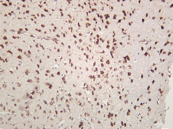
Real IHC data Immunohistochemical analysis of paraffin-embedded Human non-Hodgkin's lymphoma, using the Antibody at 1:100 dilution.
Anti-GRP78 BiP HSPA5 Rabbit Monoclonal Antibody
Cat # M00955
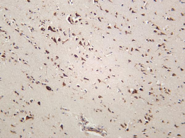
Real IHC data Rat brain was stained with anti-GRP78/BiP rabbit antibody
Anti-GRP78/BiP Rabbit Monoclonal Antibody
Cat # M00955-4
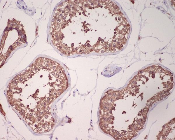
Real IHC data Immunohistochemical analysis of paraffin-embedded human testis, using GRP78 BiP Antibody.
Anti-GRP78 BiP HSPA5 Rabbit Monoclonal Antibody
Cat # M00955-1

M00955 has an IHC image from paraffin-embedded human non-Hodgkin’s lymphoma at 1:100 and an IF image from HepG2 cells (M00955 image captions). M00955-4 has an IHC image from rat brain (M00955-4 IHC caption), while M00955-1 has one from paraffin-embedded human testis (M00955-1 IHC caption).

Which to pick: Choose M00955 for paraffin-section tissue IHC when its illustrated human sample is relevant; the caption reports paraffin embedding but does not report the fixative (M00955 IHC caption). For IF/ICC, choose M00955: both applications are listed, and IF imaging is shown in HepG2 cells (M00955 applications; M00955 IF caption). For IHC across human, mouse and rat, consider M00955-4 because its reactivity list covers all three and its IHC captions show tissue from each species; the rat-brain caption does not report processing or fixative (M00955-4 reactivity; M00955-4 IHC captions).

Each figure is that product's own IHC / IF validation image from its datasheet.

References

  1. UniProt Consortium. UniProt entry P11021 (BIP_HUMAN, Endoplasmic reticulum chaperone BiP).
  2. Human Protein Atlas. HSPA5 tissue IHC expression (reliability: Enhanced).
  3. Human Protein Atlas. HSPA5 subcellular location (ICC-IF): Mainly localized to the cytosol and annulus. In addition localized to the mid piece, principal piece and end piece..
  4. Human Protein Atlas. HSPA5 antibody validation summary (3 antibodies).
  5. Elevated human placental heat shock protein 5 is associated with spontaneous preterm birth. Pediatric research 2023 — PMC9926443.
  6. Genetic and immunohistochemical analysis of HSPA5 in mouse and human retinas. Molecular vision 2016 — PMC5108462.
  7. Heat shock proteins related signature characterizes immune status and predicts prognosis of gliomas. Scientific reports 2025 — PMC12325653.
  8. Identification of robust diagnostic and prognostic gene signatures in different grades of gliomas: a retrospective study. PeerJ 2021 — PMC8121073.
  9. PubMed PMID:2840249 — UniProt-cited evidence.
  10. PubMed PMID:15164053 — UniProt-cited evidence.
  11. PubMed PMID:15489334 — UniProt-cited evidence.