This website uses cookies to ensure you get the best experience on our website.


- Table of Contents
Plan paraffin-section IFIT1 IHC around the cytoplasmic staining reported in most tissues (HPA tissue IHC). Use consistent fixation and appropriate controls when comparing intensity, since staining and RNA show low consistency (HPA tissue IHC).

Expected localisation, validated protocols, controls and antibodies — the at-a-glance facts below, then the full design guide.
| Expected localisation | Cytoplasm (HPA tissue IHC) | |
| Staining pattern | Cytoplasmic staining in most tissues (HPA tissue IHC) | |
| Antigen retrieval | Tris-EDTA pH 8.0 HIER, heat-mediated (datasheet A02652) | |
| Positive control | Adrenal gland+4 more · see all | |
| Negative control | Adipose tissue+4 more · see all |
| Fixation | Keep fixation consistent across sections. (standard IHC practice; not target-specific) | |
| Caveat | Staining and RNA show low consistency (HPA tissue IHC) | |
| Regulation | Interferon induced (UniProt) | |
| Isoform / epitope | 2 isoforms; verify antibody epitope coverage (UniProt) |
The catalog antibody’s IHC-P protocol (datasheet A02652) is accompanied by four published IFIT1 IHC protocols (PMC8749771; PMC9664554; PMC13529458; PMC13202916).
| Sample | Paraffin-embedded Human brain tissue; fixative not specified (datasheet A02652) |
| Fixation | Image fixative and duration unreported (datasheet A02652); verify before use. |
| Sectioning | 4–5 µm sections on charged slides (standard) |
| Deparaffinisation | Xylene, graded ethanol series to water (standard) |
| Antigen retrieval | Heat retrieval: Tris-EDTA pH 8.0 (datasheet A02652); 20 min, 95–100 °C (standard) |
| Peroxidase block | 3% H2O2, 10 min, room temperature (standard) |
| Blocking | 10% normal serum of the secondary host, 30 min, room temperature (standard) |
| Primary antibody | Rabbit anti-IFIT1, 1:100 - 1:300 (datasheet A02652) |
| Primary incubation | Overnight at 4 °C (standard) |
| Detection | HRP-polymer secondary, DAB chromogen 5–10 min (standard) |
| Counterstain | Hematoxylin, blue, dehydrate and mount (standard) |
| Expected result | IFIT1-positive staining in glandular cells of adrenal gland (HPA tissue IHC: Medium). HPA tissue profile: Cytoplasmic expression in most tissues. No signal in the no-primary control. |
IFIT1 is cytoplasmic, with ICC-IF showing a cytosolic pattern (UniProt P09914; HPA: cytosol, enhanced). In paraffin-section IHC, expect staining in the cell populations HPA reports as positive, including lung macrophages, liver hepatocytes and heart cardiomyocytes (HPA: medium in each). HPA rates its tissue pattern Supported, but reports low agreement with RNA expression and says external verification is pending (HPA: tissue IHC reliability).
| Cytoplasmic stain in lung macrophages, liver hepatocytes or heart cardiomyocytes, with identifiable cell boundaries and a clear counterstain. | This matches the reported compartment and cell populations; HPA calls staining medium in each (UniProt P09914: cytoplasm; HPA: tissue IHC). Compare cells within the same section rather than treating all tissue as uniformly positive. |
| Predominantly nuclear, membranous or extracellular stain, without convincing cytoplasmic signal. | That distribution conflicts with the reported cytoplasmic location (UniProt P09914; HPA: cytosol). Check morphology and controls before calling it IFIT1; misplaced signal can reflect nonspecific staining or a slide artefact. |
| Strong stain in an unexpected cell population while the expected population is unstained. | Assess that exact tissue and cell type against HPA, which also reports low or undetected staining in some populations (HPA: tissue IHC). Cross-reactivity or endogenous detection activity is possible; appearance alone cannot distinguish them. |
| Diffuse brown haze over cells, stroma or blank areas, with little separation between compartments. | The haze cannot establish cytoplasmic IFIT1 localisation (UniProt P09914: cytoplasm). General IHC causes include excessive background from detection reagents, incomplete blocking or insufficient washing; inspect a no-primary control. |
| No discernible stain in a section containing a reported medium-positive cell population. | HPA reports, for example, medium staining in kidney glomerular cells and pancreatic endocrine cells (HPA: tissue IHC). A blank result calls for assay checks, but HPA's Supported rating and pending external verification limit claims about any individual specimen. |
| Compartment and topology | IFIT1 is cytoplasmic and has no annotated transmembrane segment (UniProt P09914). Expect an intracellular pattern; membrane outlining alone does not match that record. HPA independently places ICC-IF signal in the cytosol (HPA: subcellular). |
| Choice of tissue and cell population | HPA reports medium staining in lung macrophages and liver hepatocytes, but no detection in adipocytes or bone-marrow hematopoietic cells (HPA: tissue IHC). Use the annotated cell population, not the tissue name alone, when judging a section. |
| Strength of evidence | HPA rates the tissue pattern Supported while noting low consistency with RNA and pending external verification (HPA: tissue IHC reliability). HPA lists IHC as Supported for 3 antibodies: HPA055380, CAB045980 and CAB080338 (HPA: antibody validation). |
| Isoforms and processing | UniProt lists 2 isoforms and no signal peptide or propeptide (UniProt P09914). The record does not establish which isoforms an individual antibody detects; do not infer antibody coverage or extracellular release from these annotations. |
| Detection activity | In chromogenic IHC, endogenous enzyme activity or pigment can mimic brown product (general IHC practice). A no-primary control helps assess detection-derived staining; this is a workflow consideration, not an IFIT1-specific effect. |
| Situation | Likely cause | Next action |
|---|---|---|
| Expected positive cells are blank. | The cause is unresolved from a single section; HPA reports medium staining in lung macrophages, among other cells, but flags limited tissue-pattern certainty (HPA: tissue IHC). | Confirm that the annotated cell type is present, then review the catalog antibody's IHC-P instructions and run a documented positive section alongside the sample. Do not infer IFIT1 absence from the blank slide alone. |
| Brown signal is mainly nuclear or outlines membranes. | The pattern disagrees with cytoplasmic IFIT1 and HPA's cytosolic ICC-IF result (UniProt P09914; HPA: subcellular). It may reflect nonspecific staining or misread morphology. | Use the counterstain to identify nuclei and cell borders, inspect a no-primary control, and score only signal whose compartment can be assigned confidently. |
| Unexpected cell types stain strongly. | HPA's observations vary by cell population, including no detection in adipocytes and bone-marrow hematopoietic cells (HPA: tissue IHC). Cross-reactivity or endogenous detection activity may explain discordant stain. | Compare the exact population with HPA's tissue entry and the no-primary control. If the unexpected pattern persists, seek independent antibody or orthogonal evidence before assigning IFIT1. |
| The whole section has diffuse brown background. | Excess detection signal, endogenous activity or inadequate washing can obscure cell-specific staining (general chromogenic IHC practice). Haze by itself does not establish the UniProt cytoplasmic pattern (UniProt P09914). | Review blocking and wash steps, inspect a no-primary control, and judge the sample only when cytoplasmic signal can be separated from surrounding background. |
| A reported negative population shows a few stained cells. | The HPA label applies to its named population and observations, not necessarily every neighboring cell or specimen (HPA: tissue IHC). Mixed cells, pigment or nonspecific signal may account for isolated positives. | Identify the stained cells morphologically, compare an adjacent section if available, and avoid calling a tissue-wide positive result from scattered unassigned cells. |
| IF/ICC shows a different compartment from the IHC section. | HPA reports cytosol as the enhanced ICC-IF location, with images from A-431, HeLa and U2OS (HPA: subcellular). Different specimens and detection methods can complicate direct comparison (general practice). | For this IHC interpretation, check compartment assignment and controls first; use the separate IF/ICC guide for its method. Treat a reproducible compartment conflict as unresolved until independently checked. |


Comprehensive Human Protein Atlas IHC scoring per tissue (reliability: Supported — Low consistency between antibody staining and RNA expression data. Pending external verification.). Rows are taken directly from the HPA tissue chart — click any row's HPA link to view the source.
| Tissue | Cell type | Level | Evidence | Source |
|---|---|---|---|---|
| Adrenal gland | Glandular cells | Medium | Protein (IHC) | HPA → |
| Duodenum | Glandular cells | Medium | Protein (IHC) | HPA → |
| Gallbladder | Glandular cells | Medium | Protein (IHC) | HPA → |
| Heart muscle | Cardiomyocytes | Medium | Protein (IHC) | HPA → |
| Kidney | Cells in glomeruli | Medium | Protein (IHC) | HPA → |
| Tissue | Cell type | Level | Evidence | Source |
|---|---|---|---|---|
| Adipose tissue | Adipocytes | Not detected | Protein (IHC) | HPA → |
| Bone marrow | Hematopoietic cells | Not detected | Protein (IHC) | HPA → |
| Caudate | Glial cells | Not detected | Protein (IHC) | HPA → |
| Cerebellum | Cells in granular layer | Not detected | Protein (IHC) | HPA → |
| Cervix | Glandular cells | Not detected | Protein (IHC) | HPA → |
Troubleshoot IFIT1 staining in paraffin sections by checking retrieval, cytoplasmic localisation, controls and cell-specific scoring (UniProt P09914; HPA tissue IHC).
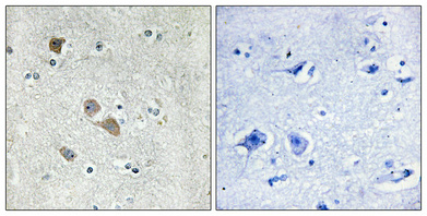
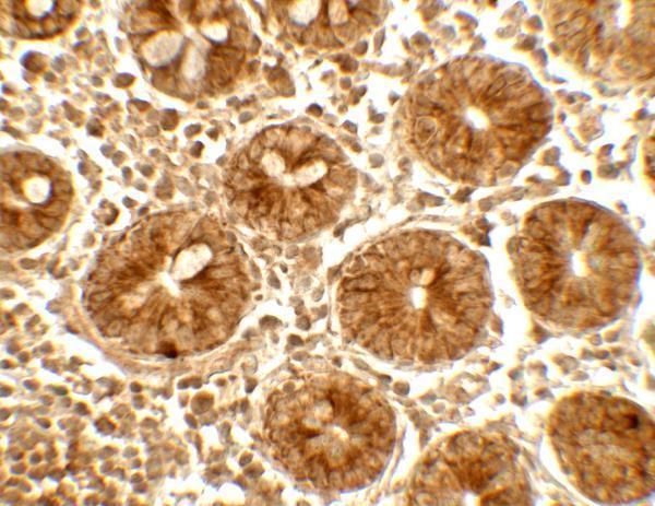
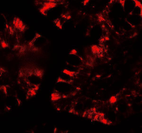
Two anti-IFIT1 antibodies have IHC images: A02652 in human brain paraffin sections and A02652-1 in human small intestine; A02652-1 also has an IF image in human small intestine (catalog image captions).
A02652 has an IHC image from human brain paraffin sections at 1:100 and lists IF and ICC applications (A02652 image caption; catalog applications). A02652-1 has IHC and IF images from human small intestine at 5 μg/mL and 20 μg/mL, respectively (A02652-1 image captions).
Which to pick: For paraffin-section IHC, choose A02652 when its documented human brain example and Tris-EDTA pH 8.0 retrieval fit the experiment; its caption does not report the fixative (A02652 IHC image caption). For IF/ICC, A02652 lists both applications, while A02652-1 has a human small-intestine IF image but does not list ICC (catalog applications; A02652-1 IF image caption). Both list human, mouse and rat reactivity, but the supplied IHC images document human tissue only; A02652 is described as polyclonal, and A02652-1’s clonality is unreported (catalog reactivity; IHC image captions; A02652 dilution data).