This website uses cookies to ensure you get the best experience on our website.
- Table of Contents
3 Citations 11 Q&As


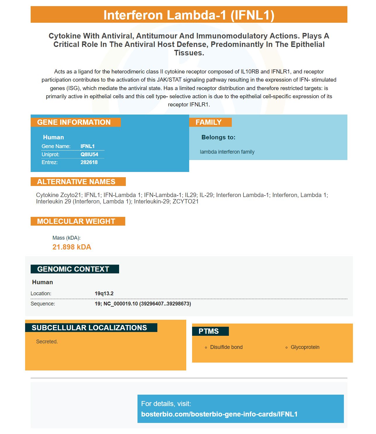
Facts about Interferon lambda-1.

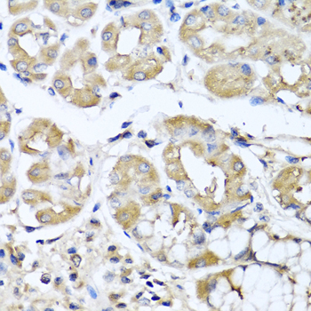
Acts as a ligand for the heterodimeric class II cytokine receptor composed of IL10RB and IFNLR1, and receptor participation contributes to the activation of this JAK/STAT signaling pathway resulting in the expression of IFN- stimulated genes (ISG), which mediate the antiviral state. Has a limited receptor distribution and therefore restricted targets: is primarily active in epithelial cells and this cell type- selective action is due to the epithelial cell-specific expression of its receptor IFNLR1.
| Human | |
|---|---|
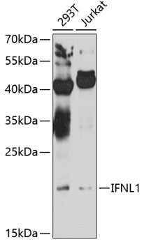
| Gene Name: | IFNL1 |
| Uniprot: | Q8IU54 |
| Entrez: | 282618 |

| Belongs to: |
|---|
| lambda interferon family |

cytokine Zcyto21; IFNL1; IFN-lambda 1; IFN-lambda-1; IL29; IL-29; interferon lambda-1; interferon, lambda 1; interleukin 29 (interferon, lambda 1); interleukin-29; ZCYTO21
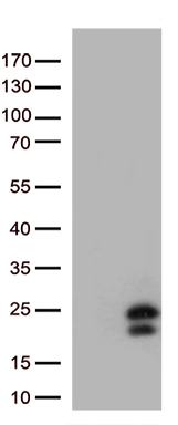
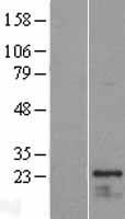
Mass (kDA):
21.898 kDA

| Human | |
|---|---|
| Location: | 19q13.2 |
| Sequence: | 19; NC_000019.10 (39296407..39298673) |
Secreted.

PMID: 12469119 by Sheppard P., et al. IL-28, IL-29 and their class II cytokine receptor IL-28R.
PMID: 12483210 by Kotenko S.V., et al. IFN-lambdas mediate antiviral protection through a distinct class II cytokine receptor complex.
*More publications can be found for each product on its corresponding product page