This website uses cookies to ensure you get the best experience on our website.


- Table of Contents
Plan chromogenic KRT8 IHC on paraffin sections using catalog antibody conditions (datasheet M01421-2). Compare cytoplasmic and membranous epithelial staining with cell-specific positive and negative tissue controls (HPA tissue IHC).

Expected localisation, validated protocols, controls and antibodies — the at-a-glance facts below, then the full design guide.
| Expected localisation | Cytoplasmic and membranous tissue staining (HPA tissue IHC) | |
| Staining pattern | Glandular epithelia show cytoplasmic and membranous staining (HPA tissue IHC) | |
| Antigen retrieval | EDTA pH 8.0 HIER, heat-mediated (datasheet M01421-2) | |
| Positive control | Appendix+4 more · see all | |
| Negative control | Adipose tissue+4 more · see all |
| Fixation | Keep fixation consistent across paraffin sections. (standard IHC practice; not target-specific) | |
| Caveat | Staining and RNA expression show medium consistency (HPA tissue IHC) | |
| Regulation | Intestine/stomach tissue-enhanced RNA (HPA tissue RNA) | |
| Isoform / epitope | 2 isoforms; antibody epitope coverage is unspecified (UniProt; catalog datasheets) |
The catalog antibody protocol uses EDTA pH 8.0 retrieval (datasheet: M01421-2). One published paraffin-section KRT8 protocol provides an additional starting point (PMC5855799).
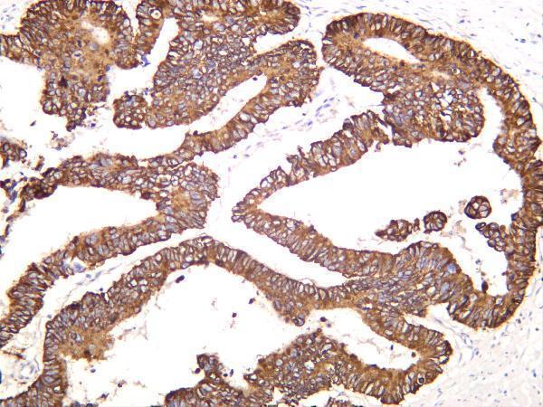
| Sample | Paraffin-embedded human lung cancer tissue; fixative not specified (datasheet M01421-2) |
| Fixation | Image fixative and duration unreported (datasheet M01421-2); verify before use. |
| Sectioning | 4–5 µm sections on charged slides (standard) |
| Deparaffinisation | Xylene, graded ethanol series to water (standard) |
| Antigen retrieval | Heat retrieval: EDTA pH 8.0 (datasheet M01421-2); 20 min, 95–100 °C (standard) |
| Peroxidase block | 3% H2O2, 10 min, room temperature (standard) |
| Blocking | 10% goat serum (datasheet M01421-2) |
| Primary antibody | Rabbit monoclonal (clone CIG-11) anti-KRT8, 1:50 (datasheet M01421-2) |
| Primary incubation | Overnight at 4 °C (datasheet M01421-2) |
| Detection | HRP-conjugated secondary, DAB chromogen (datasheet M01421-2) |
| Counterstain | Hematoxylin, blue, dehydrate and mount (standard) |
| Expected result | KRT8-positive staining in endocrine cells of appendix (HPA tissue IHC: High). HPA tissue profile: Cytoplasmic and membranous expression in most glandular epithelia, renal tubules and pneumocytes. No signal in the no-primary control. |
KRT8 is an intracellular intermediate filament protein with no transmembrane segment (UniProt P05787). In paraffin section IHC, expect cytoplasmic and sometimes membranous staining in glandular epithelia, renal tubules and pneumocytes (HPA tissue IHC). HPA rates its tissue staining reliability Enhanced, while reporting medium consistency between antibody staining and RNA expression (HPA tissue IHC).
| Cytoplasmic staining in breast glandular cells or bronchial respiratory epithelial cells. | This fits the reported high staining in those cell types (HPA tissue IHC). A filamentous pattern is biologically plausible because KRT8 contributes to the epithelial intermediate filament network (UniProt P05787). |
| Membranous staining accompanies epithelial cytoplasmic staining. | This can fit the HPA tissue profile, which describes cytoplasmic and membranous expression (HPA tissue IHC). KRT8 has no transmembrane segment, so a membrane-associated appearance does not establish that it spans the membrane (UniProt P05787). |
| Strong nucleus-only staining, with little epithelial cytoplasmic signal. | Treat this as discordant with the usual tissue IHC profile and check controls and detection background (HPA tissue IHC; general IHC practice). UniProt also annotates nucleoplasm and nuclear matrix, so nuclear staining alone cannot prove an artefact (UniProt P05787). |
| Staining predominates in adipocytes or esophageal squamous epithelial cells. | Those cells are reported as not detected (HPA tissue IHC). Review morphology and controls; antibody cross-reactivity or endogenous detection activity are possible general IHC explanations, not established KRT8-specific causes (general IHC practice). |
| Diffuse color covers cells and surrounding tissue, or a known-positive section has no signal. | Neither appearance demonstrates the expected cell-restricted pattern (HPA tissue IHC). Diffuse color warrants background controls; absent signal in a reported high-staining cell type warrants a run and reagent check (general IHC practice). |
| Cell type and tissue context | HPA reports high staining in breast glandular and bronchial respiratory epithelial cells, but not detected in adipocytes or esophageal squamous epithelial cells (HPA tissue IHC). Score the named cells rather than assuming every cell in a section should stain (general IHC practice). |
| Filament organization and partner | KRT8 is a structural component of simple epithelial intermediate filaments and forms a heterodimer with KRT18 (UniProt P05787). This supports interpreting epithelial cytoplasmic structure in context; it does not make KRT18 staining a substitute for KRT8 detection (UniProt P05787; general IHC practice). |
| Antibody validation and limits | Three listed antibodies have Enhanced IHC validation (HPA antibodies). The tissue profile still reports medium consistency with RNA expression (HPA tissue IHC). These summaries support pattern comparison, but do not establish how a particular catalog antibody performs under your staining conditions (general IHC practice). |
| Isoforms, modifications and processing | UniProt lists two isoforms, modified residues, and a single chain spanning residues 1–483, with no signal peptide or propeptide (UniProt P05787). The supplied record gives no antibody epitope or isoform specificity, so it cannot predict epitope-dependent staining differences (UniProt P05787). |
| IF/ICC Q&A: What should fluorescence show? | HPA reports supported localization to intermediate filaments in ICC-IF (HPA subcellular). That observation can inform compartment interpretation, while this page’s positive tissue calls come from paraffin section IHC (HPA tissue IHC). |
| Situation | Likely cause | Next action |
|---|---|---|
| No color in breast glandular cells or bronchial respiratory epithelial cells. | These are reported high-staining cells (HPA tissue IHC); an empty control may indicate a failed run (general IHC practice). | Check a known-positive section, antibody and detection reagents, and the counterstained section’s morphology (general IHC practice). |
| Color appears throughout the section, including cell-free areas. | Non-specific reagent deposition or incomplete washing can produce diffuse background (general IHC practice). | Review the negative reagent control, washing, blocking and detection steps before scoring cells (general IHC practice). |
| Reported negative cell types stain strongly. | Cross-reactivity or endogenous detection activity may contribute (general IHC practice); HPA reports adipocytes and esophageal squamous cells as not detected (HPA tissue IHC). | Verify cell identity and inspect an appropriate negative control and endogenous activity control (general IHC practice). |
| Signal appears only in nuclei. | This differs from HPA’s usual cytoplasmic and membranous tissue profile, although UniProt includes nuclear locations (HPA tissue IHC; UniProt P05787). | Check epithelial cytoplasm in a known-positive section and compare controls before assigning the nuclear signal to KRT8 (general IHC practice). |
| Signal is weak in an expected positive section. | The run may have limited sensitivity; no KRT8-specific fixation or retrieval effect is established by these sources (general IHC practice; HPA tissue IHC; UniProt P05787). | Check control performance and the validated IHC settings for the antibody in use; optimize retrieval only as a general assay step (general IHC practice). |
| Staining varies among sections or cell populations. | KRT8 distribution depends on the cell type sampled, and HPA reports medium antibody–RNA consistency for its tissue profile (HPA tissue IHC). | Compare matched cell types and section quality, then score localization and intensity within those cells (general IHC practice). |
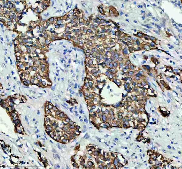
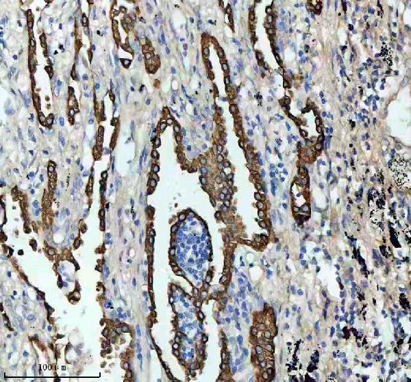
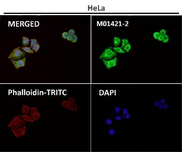


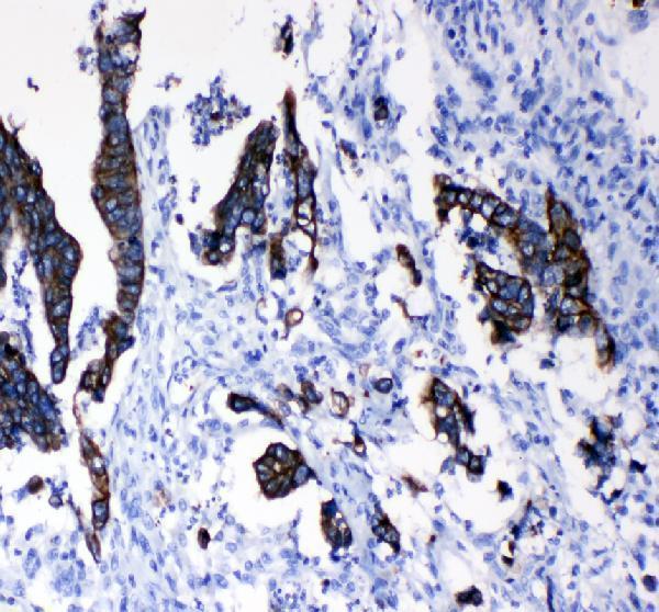
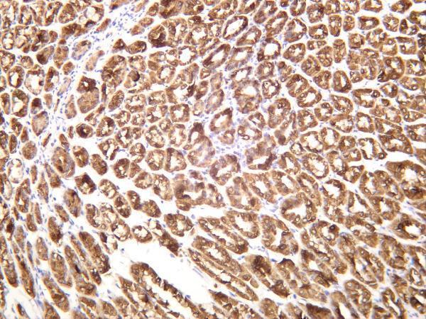


Comprehensive Human Protein Atlas IHC scoring per tissue (reliability: Enhanced — Medium consistency between antibody staining and RNA expression data.). Rows are taken directly from the HPA tissue chart — click any row's HPA link to view the source.
| Tissue | Cell type | Level | Evidence | Source |
|---|---|---|---|---|
| Appendix | Endocrine cells | High | Protein (IHC) | HPA → |
| Breast | Glandular cells | High | Protein (IHC) | HPA → |
| Bronchus | Respiratory epithelial cells | High | Protein (IHC) | HPA → |
| Cervix | Glandular cells | High | Protein (IHC) | HPA → |
| Colon | Endocrine cells | High | Protein (IHC) | HPA → |
| Tissue | Cell type | Level | Evidence | Source |
|---|---|---|---|---|
| Adipose tissue | Adipocytes | Not detected | Protein (IHC) | HPA → |
| Adrenal gland | Glandular cells | Not detected | Protein (IHC) | HPA → |
| Bone marrow | Hematopoietic cells | Not detected | Protein (IHC) | HPA → |
| Caudate | Glial cells | Not detected | Protein (IHC) | HPA → |
| Cerebellum | Cells in granular layer | Not detected | Protein (IHC) | HPA → |
Troubleshoot KRT8 chromogenic IHC in paraffin sections using the catalog antibody’s tissue image and known intermediate filament localisation.
Anti-KRT8 entries include human paraffin-section IHC and human tissue IF images; catalog reactivity spans human, mouse and rat (M01421-2/3 IHC captions; M01421-3 IF caption and catalog).
M01421-2 shows human lung cancer paraffin-section IHC, and M01421-3 shows human rectal cancer paraffin-section IHC (respective image captions). M01421-6 shows human colon carcinoma staining, while PA1240’s IHC caption identifies its target as ITGB3, so that image does not establish KRT8 staining (respective image captions).
Which to pick: For tissue IHC, M01421-2 has a rabbit monoclonal antibody and an IHC caption documenting paraffin sections, EDTA pH 8 retrieval and 1:50 dilution; the fixative is unreported (M01421-2 catalog and IHC caption). For IF/ICC, M01421-3 lists both applications and has tissue IF images; its own IHC caption documents paraffin sections and EDTA pH 8 retrieval, with fixative unreported (M01421-3 catalog, IF captions and IHC caption). For human, mouse and rat coverage, M01421-6 lists IHC and IF for a rabbit monoclonal antibody, but its human colon carcinoma IHC caption does not report section processing or fixative (M01421-6 catalog and IHC caption).