LIN28A / Protein lin-28 homolog A · IHC design guide

Design Immunohistochemistry for LIN28A

Plan chromogenic LIN28A IHC on paraffin sections using the IHC-validated antibody (datasheet A01966-2). Testis offers a positive control, with nuclear staining reported in pachytene spermatocytes (HPA tissue IHC).

Evidence assembled Oct 2026 · For research use; verify linked source records and product datasheet before use
Immunohistochemistry protocol sheet for LIN28A (IHC for LIN28A): expected localisation Tissue IHC shows nuclear subsets; UniProt describes a predominantly cytoplasmic protein (HPA tissue IHC; UniProt), antibody A01966-2, validated IHC image, and IHC protocol steps
Printable LIN28A IHC protocol sheet — expected localisation Tissue IHC shows nuclear subsets; UniProt describes a predominantly cytoplasmic protein (HPA tissue IHC; UniProt), antibody A01966-2, controls and protocol steps. Open the full LIN28A IHC guide →

LIN28A Immunohistochemistry Experimental Design Guide

Expected localisation, validated protocols, controls and antibodies — the at-a-glance facts below, then the full design guide.

Must know before staining
Expected localisation Tissue IHC shows nuclear subsets; UniProt describes a predominantly cytoplasmic protein (HPA tissue IHC; UniProt)
Staining pattern Nuclear staining in subsets of seminiferous duct and placental cells (HPA tissue IHC)
Antigen retrieval EDTA pH 8.0 HIER, heat-mediated (datasheet A01966-2)
Positive control ⓘ Testis
Negative control ⓘ Adipose tissue+4 more · see all
Important caveats
Reasons your staining may differ from the expected pattern.
Fixation Keep fixation conditions consistent across sections. (standard IHC practice; not target-specific)
Caveat Nuclear tissue staining differs from the cytoplasmic molecular expectation (HPA tissue IHC; UniProt)
Regulation May rise in HER2-overexpressing breast tumors (UniProt)
Isoform / epitope No annotated isoforms or transmembrane segment (UniProt)
Section 1

Recommended LIN28A IHC & IF Protocols

Start with the catalog antibody’s IHC-P protocol (datasheet: A01966-2), then compare published LIN28A staining of pediatric brain tumors, injured iliac artery, and marmoset ovary (PMC3508282; PMC7520293; PMC4086814).

Recommended immunohistochemistry (IHC-P) protocol parameters
SampleParaffin-embedded human testis cancer tissue; fixative not specified (datasheet A01966-2)
FixationImage fixative and duration unreported (datasheet A01966-2); verify before use.
Sectioning4–5 µm sections on charged slides (standard)
DeparaffinisationXylene, graded ethanol series to water (standard)
Antigen retrievalHeat retrieval: EDTA pH 8.0 (datasheet A01966-2); 20 min, 95–100 °C (standard)
Peroxidase block3% H2O2, 10 min, room temperature (standard)
Blocking10% goat serum (datasheet A01966-2)
Primary antibodyRabbit anti-LIN28A, 0.5-1μg/ml (datasheet A01966-2)
Primary incubationOvernight at 4 °C (datasheet A01966-2)
DetectionStreptavidin-biotin complex (SABC), DAB chromogen (datasheet A01966-2)
CounterstainHematoxylin, blue, dehydrate and mount (standard)
Expected resultLIN28A-positive staining in pachytene spermatocytes of testis (HPA tissue IHC: Medium). HPA tissue profile: Nuclear expression in subsets of cells in seminiferous ducts and in placenta. No signal in the no-primary control.
💡Decision noteTry heat-mediated EDTA pH 8.0 retrieval first (datasheet: A01966-2). For comparison, one published paraffin protocol used citrate pH 6.0 (PMC4086814).
Section 2

What Is the Expected LIN28A Staining Pattern?

LIN28A is predominantly cytoplasmic and has no transmembrane segment (UniProt Q9H9Z2). In tissue IHC, expect nuclear staining in subsets of seminiferous duct cells and placenta, with medium staining reported in pachytene spermatocytes (HPA: tissue IHC). HPA rates its tissue pattern Enhanced, while reporting medium consistency with RNA expression (HPA: tissue IHC). Interpret compartment differences in light of the assay used.

What am I looking at on my slide?
Medium nuclear staining in a subset of pachytene spermatocytes, with nearby cells less stained (HPA: testis IHC).This matches the reported testis pattern; score the named cell population and compartment rather than treating every seminiferous duct cell as an expected positive (HPA: testis IHC).
Cytoplasmic signal accompanies or predominates over nuclear signal (UniProt Q9H9Z2; HPA: subcellular ICC-IF).Cytoplasmic LIN28A is biologically plausible, although HPA describes nuclear expression in its tissue IHC profile (UniProt Q9H9Z2; HPA: tissue IHC). Investigate an exclusively membranous outline as possible nonspecific staining: LIN28A has no transmembrane segment (UniProt Q9H9Z2).
Strong staining appears broadly in HPA-listed negative cell populations, such as adipocytes in adipose tissue (HPA: tissue IHC).The pattern conflicts with that reference observation and raises concern for cross-reactivity or endogenous detection activity; it does not establish either cause by itself (HPA: tissue IHC; general IHC practice). Compare with a no-primary control (general IHC practice).
Diffuse color covers tissue and blank spaces, obscuring cell boundaries (general IHC practice).A distribution that cannot be assigned to cells cannot be scored as LIN28A localization; excess reagent, inadequate washing, or detection background are possible workflow causes (general IHC practice).
Pachytene spermatocytes show no discernible signal in a testis section (HPA: testis IHC).This fails to reproduce HPA's medium staining in that cell population (HPA: testis IHC). Check section identity and assay controls before calling the specimen LIN28A-negative (general IHC practice).
💡Expected LIN28A appearanceA convincing IHC positive is cell-resolved, medium nuclear staining in a subset of pachytene spermatocytes (HPA: testis IHC); broad staining of HPA-listed negative cells or blank spaces warrants a background check (HPA: tissue IHC; general IHC practice).
How each factor affects the staining
Tissue and cell selectionTestis pachytene spermatocytes provide a reported medium-staining reference; placenta has nuclear expression in cell subsets without a supplied intensity grade (HPA: tissue IHC). HPA also lists several cell populations as low or not detected, so identify the cells being compared (HPA: tissue IHC).
Compartment and topologyUniProt describes LIN28A as predominantly cytoplasmic, with possible nucleolar localization, and reports no transmembrane segment (UniProt Q9H9Z2). HPA tissue IHC emphasizes nuclear subsets; use the reference appropriate to the assay when interpreting mixed staining (HPA: tissue IHC).
Antibody validationHPA lists CAB020785 as IHC Enhanced, a status based on reproduction by independent antibodies or orthogonal data (HPA: antibodies). The supplied record does not establish that every antibody will reproduce the same pattern (HPA: antibodies).
IF/ICC Q: where should signal appear?A: Mainly in the cytosol, which HPA marks supported; additional nucleolar and rods-and-rings locations are marked uncertain (HPA: subcellular ICC-IF). This IF/ICC observation should not replace the tissue IHC reference pattern (HPA: tissue IHC; HPA: subcellular ICC-IF).
Why is my staining missing, weak or wrong?
SituationLikely causeNext action
Testis control has no signal in pachytene spermatocytes (HPA: testis IHC).The control may be misidentified, or an assay step may have failed; the staining result alone cannot distinguish them (general IHC practice).Confirm the section and cell identity, then check primary-antibody application, detection reagents, and a concurrently processed positive control (general IHC practice).
Strong signal appears in HPA-listed negative cells (HPA: tissue IHC).Cross-reactivity or endogenous detection activity is possible; HPA's negative observation does not identify the mechanism (HPA: tissue IHC; general IHC practice).Compare a no-primary control and review detection blocking and cell identity before scoring the stain as LIN28A (general IHC practice).
Color is diffuse across the section or present outside cells (general IHC practice).High detection background, excess reagent, or insufficient washing can obscure cell-localized signal (general IHC practice).Inspect the no-primary control; adjust blocking, reagent concentration, or washes one variable at a time (general IHC practice).
Only cell borders stain, with no convincing intracellular pattern (general IHC practice).This conflicts with UniProt's intracellular locations and lack of a transmembrane segment; nonspecific edge staining is possible (UniProt Q9H9Z2; general IHC practice).Compare the no-primary control and inspect whether the apparent border signal tracks section edges or damaged areas (general IHC practice).
Nuclear and cytoplasmic signals disagree with a single expected-compartment rule (UniProt Q9H9Z2; HPA: tissue IHC).The references describe different observations: predominantly cytoplasmic localization and nuclear expression in tissue subsets (UniProt Q9H9Z2; HPA: tissue IHC).Record each compartment and the stained cell population separately; compare tissue IHC with the HPA tissue pattern (HPA: tissue IHC; general IHC practice).
Placenta shows sparse positive cells while the whole-section score appears low (HPA: tissue IHC).HPA describes nuclear expression in subsets of placental cells, so an aggregate score can conceal localized staining (HPA: tissue IHC).Inspect cell-resolved nuclear staining and report the positive subset; do not assign a placenta intensity grade from the supplied HPA record (HPA: tissue IHC; general IHC practice).

Sample controls for LIN28A IHC & IF

🧪Run testis first: pachytene spermatocytes should stain at a Medium level (HPA: Testis, pachytene spermatocytes, Medium). Run adipose tissue as the negative, where adipocytes are Not detected (HPA: Adipose tissue, adipocytes, Not detected); cells without specific staining on the testis slide should remain chromogen-free, but the supplied HPA row does not identify a named internal-negative cell type (HPA: Testis row).
Positive control tissue: Testis (Pachytene spermatocytes, HPA Medium)
Negative control tissue: Adipose tissue (HPA Not detected)
ICC-IF cell lines (HPA subcellular resource): HPA ICC-IF images show LIN28A in AF22, CACO-2, with annotated localisation: Cytosol (supported) (HPA subcellular).
Technical controls: Include a no-primary, secondary-only control; a concentration-matched rabbit IgG isotype control; and a confirmed LIN28A knockout specimen as a biological negative (caption: rabbit anti-LIN28A; standard IHC practice). Check endogenous peroxidase and biotin background in the testis section because the reported detection uses a biotinylated secondary, streptavidin–biotin complex, and DAB (caption: A01966-2 detection).
⚠️Feasibility: A target-specific fixation window and fixation effect are unreported, and the selected paraffin-section caption does not state its fixative (caption: A01966-2). The reported tissue IHC uses heat retrieval in EDTA, pH 8.0 before staining; whether retrieval is required has not been established by the supplied evidence (caption: A01966-2). The supplied evidence does not establish whether frozen sections or IF are easier; assess cytoplasmic signal against testis background and treat nucleolar signal cautiously because cytosol is the supported ICC-IF location while nucleoli are uncertain (HPA: subcellular localization).

HPA tissue IHC evidence for LIN28A

Comprehensive Human Protein Atlas IHC scoring per tissue (reliability: Enhanced — Medium consistency between antibody staining and RNA expression data.). Rows are taken directly from the HPA tissue chart — click any row's HPA link to view the source.

Positive expression · recommended positive controls

TissueCell typeLevelEvidenceSource
Testis Pachytene spermatocytes Medium Protein (IHC) HPA →

Undetected expression · recommended negative controls

TissueCell typeLevelEvidenceSource
Adipose tissue Adipocytes Not detected Protein (IHC) HPA →
Adrenal gland Glandular cells Not detected Protein (IHC) HPA →
Appendix Glandular cells Not detected Protein (IHC) HPA →
Bone marrow Hematopoietic cells Not detected Protein (IHC) HPA →
Breast Adipocytes Not detected Protein (IHC) HPA →
Section 3

Advanced LIN28A IHC Tips

Troubleshoot LIN28A staining in paraffin sections by checking retrieval, compartment, cell identity and detection controls before scoring chromogenic signal.

Which retrieval conditions should I try first for LIN28A in paraffin sections?
Start with heat-mediated antigen retrieval in EDTA at pH 8.0 for paraffin sections (datasheet A01966-2). The selected image used that retrieval before incubation with 1 μg/mL catalog antibody overnight at 4°C, so keep those conditions together when reproducing its staining (datasheet A01966-2). If staining is weak, adjust heating time or cooling gradually while keeping a known positive section in every run; excessive heating can damage tissue morphology (general IHC practice). Compare signal in the expected cells with no-primary controls, because stronger staining alone does not establish LIN28A specificity (HPA: medium staining in pachytene spermatocytes; general IHC practice).
How should I troubleshoot fixation-dependent loss of LIN28A staining?
Target-specific fixation sensitivity is unknown from the supplied evidence, and the selected paraffin-section caption does not state a fixative (datasheet A01966-2). Record the actual fixative, fixation duration and processing history for each section, then compare a suspect sample with a consistently processed positive control (general IHC practice). Keep retrieval and antibody incubation constant during that comparison; the selected image used EDTA at pH 8.0 and 1 μg/mL antibody overnight at 4°C (datasheet A01966-2). If morphology and staining both deteriorate, evaluate processing quality before attributing the result to LIN28A biology (general IHC practice).
Should LIN28A staining be cytoplasmic, nuclear or both?
Assess cytoplasmic staining first: LIN28A is predominantly cytoplasmic, with reported association with rough endoplasmic reticulum, P-bodies and stress granules (UniProt Q9H9Z2), and HPA supports a mainly cytosolic location (HPA subcellular). Nucleolar localisation is reported, although HPA labels its additional nucleolar signal uncertain (UniProt Q9H9Z2; HPA subcellular). Nuclear staining in subsets of seminiferous-duct cells and placenta is also reported in tissue IHC (HPA tissue IHC). Score nuclear, nucleolar and cytoplasmic compartments separately against a positive section and no-primary control; do not classify diffuse staining across every compartment as specific solely because it is strong (general IHC practice).
Could isoforms or epitope accessibility explain discordant LIN28A staining?
The supplied record lists 0 isoforms and a cold-shock domain at residues 39–112, but it does not identify the catalog antibody's epitope (UniProt Q9H9Z2). LIN28A has no transmembrane segment and has recorded modifications at residues 2, 3, 120 and 200; their effect on this antibody's staining is unknown (UniProt Q9H9Z2). Accordingly, do not assign discordant compartment staining to an isoform or modification without independent epitope evidence (UniProt Q9H9Z2; general IHC practice). Compare retrieval conditions and include appropriate positive and no-primary sections before considering an epitope-accessibility explanation (datasheet A01966-2; general IHC practice).
How can IF help resolve ambiguous LIN28A localisation in tissue?
Use IF as a separate localisation check, multiplexing LIN28A with a validated marker for the expected pachytene spermatocytes in testis and a nuclear counterstain (HPA tissue IHC; general IF practice). Choose spectrally separated fluorophores and, where tissue autofluorescence interferes, place the weaker target signal in a far-red channel with matched unstained controls (general IF practice). LIN28A has no transmembrane segment and is mainly cytosolic, with possible nucleolar signal; use controlled permeabilisation that permits antibody access to these intracellular compartments (UniProt Q9H9Z2; HPA subcellular; general IF practice). Compare compartment patterns with the chromogenic section without assuming identical sensitivity across detection methods (general IHC/IF practice).
What should I check when DAB background obscures LIN28A staining?
Inspect a no-primary section to separate secondary-reagent or detection-system staining from signal dependent on the primary antibody (general IHC practice). The selected workflow used 10% goat serum, biotinylated goat anti-rabbit secondary antibody, a streptavidin–biotin complex and DAB; reproduce its blocking step when comparing runs (datasheet A01966-2). Apply a peroxidase block and examine endogenous biotin background with an appropriate control when using this detection chemistry (general IHC practice). Reduce nonspecific staining by checking primary-antibody concentration, wash adequacy and DAB development time, while retaining a positive section to detect any loss of useful signal (general IHC practice).
How should I score LIN28A staining across paraffin sections? ⚠ ANSWER MARKED FOR VERIFICATION
Define the cell population and compartment before scoring, because HPA reports medium staining in pachytene spermatocytes and nuclear expression in subsets of seminiferous-duct cells (HPA tissue IHC). Report the percentage of positive eligible cells and an intensity-based H-score from 0–300, with cytoplasmic and nuclear scores recorded separately (general IHC practice). For spatial analyses, report positive-cell density per mm² of evaluable tissue and exclude folds, necrosis and tissue edges using the same rules across slides (general IHC practice). Normalise counts to the number of eligible cells or evaluable tissue area, and use identically processed controls to compare batches (general IHC practice).
How do I distinguish true LIN28A positivity from staining artefact?
A credible result has staining in an anatomically appropriate cell population: HPA reports medium signal in pachytene spermatocytes, while its listed adipocytes in adipose tissue are not detected (HPA tissue IHC). Predominantly cytoplasmic signal is biologically plausible, and limited nuclear or nucleolar signal can occur; broad staining in implausible compartments needs control-based review (UniProt Q9H9Z2; HPA subcellular; HPA tissue IHC). Check whether apparent positivity clusters at cut edges, tissue folds or necrotic regions, where staining can be misleading (general IHC practice). Finally, compare no-primary and peroxidase-block controls to identify detection background or endogenous enzyme activity before calling faint DAB deposits LIN28A positive (general IHC practice).
Boster reagents

Best LIN28A / Protein lin-28 homolog A IHC Antibodies

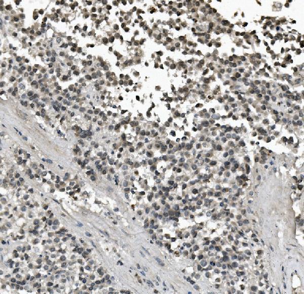
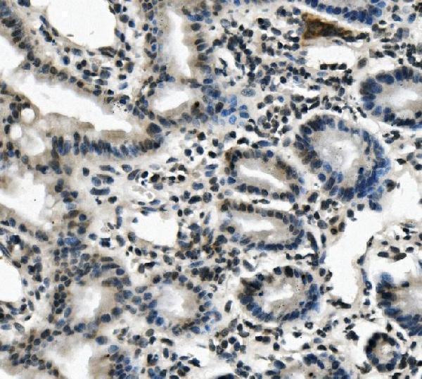
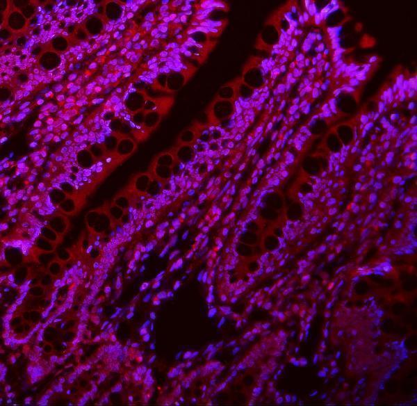
The catalog includes anti-LIN28A antibodies with IHC images from human testis cancer and mouse and rat intestine, plus IF images from human mammary cancer and rat intestine (A01966-2 image captions).

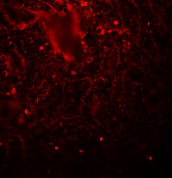
Real IHC data IHC analysis of LIN28A using anti-LIN28A antibody (A01966-2). LIN28A was detected in paraffin-embedded section of human testis cancer tissue. Heat mediated antigen retrieval was performed in EDTA buffer (pH8.0, epitope retrieval solution). The tissue section was blocked with 10% goat serum. The tissue section was then incubated with 1μg/ml rabbit anti-LIN28A Antibody (A01966-2) overnight at 4°C. Biotinylated goat anti-rabbit IgG was used as secondary antibody and incubated for 30 minutes at 37°C. The tissue section was developed using Strepavidin-Biotin-Complex (SABC) (Catalog # SA1022) with DAB as the chromogen.
Anti-Lin28/LIN28A Antibody ®
Cat # A01966-2
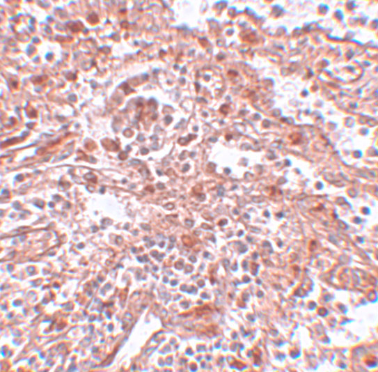
Real IHC data Immunohistochemistry of KLF4 in human spleen tissue with KLF4 antibody at 5 μg/mL.
Anti-Lin28 Antibody
Cat # A01966

A01966-2 has paraffin-section IHC images from human testis cancer and mouse and rat intestine, plus IF images from human mammary cancer and rat intestine (A01966-2 image captions). A01966 lists human IHC-P and IF; its IF caption shows LIN28 in human spleen, but its IHC caption names KLF4, so that image cannot establish LIN28A staining (A01966 applications and image captions).

Which to pick: Choose A01966-2 for tissue IHC, especially across human, mouse and rat samples: its paraffin-section captions document LIN28A staining in all three species, while the fixative is unreported (A01966-2 reactivity and IHC image captions). For IF, A01966-2 has tissue images; M01966-1 lists ICC/IF and is a rabbit monoclonal, but has no supplied ICC/IF image (A01966-2 IF image captions; M01966-1 catalog entry). A01966 lists human IHC-P and IF, but its mismatched KLF4 IHC caption warrants checking target identity before using that image as LIN28A evidence (A01966 applications and IHC image caption).

Each figure is that product's own IHC / IF validation image from its datasheet.

References

  1. UniProt Consortium. UniProt entry Q9H9Z2 (LN28A_HUMAN, Protein lin-28 homolog A).
  2. Human Protein Atlas. LIN28A tissue IHC expression (reliability: Enhanced).
  3. Human Protein Atlas. LIN28A subcellular location (ICC-IF): Mainly localized to the cytosol. In addition localized to the nucleoli and rods & rings..
  4. Human Protein Atlas. LIN28A antibody validation summary (3 antibodies).
  5. LIN28A immunoreactivity is a potent diagnostic marker of embryonal tumor with multilayered rosettes (ETMR). Acta neuropathologica 2012 — PMC3508282.
  6. Analysis of LIN28A in early human ovary development and as a candidate gene for primary ovarian insufficiency. Molecular and cellular endocrinology 2012 — PMC3314903.
  7. Lin28a up-regulation is associated with the formation of restenosis via promoting proliferation and migration of vascular smooth muscle cells. Journal of cellular and molecular medicine 2020 — PMC7520293.
  8. The neonatal marmoset monkey ovary is very primitive exhibiting many oogonia. Reproduction (Cambridge, England) 2014 — PMC4086814.
  9. PubMed PMID:12798299 — UniProt-cited evidence.
  10. PubMed PMID:14702039 — UniProt-cited evidence.
  11. PubMed PMID:16710414 — UniProt-cited evidence.