This website uses cookies to ensure you get the best experience on our website.


- Table of Contents
Plan chromogenic LY9 IHC in paraffin sections with the IHC-validated antibody A05830-1 (datasheet A05830-1). Use lymphoid cell subsets as staining references (HPA tissue IHC), and interpret their observed cytoplasmic signal alongside LY9’s expected cell membrane location (HPA tissue IHC; UniProt).

Expected localisation, validated protocols, controls and antibodies — the at-a-glance facts below, then the full design guide.
| Expected localisation | Cell membrane expected (UniProt); lymphoid cytoplasm observed (HPA tissue IHC) | |
| Staining pattern | Cytoplasmic staining in subsets of lymphoid cells (HPA tissue IHC) | |
| Antigen retrieval | Citrate pH 6 HIER, heat-mediated (datasheet A05830-1) | |
| Positive control | Appendix+3 more · see all | |
| Negative control | Adipose tissue+4 more · see all |
| Fixation | Keep section fixation consistent (standard IHC practice; not target-specific); Matched tissue-IHC evidence does not establish the fixation claim. Validate the specimen-specific method before use. (selected-SKU IHC image A05830-1) | |
| Caveat | A secreted variant may alter tissue staining (HPA tissue IHC) | |
| Regulation | Surface LY9 increases on SLE T cells (UniProt) | |
| Isoform / epitope | Four isoforms; check whether the epitope is extracellular or cytoplasmic (UniProt) |
The catalog antibody’s IHC-P protocol (datasheet A05830-1) is accompanied by published LY9 IHC methods for FFPE tissue (PMC10901781) and lung adenocarcinoma tissue (PMC11348898).
| Sample | Paraffin-embedded mouse spleen tissues; fixative not specified (datasheet A05830-1) |
| Fixation | Image fixative and duration unreported (datasheet A05830-1); verify before use. |
| Sectioning | 4–5 µm sections on charged slides (standard) |
| Deparaffinisation | Xylene, graded ethanol series to water (standard) |
| Antigen retrieval | Heat retrieval: Citrate pH 6, 20 min (datasheet A05830-1) |
| Peroxidase block | 3% H2O2, 10 min, room temperature (standard) |
| Blocking | 10% goat serum (datasheet A05830-1) |
| Primary antibody | Rabbit anti-LY9, 0.5-1μg/ml (datasheet A05830-1) |
| Primary incubation | Overnight at 4 °C (datasheet A05830-1) |
| Detection | Streptavidin-biotin complex (SABC), DAB chromogen (datasheet A05830-1) |
| Counterstain | Hematoxylin, blue, dehydrate and mount (standard) |
| Expected result | LY9-positive staining in lymphoid tissue of appendix (HPA tissue IHC: Medium). HPA tissue profile: Cytoplasmic expression mainly in subsets of lymphoid cells. No signal in the no-primary control. |
LY9 is a cell membrane protein with an extracellular region and a cytoplasmic tail (UniProt Q9HBG7 topology). In paraffin tissue IHC, HPA reports chiefly cytoplasmic staining in subsets of lymphoid cells, with medium staining in appendix lymphoid tissue, splenic white pulp and tonsil non-germinal center cells (HPA tissue IHC). HPA rates its tissue IHC profile Approved, with medium consistency between staining and RNA data and external verification pending (HPA tissue IHC).
| Discrete staining in lymphoid cell subsets, especially in appendix, splenic white pulp or tonsil non-germinal center cells (HPA tissue IHC). | This fits the reported IHC distribution; medium is the recorded level for those cell populations, so assess the named cells rather than assigning one intensity to the whole section (HPA tissue IHC). |
| Predominantly cytoplasmic signal in a subset of lymphoid cells (HPA tissue IHC). | This can match the paraffin IHC profile even though LY9 is assigned to the cell membrane by UniProt and mainly to the plasma membrane by HPA ICC-IF (UniProt Q9HBG7 subcellular location; HPA tissue IHC; HPA subcellular ICC-IF). Interpret the two assay readouts separately. |
| Strong, isolated nuclear staining, without the reported lymphoid cell pattern (HPA tissue IHC). | Nuclear localization is absent from the supplied LY9 locations; treat this as suspect staining and compare with an appropriate negative control before scoring it as LY9 (UniProt Q9HBG7 subcellular location; HPA subcellular ICC-IF; standard IHC practice). |
| Prominent staining of adipocytes or bronchial respiratory epithelial cells (HPA tissue IHC). | HPA reports LY9 as not detected in these cell populations; investigate antibody cross-reactivity or endogenous chromogenic activity if they stain, especially when the expected lymphoid distribution is absent (HPA tissue IHC; standard IHC practice). |
| Diffuse color across cells and surrounding tissue, or no signal in appendix lymphoid tissue (HPA tissue IHC). | Diffuse color can obscure cell-specific scoring; absent signal in a reported medium-positive population makes a negative test section difficult to interpret (HPA tissue IHC; standard IHC practice). Check controls and the staining workflow before making a biological call. |
| Compartment and epitope position (UniProt Q9HBG7 topology). | LY9 spans residues 455–476, with extracellular residues 48–454 and a cytoplasmic region at 477–655 (UniProt Q9HBG7 topology). The supplied record gives no antibody epitope, so topology alone cannot predict which part this IHC antibody detects. |
| Protein variants and tissue correlation (UniProt Q9HBG7 isoforms; HPA tissue IHC). | UniProt lists four LY9 isoforms; HPA notes that at least one protein variant is secreted and that RNA and protein locations may differ (UniProt Q9HBG7 isoforms; HPA tissue IHC). Do not infer isoform identity from an IHC pattern. |
| Strength of tissue evidence (HPA tissue IHC; HPA antibodies). | The tissue profile is Approved but has medium staining–RNA consistency and awaits external verification; the listed antibody HPA050917 has IHC Approved status (HPA tissue IHC; HPA antibodies). Treat agreement with the reported pattern as support, not proof of antibody specificity. |
| Reported levels vary by cell population (HPA tissue IHC). | HPA reports low staining in lymph node germinal center cells and bone marrow hematopoietic cells, versus medium in appendix lymphoid tissue and splenic white pulp (HPA tissue IHC). A weak result in a low-level population needs a stronger reference population for comparison. |
| Antigen retrieval and chromogenic detection (standard IHC practice). | These are general assay variables to check when controls fail (standard IHC practice). The supplied UniProt and HPA records do not establish an LY9-specific retrieval setting or fixation sensitivity, so do not attribute a tissue result to either without separate evidence. |
| Situation | Likely cause | Next action |
|---|---|---|
| No staining in appendix lymphoid tissue, where HPA reports medium signal (HPA tissue IHC). | A run-level failure is possible; absence alone cannot distinguish assay failure from a different sample pattern (standard IHC practice). | Check the positive control, reagent omission control, retrieval run, primary antibody step and chromogenic detection before scoring the specimen negative (standard IHC practice). |
| Broad, low-level color obscures lymphoid cells (HPA tissue IHC). | Background from detection reagents, blocking or washing can reduce cell-level contrast (standard IHC practice). | Review the reagent omission control and the blocking, washing and detection steps; score only interpretable cells against the reported lymphoid distribution (standard IHC practice; HPA tissue IHC). |
| Adipocytes or bronchial respiratory epithelial cells stain prominently (HPA tissue IHC). | These cell types are reported as not detected; cross-reactivity or endogenous detection activity is plausible (HPA tissue IHC; standard IHC practice). | Compare with an omission control and a reported positive lymphoid population, then check endogenous activity controls appropriate to the chromogen (standard IHC practice; HPA tissue IHC). |
| Only nuclei show a sharp signal (UniProt Q9HBG7 subcellular location). | An isolated nuclear pattern lacks support from the supplied membrane and IHC distribution records (UniProt Q9HBG7; HPA tissue IHC). | Recheck morphology, counterstain and controls; withhold a positive LY9 call unless cell-associated staining agrees with the reported tissue pattern (standard IHC practice; HPA tissue IHC). |
| A lymph node germinal center looks weaker than splenic white pulp (HPA tissue IHC). | HPA records low staining in lymph node germinal center cells and medium staining in splenic white pulp cells (HPA tissue IHC). | Compare like cell populations and their reported levels; do not use equal intensity in these two sites as a pass criterion (HPA tissue IHC). |
| Q: Should ICC-IF look exactly like paraffin IHC? (HPA tissue IHC; HPA subcellular ICC-IF). | HPA reports mainly plasma membrane and additional centriolar satellite localization in ICC-IF, while its tissue IHC profile is chiefly cytoplasmic in lymphoid subsets (HPA subcellular ICC-IF; HPA tissue IHC). | A: Interpret ICC-IF on its own guide page using its localization evidence; use the reported tissue IHC pattern to judge paraffin sections (HPA subcellular ICC-IF; HPA tissue IHC). |


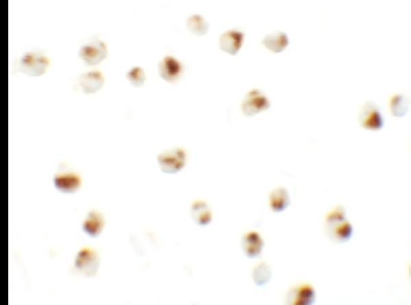


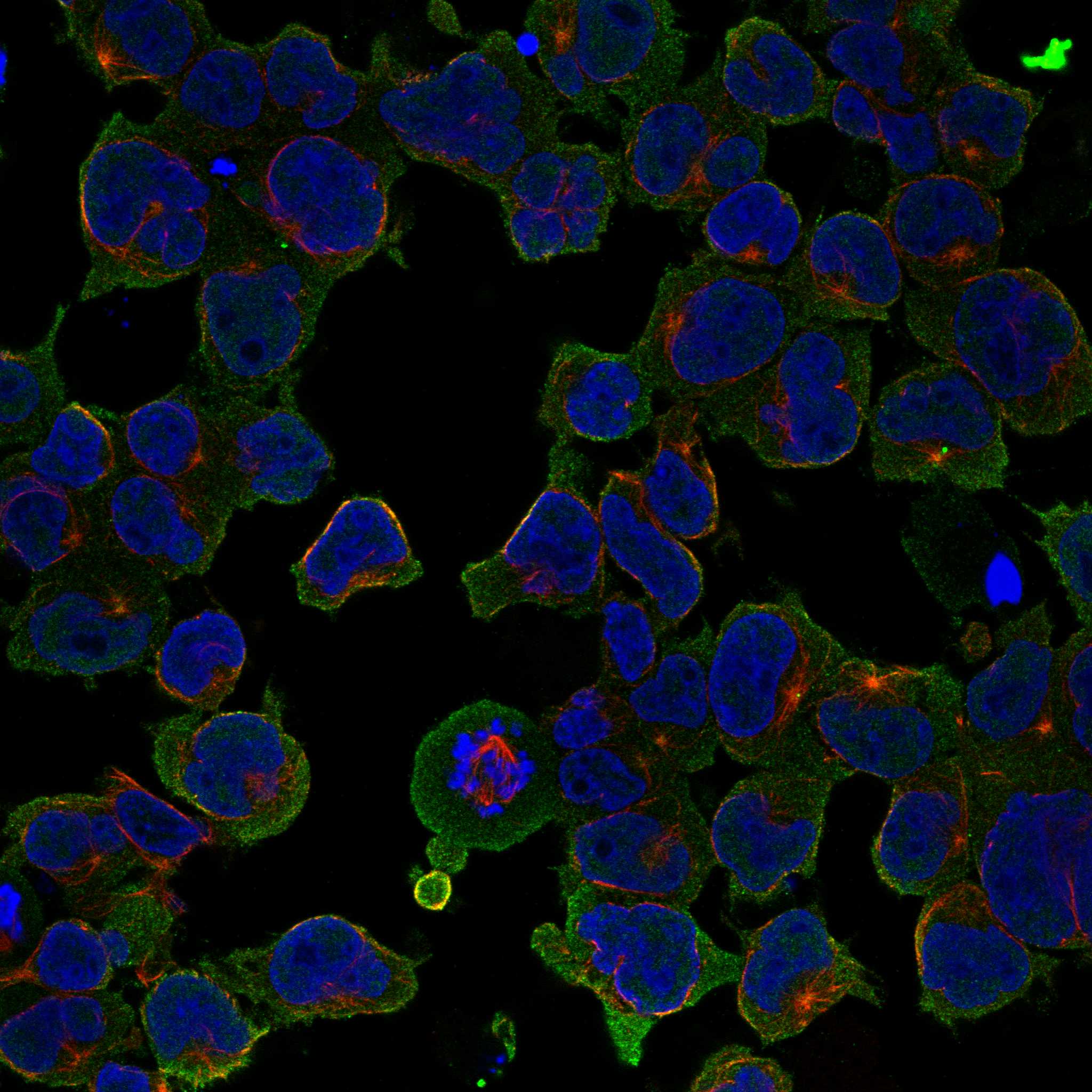
Comprehensive Human Protein Atlas IHC scoring per tissue (reliability: Approved — Medium consistency between antibody staining and RNA expression data. At least one protein variant secreted, tissue location of RNA and protein might differ and correlation is complex. Pending external verification.). Rows are taken directly from the HPA tissue chart — click any row's HPA link to view the source.
| Tissue | Cell type | Level | Evidence | Source |
|---|---|---|---|---|
| Appendix | Lymphoid tissue | Medium | Protein (IHC) | HPA → |
| Spleen | Cells in white pulp | Medium | Protein (IHC) | HPA → |
| Testis | Spermatogonia cells | Medium | Protein (IHC) | HPA → |
| Tonsil | Non-germinal center cells | Medium | Protein (IHC) | HPA → |
| Tissue | Cell type | Level | Evidence | Source |
|---|---|---|---|---|
| Adipose tissue | Adipocytes | Not detected | Protein (IHC) | HPA → |
| Adrenal gland | Glandular cells | Not detected | Protein (IHC) | HPA → |
| Breast | Adipocytes | Not detected | Protein (IHC) | HPA → |
| Bronchus | Respiratory epithelial cells | Not detected | Protein (IHC) | HPA → |
| Caudate | Glial cells | Not detected | Protein (IHC) | HPA → |
Troubleshoot LY9 chromogenic IHC by checking retrieval, staining compartment, cell identity, and controls before interpreting signal (UniProt Q9HBG7; HPA tissue IHC).
Anti-LY9 antibodies have IHC images from mouse and rat paraffin sections and IF/ICC images from 293 cells; both catalog entries list human, mouse and rat reactivity (catalog images and reactivity).
A05830-1 lists IHC and ICC and shows LY9 staining in mouse and rat paraffin spleen sections (A05830-1 applications and IHC captions). A05830-2 lists IF and ICC and shows staining in 293 cells, with no tissue IHC image supplied (A05830-2 applications and image captions).
Which to pick: Choose A05830-1 for paraffin-section IHC: its captions document heat retrieval in citrate buffer at pH 6 for 20 minutes in mouse and rat spleen, but do not report the fixative (A05830-1 IHC captions). Choose A05830-2 for IF/ICC in cells; its images use 293 cells, and IHC is absent from its application list (A05830-2 applications and IF/ICC captions). For cross-species tissue IHC, A05830-1 has mouse and rat image evidence and listed human reactivity, but no human tissue image; clonality is unreported for both SKUs (catalog reactivity, images and clone fields).