This website uses cookies to ensure you get the best experience on our website.


- Table of Contents
Plan chromogenic LYZ IHC in paraffin sections using Paneth cells and lung macrophages as high-staining references (HPA tissue IHC). Score cytoplasmic staining and interpret its distribution with LYZ secretion in mind (HPA tissue IHC; UniProt).

Expected localisation, validated protocols, controls and antibodies — the at-a-glance facts below, then the full design guide.
| Expected localisation | Cytoplasmic tissue staining; LYZ is secreted (HPA tissue IHC; UniProt) | |
| Staining pattern | High cytoplasmic staining in marrow cells, Paneth cells and lung macrophages (HPA tissue IHC) | |
| Antigen retrieval | EDTA pH 8.0 HIER, heat-mediated (datasheet PB9663) | |
| Positive control | Bone marrow+4 more · see all | |
| Negative control | Adipose tissue+4 more · see all |
| Fixation | Matched tissue-IHC evidence does not establish the fixation claim. Validate the specimen-specific method before use. (selected-SKU IHC image PB9663) | |
| Caveat | Secreted LYZ may appear away from expressing cells (HPA tissue IHC; UniProt) | |
| Regulation | No expression regulator specified (UniProt) | |
| Isoform / epitope | No isoforms annotated; mature chain spans residues 19–148 (UniProt) |
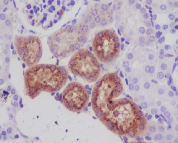
The catalog antibody’s IHC-P protocol is accompanied by published LYZ staining methods for porcine intestine, mouse kidney, and human intestinal biopsies (PMC13317171; PMC11101254; PMC2776509).
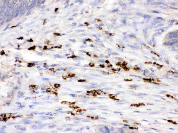
| Sample | Paraffin-embedded human intestinal cancer tissue; fixative not specified (datasheet PB9663) |
| Fixation | Image fixative and duration unreported (datasheet PB9663); verify before use. |
| Sectioning | 4–5 µm sections on charged slides (standard) |
| Deparaffinisation | Xylene, graded ethanol series to water (standard) |
| Antigen retrieval | Heat retrieval: EDTA pH 8.0 (datasheet PB9663); 20 min, 95–100 °C (standard) |
| Peroxidase block | 3% H2O2, 10 min, room temperature (standard) |
| Blocking | 10% goat serum (datasheet PB9663) |
| Primary antibody | Rabbit anti-LYZ, 0.5-1μg/ml (datasheet PB9663) |
| Primary incubation | Overnight at 4 °C (datasheet PB9663) |
| Detection | Streptavidin-biotin complex (SABC), DAB chromogen (datasheet PB9663) |
| Counterstain | Hematoxylin, blue, dehydrate and mount (standard) |
| Expected result | LYZ-positive staining in hematopoietic cells of bone marrow (HPA tissue IHC: High). HPA tissue profile: High cytoplasmic expression in selected cells types including cells in bone marrow, Paneth cells and a subset of cells in salivary gland. No signal in the no-primary control. |
In paraffin section IHC, LYZ should stain the cytoplasm of selected hematopoietic cells, Paneth cells, lung macrophages, and some glandular cells (HPA: tissue IHC). LYZ is secreted and has no transmembrane segment (UniProt P61626: subcellular location and topology). HPA rates the tissue staining evidence Enhanced, while reporting medium consistency between staining and RNA data because secreted protein can appear away from its site of synthesis (HPA: tissue IHC reliability).
| Strong cytoplasmic staining in duodenal or small intestinal Paneth cells, lung macrophages, or bone marrow hematopoietic cells (HPA: High in these cells). | This matches the reported positive pattern. Judge the named cell population and its cytoplasm, rather than treating uniform staining across every cell in the section as the expected result (HPA: tissue IHC profile). |
| Predominantly nuclear staining, with little cytoplasmic signal, in a tissue chosen for positive IHC (HPA: cytoplasmic tissue IHC profile). | This does not match the reported tissue IHC pattern; review morphology, controls, and detection background before calling the cells LYZ positive (HPA: tissue IHC profile). The separate ICC-IF record lists nucleoplasm only as an uncertain additional location (HPA: subcellular ICC-IF). |
| Strong staining in adipocytes or another cell type reported as not detected (HPA: adipose tissue and breast adipocytes; tissue IHC). | Treat an unexpected cell pattern as provisional: cross-reactivity or endogenous detection activity is possible (general IHC practice). Confirm cell identity and compare a no-primary control; secretion means protein location need not mirror cellular RNA expression (HPA: reliability description; UniProt P61626: Secreted). |
| Diffuse color across tissue, including cells expected to have little or no staining (HPA: low and not-detected tissue IHC entries). | The contrast needed to recognize selected positive cells has been lost (HPA: tissue IHC profile). General IHC practice: check no-primary background, blocking, washes, detection conditions, and counterstain before interpreting faint cell-specific signal. |
| No signal in correctly identified Paneth cells, lung macrophages, or bone marrow hematopoietic cells (HPA: High in these populations). | An expected positive population is missing, so a negative result elsewhere is difficult to interpret (HPA: tissue IHC). General IHC practice: verify tissue identity and controls, then review antibody dilution, retrieval, detection, and reagent performance. |
| Cell population and tissue (HPA: tissue IHC) | High staining is reported in bone marrow hematopoietic cells, duodenal and small intestinal Paneth cells, and lung macrophages; appendix, kidney, salivary gland, and stomach have specified medium-staining populations (HPA: tissue IHC). Choose and score the relevant cells, not the whole section. |
| Secretion and interpretation (UniProt P61626: Secreted; HPA: reliability description) | LYZ lacks a transmembrane segment and is secreted (UniProt P61626: topology and location). HPA cautions that protein and RNA tissue locations can differ for this reason, and describes medium staining–RNA consistency despite Enhanced tissue IHC reliability (HPA: tissue IHC). |
| Processing and epitope information (UniProt P61626: sequence features) | The precursor is 148 amino acids, with a signal sequence at residues 1–18 and a mature chain at 19–148 (UniProt P61626: processing). The supplied record does not locate this antibody’s epitope; these sequence features alone cannot predict its retrieval conditions. |
| Antibody evidence (HPA: antibody validation and tissue IHC reliability) | HPA lists Enhanced IHC status for HPA048284 and CAB000055 (HPA: antibody validation). That supports the reported tissue pattern, while the medium RNA–staining consistency remains a limitation when interpreting individual unexpected cells (HPA: reliability description). |
| IF/ICC Q&A: where can LYZ appear? (HPA: subcellular ICC-IF) | HPA lists Golgi apparatus as supported, actin filaments as uncertain, and nucleoplasm as an uncertain additional ICC-IF location (HPA: subcellular ICC-IF). These cell imaging observations should be interpreted separately from the cytoplasmic tissue IHC pattern (HPA: tissue IHC profile). |
| Situation | Likely cause | Next action |
|---|---|---|
| A known positive population is blank (HPA: High in Paneth cells, lung macrophages, and bone marrow hematopoietic cells). | General IHC practice: an inactive detection step, unsuitable working conditions, or an incorrectly identified cell population could explain the result; the supplied sources do not establish LYZ-specific fixation sensitivity. | General IHC practice: confirm morphology and positive control performance; then review reagent function, antibody dilution, and retrieval settings against the antibody’s IHC instructions. |
| Only weak signal appears in a medium or low HPA population (HPA: tissue IHC levels). | The selected population has less reported staining than the High reference populations (HPA: tissue IHC). A weak section alone cannot distinguish low target abundance from a workflow problem. | Include a High reference population from the HPA tissue IHC record and compare matched sections under the same general IHC detection conditions (HPA: High populations; general IHC practice). |
| Unexpected widespread nuclear staining appears in tissue IHC (HPA: cytoplasmic tissue IHC profile). | The pattern conflicts with HPA tissue IHC; background or nonspecific binding is possible (HPA: tissue IHC profile; general IHC practice). HPA’s uncertain nucleoplasmic ICC-IF entry does not validate widespread nuclear tissue staining (HPA: subcellular ICC-IF). | General IHC practice: compare no-primary and positive controls, inspect counterstain and cell boundaries, and reassess before assigning nuclear LYZ expression. |
| Color develops in a no-primary control (general IHC practice). | Signal without the primary antibody points to detection background, which can include endogenous enzyme activity in chromogenic IHC (general IHC practice). | General IHC practice: review the chromogen system, endogenous activity blocking where applicable, secondary detection, and wash conditions before interpreting the test section. |
| Adipocytes stain strongly despite their not-detected HPA entries (HPA: adipose tissue and breast adipocytes). | The observed cell pattern differs from those HPA tissue IHC entries; cross-reactivity, detection background, or mistaken cell identification is possible (HPA: tissue IHC; general IHC practice). | General IHC practice: verify morphology, compare a no-primary control, and seek agreement with an independent antibody or another appropriate specificity control. |
| Staining and RNA results seem to disagree across tissues (HPA: medium staining–RNA consistency). | LYZ is secreted (UniProt P61626: subcellular location); HPA says the tissue locations of its RNA and protein may differ (HPA: reliability description). | Interpret the stained cell population against HPA’s tissue IHC observations and the section’s controls; avoid using RNA abundance alone to declare IHC staining correct or erroneous (HPA: tissue IHC reliability; general IHC practice). |
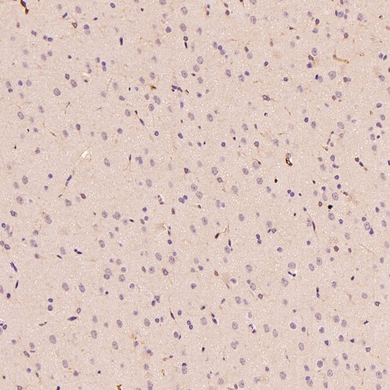


Comprehensive Human Protein Atlas IHC scoring per tissue (reliability: Enhanced — Medium consistency between antibody staining and RNA expression data. Secreted protein, tissue location of RNA and protein is expected to differ.). Rows are taken directly from the HPA tissue chart — click any row's HPA link to view the source.
| Tissue | Cell type | Level | Evidence | Source |
|---|---|---|---|---|
| Bone marrow | Hematopoietic cells | High | Protein (IHC) | HPA → |
| Duodenum | Paneth cells | High | Protein (IHC) | HPA → |
| Lung | Macrophages | High | Protein (IHC) | HPA → |
| Small intestine | Paneth cells | High | Protein (IHC) | HPA → |
| Appendix | Non-germinal center cells | Medium | Protein (IHC) | HPA → |
| Tissue | Cell type | Level | Evidence | Source |
|---|---|---|---|---|
| Adipose tissue | Adipocytes | Not detected | Protein (IHC) | HPA → |
| Adrenal gland | Glandular cells | Not detected | Protein (IHC) | HPA → |
| Breast | Adipocytes | Not detected | Protein (IHC) | HPA → |
| Caudate | Glial cells | Not detected | Protein (IHC) | HPA → |
| Cerebellum | Cells in granular layer | Not detected | Protein (IHC) | HPA → |
Troubleshoot LYZ staining in paraffin sections by checking retrieval, tissue handling, cell identity, and the distribution of this secreted protein (UniProt P61626; HPA tissue IHC).
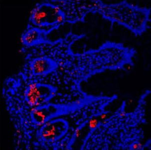
Anti-LYZ antibodies have IHC images from paraffin sections of human intestinal cancer and mouse kidney (catalog IHC captions), plus IF images from human and mouse samples (catalog IF captions).
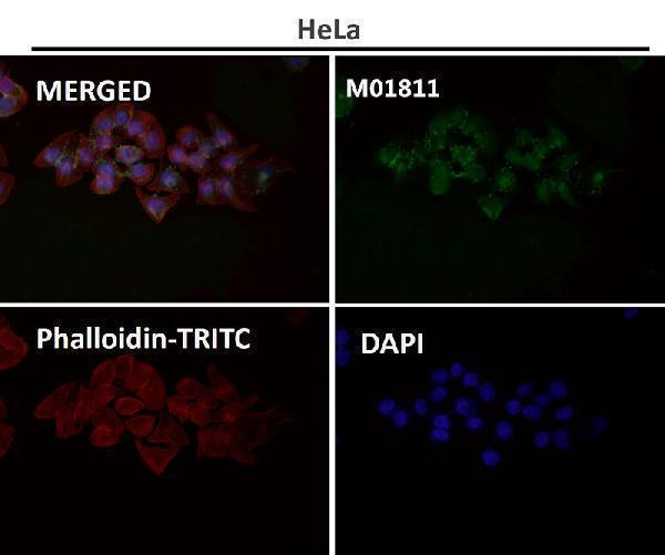
PB9663 has an IHC image from paraffin-embedded human intestinal cancer tissue and IF images from human ileum, human colon organoids, mouse ileum, and mouse ileum organoids (PB9663 image captions). M01811 has an IHC image from paraffin-embedded mouse kidney and an IF image from mouse colon; its application list also includes ICC (M01811 image captions; catalog applications).
Which to pick: For human tissue IHC, PB9663 has a paraffin-section image using 1 μg/ml antibody; the caption does not report the fixative (PB9663 IHC caption). For IF/ICC, M01811 is the rabbit monoclonal option with both applications listed, while PB9663 has IF images from human and mouse samples (M01811 catalog applications and clone; PB9663 IF captions). For work across human, mouse, and rat, both list those species as reactive, but their own IHC figure captions document only human intestinal cancer for PB9663 and mouse kidney for M01811; neither caption reports the fixative (catalog reactivity; PB9663 and M01811 IHC captions).