Click image to see more details
-

-
-
-
-
+11
Product Info Summary
| SKU: | M01811 |
|---|---|
| Size: | 100 μl |
| Reactive Species: | Human, Mouse, Rat |
| Host: | Rabbit |
| Application: | IP, IF, IHC, ICC, WB |
Customers Who Bought This Also Bought

Product info
Product Name
Anti-Lysozyme LYZ Rabbit Monoclonal Antibody
SKU/Catalog Number
M01811
BM4383 is an alternative SKU for this antibody, used in previous lots.
Size
100 μl
Form
Liquid
Description
Boster Bio Anti-Lysozyme LYZ Rabbit Monoclonal Antibody catalog # M01811. Tested in WB, IHC, ICC/IF, IP applications. This antibody reacts with Human, Mouse, Rat.
Storage & Handling
Store at -20°C for one year. For short term storage and frequent use, store at 4°C for up to one month. Avoid repeated freeze-thaw cycles.
Cite This Product
Anti-Lysozyme LYZ Rabbit Monoclonal Antibody (Boster Biological Technology, Pleasanton CA, USA, Catalog # M01811)
Host
Rabbit
Contents
Rabbit IgG in stabilizing components, phosphate buffered saline, pH 7.4, 150mM NaCl, 0.02% sodium azide and 50% glycerol.
*This antibody is supplied in a stabilized formulation.
Compatibility with conjugation reactions depends on the chemistry of the conjugation method used.
For conjugation methods that are not compatible with the stabilizing components present in this formulation, a carrier-free antibody format is required.
Clonality
Monoclonal
Clone Number
EBO-12
Isotype
Rabbit IgG
Immunogen
A synthesized peptide derived from human Lysozyme
Reactive Species
M01811 is reactive to LYZ in Human, Mouse, Rat
Observed Molecular Weight
15 kDa
Calculated molecular weight
16.5 kDa
Antibody Validation
Boster validates all antibodies on WB, IHC, ICC, Immunofluorescence, and ELISA with known positive control and negative samples to ensure specificity and high affinity, including thorough antibody incubations.
Application & Images
Applications
M01811 is guaranteed for IP, IF, IHC, ICC, WB Boster Guarantee
Recommend Dilution
WB 1:1000-5000
IHC 1:50-200
ICC/IF 1:50-200
IP 1:20
Tested application
Suggested blocking solution with 5% non-fat milk or BSA; (*)Recommended protein loading: 20-40 µg per lane
Use TE buffer pH 9.0 for antigen retrieval; (*) citrate buffer pH 6.0 is an alternative.
Validation Images & Assay Conditions
Click image to see more details
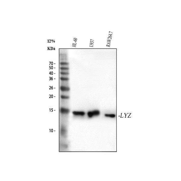
Western blot analysis of Lysozyme using anti-Lysozyme antibody (M01811).
Electrophoresis was performed on a 5-20% SDS-PAGE gel at 70V (Stacking gel) / 90V (Resolving gel) for 2-3 hours. The sample well of each lane was loaded with 30 ug of sample under reducing conditions.
Lane 1: human HL-60 whole cell lysates,
Lane 2: human U937 whole cell lysates,
Lane 3: mouse RAW264.7 whole cell lysates.
After electrophoresis, proteins were transferred to a nitrocellulose membrane at 150 mA for 50-90 minutes. Blocked the membrane with 5% non-fat milk/TBS for 1.5 hour at RT. The membrane was incubated with rabbit anti-Lysozyme antigen affinity purified monoclonal antibody (Catalog # M01811) at 1:5000 overnight at 4°C, then washed with TBS-0.1%Tween 3 times with 5 minutes each and probed with a goat anti-rabbit IgG-HRP secondary antibody at a dilution of 1:5000 for 1.5 hour at RT. The signal is developed using an Enhanced Chemiluminescent detection (ECL) kit (Catalog # EK1002) with Tanon 5200 system. A specific band was detected for Lysozyme at approximately 15 kDa. The expected band size for Lysozyme is at 17 kDa.

Click image to see more details
IF analysis of Lysozyme using anti-Lysozyme antibody (M01811). Lysozyme was detected in a paraffin-embedded section of mouse colon tissue. Heat mediated antigen retrieval was performed in EDTA buffer (pH 8.0, epitope retrieval solution). The tissue section was blocked with 10% goat serum. The tissue section was then incubated at 1:50 rabbit anti-Lysozyme Antibody (M01811) overnight at 4°C. DyLight®488 Conjugated Goat Anti-Rabbit IgG (BA1127) was used as secondary antibody at 1:500 dilution and incubated for 30 minutes at 37°C. The section was counterstained with DAPI. Visualize using a fluorescence microscope and filter sets appropriate for the label used.

Click image to see more details
MG53 attenuates intestinal injury in mouse models. a Serum IL-6 and IL-18 levels of MG53-TG and their WT littermates at the indicated time points following DSS treatment. n = 3 for each group. Overexpression of MG53 attenuated DSS-induced IBD symptoms, including disease activity index (DAI; b ), body weight change ( c ), disruption of intestinal structure as demonstrated by H&E staining of the jejunum, ileum, and colon ( d ; scale bar, 100 μm), and shortening of the colon as evidenced by the representative images ( e ) and statistic results of colon length ( f ) in MG53-TG and WT mice. n = 7 for each group. g , h Depletion of MG53 exacerbated DSS-induced IBD symptoms. The DAI ( g ) and body weight change ( h ) of MG53-KO and the WT littermates at the indicated time points after DSS challenge. n = 5 for each group. Immunofluorescence staining of Ki-67 (red) to indicate the TA zone and the statistic results of its length in MG53-TG ( i , n = 20 for each group) or MG53-KO ( j , n = 13 for each group) as compared with their corresponding WT littermates. The nuclei were indicated by DAPI staining (blue); scale bar, 100 μm. Normal distribution was confirmed by Shapiro–Wilk test. Data were analyzed using two tailed paired t test ( a – c , f – j ). All data were presented as mean ± s.e.m. * p < 0.05, ** p < 0.01, and *** p < 0.001 as compared with the corresponding controls Full size image
Index in PubMed under a CC BY license. PMID: 40494851

Click image to see more details
PPARα mediates the effect of MG53 in promoting asymmetric division of Lgr5 + cells. Representative images ( a ) and statistic results ( b , n = 3 for each group) of asymmetric division of Lgr5 + cells with or without GW6471 (10 μM, 24 h) treatment. Lgr5 + cells were in green; all the cells were visualized by tubulin (red). Scale bar, 10 μm. c FACS analysis of Lgr5 + , Atoh1 + , and Notch1 + cells in the Lgr5 and MG53;Lgr5 organoids treated with GW6471 or vehicle. d Immunofluorescence staining of Ki-67 (red) and PPARα (green) and statistic results of PPARα positive cells ( n = 3 for each group) and the length of TA zone ( n = 18 for each group) in mice treated GW6471 or vehicle. Nuclei were stained with DAPI (blue); scale bar, 100 μm. Representative images and statistic results of PAS staining of goblet cells in the ileum ( e ; n = 10 for each group; TG, MG53-TG; scale bar, 50 μm) and colon ( f ; n = 10 for each group; scale bar, 100 μm). Normal distribution was confirmed by Shapiro–Wilk test. Data were analyzed using one-way ANOVA with Tukey post hoc test ( b , d , e , and f ) and were presented as mean ± s.e.m. ns not significant and *** p < 0.001 as compared with the corresponding controls Full size image
Index in PubMed under a CC BY license. PMID: 40494851

Click image to see more details
MG53 overexpression promotes secretory lineage commitment. a Immunofluorescence staining of BrdU (red) and EdU (yellow) in the ileum of MG53-TG and their WT littermates subjected to DSS treatment and administered BrdU 48 h and EdU 24 h before euthanasia. n = 10 for each group. Scale bar, 100 μm. b UMAP of scRNA-seq of Epcam + CD45 − intestinal epithelial cells collected from MG53-TG and their WT littermates on Day 7 of DSS treatment, n = 5 for each group. c Proportions of stem, secretory, and absorptive cells in general and each particular cell clusters. d Representative images and statistic results of PAS staining of goblet cells of MG53-TG and WT mice in the colon. n = 10 for each group. Scale bar, 100 μm. e Representative images of intestinal organoids derived from MG53;Lgr5 mice and their Lgr5 littermates at the indicated time points. f Relative mRNA levels of the marker genes in the transcriptome analysis of the crypt organoids. n = 4 for each group. g Representative images of colonic organoids derived from MG53;Lgr5 mice and their Lgr5 littermates at the indicated time points. h Representative FACS profiles and statistic results of Lgr5 + cells in the colonic organoids derived from MG53;Lgr5 and the control Lgr5 mice. n = 6 for each group. Normal distribution was confirmed by Shapiro–Wilk test. Data were analyzed using two tailed paired t test ( a , d , f , and h ), and were presented as mean ± s.e.m. * p < 0.05, ** p < 0.01, and *** p < 0.001 as compared with the corresponding controls Full size image
Index in PubMed under a CC BY license. PMID: 40494851

Click image to see more details
PPAR signaling is enhanced by MG53 overexpression. a Trajectory analysis of Epcam + CD45 − intestinal epithelial cells Using Monocle 2. b KEGG pathway enrichment analysis of the differentially expressed genes between the secretory and absorptive branches. GSEA results ( c ) and the heatmap of differentially expressed genes ( d ) related to fatty acid oxidation and PPAR signaling derived from the bulk RNAseq data of the intestinal organoids from MG53-TG and WT mice. n = 4 for each group. Representative images ( e ) and statistic results of signal intensity of the immunofluorescence staining of MG53 (green) and PPARα (red) determined by Aperio Scanscope scanner and Halo software ( f ), as well as the correlation of the levels of these two proteins ( g ) in the human small intestine of normal subjects ( n = 5) and patients with intestinal inflammation ( n = 8). Nuclei were stained with DAPI (blue); scale bars as indicated. Representative images ( h ) and statistic results of signal intensity of the immunofluorescence staining of MG53 (green) and PPARα (red) determined by Aperio Scanscope scanner and Halo software ( i ), as well as the correlation of the levels of these two proteins ( j ) in the human colon of normal subjects ( n = 4) and patients with intestinal inflammation ( n = 12). Normal distribution was confirmed by Shapiro–Wilk test. Data were analyzed using Mann–Whitney U test ( f , i ) and Pearson correlation analysis ( g , j ). Data were presented as mean ± s.e.m. * p < 0.05 as compared with the corresponding controls Full size image
Index in PubMed under a CC BY license. PMID: 40494851

Click image to see more details
Activation of PPARα signaling is required for MG53-induced alleviation of intestinal injury. Body weight change and DAI ( a ) as well as the representative images and statistic results of the intestinal length ( b ) of ISC-MG53-TG and the control MG53 tg-fl mice. n = 7 for each group. Body weight change and DAI ( c ) as well as the representative images and statistic results of the intestinal length ( d ) of the Lgr5 mice with ISC-specific inhibition of MG53 expression via infection with adeno-associated virus with inducible expression of shRNA targeting MG53 (AAV-shMG53) or the control mice infected with control shRNA (AAV-shCON). n = 6 for each group. Body weight change and DAI ( e ) as well as the intestinal length ( f ) of ISC-Ppara-KO mice with or without ISC-specific MG53 overexpression via injecting AAV-MG53 or AAV-CON. n = 8 for each group. Body weight change and DAI ( g ) as well as the intestinal length ( h ) of MG53-KO and control WT mice treated with PPARα agonist fenofibrate or vehicle following DSS treatment. n = 8 for each group. Normal distribution was confirmed by Shapiro–Wilk test. Data were analyzed using two-tailed paired t test ( a – e , g ), Mann–Whitney U test ( f , h ), and were presented as mean ± s.e.m. ns not significant, * p < 0.05 and ** p < 0.01 as compared with the corresponding controls Full size image
Index in PubMed under a CC BY license. PMID: 40494851

Click image to see more details
Enhanced PPARα activation by palmitoleic acid contributes to MG53-mediated amelioration of intestinal injury. a Heatmap of free fatty acids (FFA) in MG53-TG and WT intestinal tissues at the indicated time points during DSS treatment. n = 3 for each group. b Changes in the intestinal content of FFA in the MG53-TG and their WT littermates at day 7 after DSS treatment. n = 3 for each group. c Relative change in POA concentrations in the serum of IBD patients in remission ( n = 11) or flare ( n = 26). d Luciferase reporter activity driven by the PPARα-responsive element in the presence of different FFA in HEK293 cells. n = 3 for each group. Body weight change and DAI ( e ) as well as the colon length ( f ) of MG53-TG (TG) and their WT littermates challenged with DSS with or without POA treatment. n = 12 for each group. g Immunofluorescence staining of Ki-67 (red) and statistic results of the length of TA zone of MG53-TG and their WT littermates with or without POA treatment. n = 15 for each group. The nuclei were indicated by DAPI staining (blue); scale bar, 100 μm. h Representative images and statistic results of PAS staining of goblet cells in the colon after POA treatment. n = 10 for each group. Scale bar, 100 μm. Normal distribution was confirmed by Shapiro–Wilk test. Data were analyzed using one-way ANOVA with Tukey post hoc test ( c , e ), two tailed t test ( d , f – h ) and were presented as mean ± s.e.m. ns not significant, * p < 0.05, ** p < 0.01, and *** p < 0.001 as compared with the corresponding controls Full size image
Index in PubMed under a CC BY license. PMID: 40494851

Click image to see more details
Immunohistochemical analysis of paraffin-embedded mouse kidney, using Lysozyme Antibody.

Click image to see more details
Immunohistochemical analysis of paraffin-embedded Rat cerebral cortex, using the Antibody at 1:2000 dilution.

Click image to see more details
Immunohistochemical analysis of paraffin-embedded Human thyroid cancer, using the Antibody at 1:4000 dilution.

Click image to see more details
Immunohistochemical analysis of paraffin-embedded Human placenta, using the Antibody at 1:4000 dilution.

Click image to see more details
Immunohistochemical analysis of paraffin-embedded Mouse skin, using the Antibody at 1:2000 dilution.

Click image to see more details
Immunohistochemical analysis of paraffin-embedded Mouse heart, using the Antibody at 1:2000 dilution.

Click image to see more details
Immunofluorescent analysis using the Antibody at 1:50 dilution.
Specific Publications For Anti-Lysozyme LYZ Rabbit Monoclonal Antibody (M01811)
Loading publications
Recommended Resources
Here are featured tools and databases that you might find useful.
- Boster's Pathways Library
- Protein Databases
- Bioscience Research Protocol Resources
- Data Processing & Analysis Software
- Photo Editing Software
- Scientific Literature Resources
- Research Paper Management Tools
- Molecular Biology Software
- Primer Design Tools
- Bioinformatics Tools
- Phylogenetic Tree Analysis
Customer Reviews
Have you used Anti-Lysozyme LYZ Rabbit Monoclonal Antibody?
Share your experimental results or join a short interview to earn up to $1,000 in product credits or other rewards.
0 Reviews For Anti-Lysozyme LYZ Rabbit Monoclonal Antibody
Customer Q&As
Have a question?
Find answers in Q&As, reviews.
Can't find your answer?
Submit your question
16 Customer Q&As for Anti-Lysozyme LYZ Rabbit Monoclonal Antibody
Question
Does M01811 anti-Lysozyme Rabbit Monoclonal antibody work on parafin embedded sections? If so, which fixation method do you recommend we use (PFA, paraformaldehyde, other)?
Verified Customer
Verified customer
Asked: 2020-02-18
Answer
You can see on the product datasheet, M01811 anti-Lysozyme Rabbit Monoclonal antibody as been tested on ICC. It is best to use PFA for fixation because it has better tissue penetration ability. PFA needs to be prepared fresh before use. Long term stored PFA turns into formalin, as the PFA molecules congregate and become formalin.
Boster Scientific Support
Answered: 2020-02-18
Question
We have observed staining in mouse colon. Any tips? Is anti-Lysozyme Rabbit Monoclonal antibody supposed to stain colon positively?
Verified Customer
Verified customer
Asked: 2020-01-08
Answer
From literature colon does express LYZ. From Uniprot.org, LYZ is expressed in nasal cavity mucosa, colon, urine, milk, tear, among other tissues. Regarding which tissues have LYZ expression, here are a few articles citing expression in various tissues:
Colon, Pubmed ID: 15489334
Milk, Pubmed ID: 5168859, 11946553
Tear, Pubmed ID: 25946035
Urine, Pubmed ID: 5284421, 11946554
Boster Scientific Support
Answered: 2020-01-08
Question
Our team were happy with the WB result of your anti-Lysozyme Rabbit Monoclonal antibody. However we have been able to see positive staining in nasal cavity mucosa secreted. using this antibody. Is that expected? Could you tell me where is LYZ supposed to be expressed?
Verified Customer
Verified customer
Asked: 2019-12-02
Answer
According to literature, nasal cavity mucosa does express LYZ. Generally LYZ expresses in secreted. Regarding which tissues have LYZ expression, here are a few articles citing expression in various tissues:
Colon, Pubmed ID: 15489334
Milk, Pubmed ID: 5168859, 11946553
Tear, Pubmed ID: 25946035
Urine, Pubmed ID: 5284421, 11946554
Boster Scientific Support
Answered: 2019-12-02
Question
We have tried in the past anti-Lysozyme Rabbit Monoclonal antibody for IP on tear in the past. I am using human, and We want to use the antibody for IHC next. My lab would like examining tear as well as milk in our next experiment. Do you have any suggestion on which antibody would work the best for IHC?
Verified Customer
Verified customer
Asked: 2019-11-22
Answer
I looked at the website and datasheets of our anti-Lysozyme Rabbit Monoclonal antibody and it seems that M01811 has been validated on human in both IP and IHC. Thus M01811 should work for your application. Our Boster satisfaction guarantee will cover this product for IHC in human even if the specific tissue type has not been validated. We do have a comprehensive range of products for IHC detection and you can check out our website bosterbio.com to find out more information about them.
Boster Scientific Support
Answered: 2019-11-22
Question
I was wanting to use to test anti-Lysozyme Rabbit Monoclonal antibody M01811 on mouse colon for research purposes, then I may be interested in using anti-Lysozyme Rabbit Monoclonal antibody M01811 for diagnostic purposes as well. Is the antibody suitable for diagnostic purposes?
Verified Customer
Verified customer
Asked: 2019-10-28
Answer
The products we sell, including anti-Lysozyme Rabbit Monoclonal antibody M01811, are only intended for research use. They would not be suitable for use in diagnostic work. If you have the means to develop a product into diagnostic use, and are interested in collaborating with us and develop our product into an IVD product, please contact us for more discussions.
Boster Scientific Support
Answered: 2019-10-28
Question
Do you have a BSA free version of anti-Lysozyme Rabbit Monoclonal antibody M01811 available?
Verified Customer
Verified customer
Asked: 2019-07-25
Answer
Thanks for your recent telephone inquiry. I can confirm that some lots of this anti-Lysozyme Rabbit Monoclonal antibody M01811 are BSA free. For now, these lots are available and we can make a BSA free formula for you free of charge. It will take 3 extra days to prepare. If you require this antibody BSA free again in future, please do not hesitate to contact me and I will be pleased to check which lots we have in stock that are BSA free.
Boster Scientific Support
Answered: 2019-07-25
Question
See attached the WB image, lot number and protocol we used for colon using anti-Lysozyme Rabbit Monoclonal antibody M01811. Please let me know if you require anything else.
Verified Customer
Verified customer
Asked: 2019-07-16
Answer
Thank you very much for the data. Our lab team are working to resolve this as quickly as possible, and we appreciate your patience and understanding! You have provided everything we needed. Please let me know if there is anything you need in the meantime.
Boster Scientific Support
Answered: 2019-07-16
Question
Is a blocking peptide available for product anti-Lysozyme Rabbit Monoclonal antibody (M01811)?
Verified Customer
Verified customer
Asked: 2019-06-07
Answer
We do provide the blocking peptide for product anti-Lysozyme Rabbit Monoclonal antibody (M01811). If you would like to place an order for it please contact support@bosterbio.com and make a special request.
Boster Scientific Support
Answered: 2019-06-07
Question
Can you help my question with product M01811, anti-Lysozyme Rabbit Monoclonal antibody. I was wondering if it would be possible to conjugate this antibody with biotin. I would need it to be without BSA or sodium azide. I am planning on using a buffer exchange of sodium azide with PBS only. Would there be problems for me to conjugate the antibody and store it in -20 degrees in small aliquots?
Verified Customer
Verified customer
Asked: 2019-05-31
Answer
We do not recommend storing this antibody with PBS buffer only in -20 degrees. If you want to store it in -20 degrees it is best to add some cryoprotectant like glycerol. If you want carrier free M01811 anti-Lysozyme Rabbit Monoclonal antibody, we can provide it to you in a special formula with trehalose and/or glycerol. These molecules will not interfere with conjugation chemistry and provide a good level of protection for the antibody from degradation. Please be sure to specify this in your purchase order.
Boster Scientific Support
Answered: 2019-05-31
Question
Is this M01811 anti-Lysozyme Rabbit Monoclonal antibody reactive to the isotypes of LYZ?
Verified Customer
Verified customer
Asked: 2019-05-24
Answer
The immunogen of M01811 anti-Lysozyme Rabbit Monoclonal antibody is A synthesized peptide derived from human Lysozyme . Could you tell me which isotype you are interested in so I can help see if the immunogen is part of this isotype?
Boster Scientific Support
Answered: 2019-05-24
Question
Would anti-Lysozyme Rabbit Monoclonal antibody M01811 work for ICC with colon?
A. Krishna
Verified customer
Asked: 2017-10-26
Answer
According to the expression profile of colon, LYZ is highly expressed in colon. So, it is likely that anti-Lysozyme Rabbit Monoclonal antibody M01811 will work for ICC with colon.
Boster Scientific Support
Answered: 2017-10-26
Question
I was wanting to use your anti-Lysozyme Rabbit Monoclonal antibody for ICC for mouse colon on frozen tissues, but I want to know if it has been validated for this particular application. Has this antibody been validated and is this antibody a good choice for mouse colon identification?
Verified Customer
Verified customer
Asked: 2017-10-11
Answer
You can see on the product datasheet, M01811 anti-Lysozyme Rabbit Monoclonal antibody has been validated for IP, IF, IHC, ICC, WB on human, mouse tissues. We have an innovator award program that if you test this antibody and show it works in mouse colon in IHC-frozen, you can get your next antibody for free.
Boster Scientific Support
Answered: 2017-10-11
Question
I see that the anti-Lysozyme Rabbit Monoclonal antibody M01811 works with ICC, what is the protocol used to produce the result images on the product page?
W. Miller
Verified customer
Asked: 2017-10-10
Answer
You can find protocols for ICC on the "support/technical resources" section of our navigation menu. If you have any further questions, please send an email to support@bosterbio.com
Boster Scientific Support
Answered: 2017-10-10
Question
Our lab want to know about using your anti-Lysozyme Rabbit Monoclonal antibody for cellular protein metabolic process studies. Has this antibody been tested with western blotting on mouse kidney? We would like to see some validation images before ordering.
L. Krishna
Verified customer
Asked: 2014-07-29
Answer
I appreciate your inquiry. This M01811 anti-Lysozyme Rabbit Monoclonal antibody is tested on k562 cell lysate, mouse kidney. It is guaranteed to work for IP, IF, IHC, ICC, WB in human, mouse. Our Boster guarantee will cover your intended experiment even if the sample type has not been be directly tested.
Boster Scientific Support
Answered: 2014-07-29
Question
We appreciate helping with my inquiry over the phone. Here are the WB image, lot number and protocol we used for colon using anti-Lysozyme Rabbit Monoclonal antibody M01811. Let me know if you need anything else.
M. Carter
Verified customer
Asked: 2014-03-03
Answer
I appreciate the data. You have provided everything we needed. Our lab team are working to resolve your inquiry as quickly as possible, and we appreciate your patience and understanding! Please let me know if there is anything you need in the meantime.
Boster Scientific Support
Answered: 2014-03-03
Question
We are currently using anti-Lysozyme Rabbit Monoclonal antibody M01811 for human tissue, and we are happy with the ICC results. The species of reactivity given in the datasheet says human, mouse. Is it likely that the antibody can work on monkey tissues as well?
C. Bhatt
Verified customer
Asked: 2013-08-09
Answer
The anti-Lysozyme Rabbit Monoclonal antibody (M01811) has not been validated for cross reactivity specifically with monkey tissues, but there is a good chance of cross reactivity. We have an innovator award program that if you test this antibody and show it works in monkey you can get your next antibody for free. Please contact me if I can help you with anything.
Boster Scientific Support
Answered: 2013-08-09
