This website uses cookies to ensure you get the best experience on our website.


- Table of Contents
Plan ME1 chromogenic IHC in paraffin sections using cytoplasmic staining as the expected pattern (HPA tissue IHC). Adrenal glandular cells provide a strong staining reference (HPA tissue IHC); the catalog antibody M03449 has an IHC dilution of 2–5 μg/mL (datasheet M03449).

Expected localisation, validated protocols, controls and antibodies — the at-a-glance facts below, then the full design guide.
| Expected localisation | Cytoplasmic staining in several tissues (HPA tissue IHC) | |
| Staining pattern | Cytoplasmic staining, strongest in adrenal glandular cells (HPA tissue IHC) | |
| Antigen retrieval | EDTA pH 8.0 HIER, heat-mediated (datasheet M03449) | |
| Positive control | Adrenal gland+4 more · see all | |
| Negative control | Appendix+4 more · see all |
| Fixation | Keep fixation conditions consistent across sections. (standard IHC practice; not target-specific) | |
| Caveat | Staining has medium agreement with RNA data (HPA tissue IHC) | |
| Regulation | No stimulus-linked regulation specified (UniProt) | |
| Isoform / epitope | Two isoforms; check antibody epitope coverage (UniProt) |
Compare the catalog antibody’s IHC-P protocol (datasheet M03449) with four published ME1 immunohistochemistry protocols (PMC5989842; PMC11850758; PMC6628973; PMC3464285).
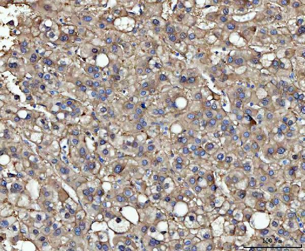
| Sample | Paraffin-embedded human liver cancer tissue; fixative not specified (datasheet M03449) |
| Fixation | Image fixative and duration unreported (datasheet M03449); verify before use. |
| Sectioning | 4–5 µm sections on charged slides (standard) |
| Deparaffinisation | Xylene, graded ethanol series to water (standard) |
| Antigen retrieval | Heat retrieval: EDTA pH 8.0 (datasheet M03449); 20 min, 95–100 °C (standard) |
| Peroxidase block | 3% H2O2, 10 min, room temperature (standard) |
| Blocking | 10% goat serum (datasheet M03449) |
| Primary antibody | Mouse monoclonal (clone 5E5F7) anti-ME1, 2-5 μg/ml (datasheet M03449) |
| Primary incubation | Overnight at 4 °C (datasheet M03449) |
| Detection | HRP-conjugated secondary, DAB chromogen (datasheet M03449) |
| Counterstain | Hematoxylin, blue, dehydrate and mount (standard) |
| Expected result | ME1-positive staining in glandular cells of adrenal gland (HPA tissue IHC: High). HPA tissue profile: Cytoplasmic expression in several tissues, most abundant in adrenal gland. No signal in the no-primary control. |
ME1 is a cytoplasmic enzyme with no transmembrane segment (UniProt P48163 localization and topology). In paraffin-section IHC, expect cytoplasmic staining in adrenal glandular cells and late spermatids, with weaker staining in several other listed cell types (HPA tissue IHC). HPA rates the tissue profile Enhanced, but describes only medium agreement with RNA data and says external verification is pending (HPA tissue IHC reliability).
| Strong cytoplasmic stain in adrenal glandular cells or elongated/late spermatids. | This matches HPA's High staining in those cell types and UniProt's cytoplasmic localization (HPA tissue IHC; UniProt P48163). Compare cells within the section: HPA levels describe observed cell types, not a uniform intensity across an entire tissue (HPA tissue IHC). |
| Predominantly nuclear or sharply membrane-bound signal replaces cytoplasmic staining. | Treat this as a localization discrepancy and check controls and morphology (UniProt P48163 localization; HPA tissue IHC). HPA reports nucleoplasm and plasma membrane in ICC-IF as uncertain, while its cytosol assignment is supported; the uncertain IF findings do not establish the expected IHC pattern (HPA subcellular ICC-IF). |
| Strong signal appears in a cell type HPA lists as Not detected, such as appendix glandular cells. | Consider cross-reactivity or endogenous chromogenic activity, then verify with assay controls (HPA tissue IHC; general IHC practice). Not detected refers to the named cell type under HPA's conditions; it does not certify that every cell in the organ is negative (HPA tissue IHC). |
| Brown stain spreads across tissue and blank areas without cell boundaries. | Diffuse background prevents a reliable cytoplasmic call (UniProt P48163 localization; general IHC practice). Examine the no-primary control and review blocking, washes, chromogen development and counterstain before assigning ME1 positivity (general IHC practice). |
| No visible signal in adrenal glandular cells on a putative positive section. | This conflicts with HPA's High result for that cell type (HPA tissue IHC). Check tissue identity, section quality, antibody and detection controls, and the catalog IHC-P instructions before calling the sample biologically negative (general IHC practice). |
| Cell-type baseline in the selected section | Adrenal glandular cells and late spermatids are High, whereas bone-marrow hematopoietic cells, Purkinje cells, cardiomyocytes and listed neuronal cells are Medium (HPA tissue IHC). Select a comparator by cell type and reported level; a negative call for appendix glandular cells or breast adipocytes applies only to those listed cells (HPA tissue IHC). |
| Strength of IHC evidence | HPA006493 is IHC Enhanced, and HPA describes the tissue profile as Enhanced with medium RNA agreement pending external verification (HPA antibodies; HPA tissue IHC). Use that profile as an observed benchmark, while checking the catalog antibody's own IHC-P validation when interpreting a new assay (general IHC practice). |
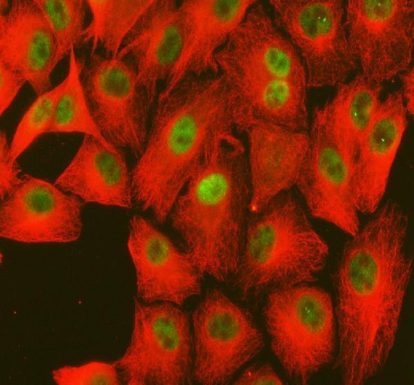
| IF/ICC Q: Should nuclear or plasma-membrane staining set the IHC expectation? | No. HPA marks those ICC-IF locations uncertain and cytosol supported; ICC validation for HPA006493 is Uncertain (HPA subcellular ICC-IF; HPA antibodies). UniProt places ME1 in the cytoplasm and lists no transmembrane segment (UniProt P48163 localization and topology). Interpret IF/ICC on its own guide page. |
| Situation | Likely cause | Next action |
|---|---|---|
| Adrenal glandular cells have no detectable chromogen. | A failed staining or detection step is possible; HPA records this cell type as High (HPA tissue IHC; general IHC practice). | Confirm the cell type and section quality, run a positive-control section, and check the catalog IHC-P protocol and detection reagents (general IHC practice). |
| Signal is chiefly nuclear or outlines cell membranes. | That distribution conflicts with UniProt's cytoplasmic localization; HPA's corresponding ICC-IF assignments are uncertain (UniProt P48163; HPA subcellular ICC-IF). | Check the no-primary control, chromogen deposits and tissue morphology; require reproducible cytoplasmic signal before scoring IHC positivity (general IHC practice). |
| Appendix glandular cells stain strongly. | HPA lists that specific cell type as Not detected; cross-reactivity or endogenous detection activity is possible, not proven (HPA tissue IHC; general IHC practice). | Compare a no-primary control, verify cell identity and review endogenous enzyme blocking appropriate to the detection chemistry (general IHC practice). |
| Every compartment and tissue area has a brown haze. | Nonspecific background or excessive chromogen development can obscure cell-level localization (general IHC practice). | Inspect the no-primary control; review blocking, antibody conditions, washes and development time against the catalog IHC-P protocol (general IHC practice). |
| A Medium HPA cell type looks weaker than adrenal glandular cells. | This may fit the reported relative staining levels rather than indicate assay failure (HPA tissue IHC). | Score the specified cells and compare them with an appropriate positive-control section under the same detection conditions (HPA tissue IHC; general IHC practice). |
| A whole organ is called ME1-negative because one listed cell type is Not detected. | HPA's negative entries identify particular cells, while UniProt reports expression across tested tissues (HPA tissue IHC; UniProt P48163 tissue specificity). | Record cell identity and compartment in the score; limit the negative conclusion to the examined cell population and assay conditions (general IHC practice). |
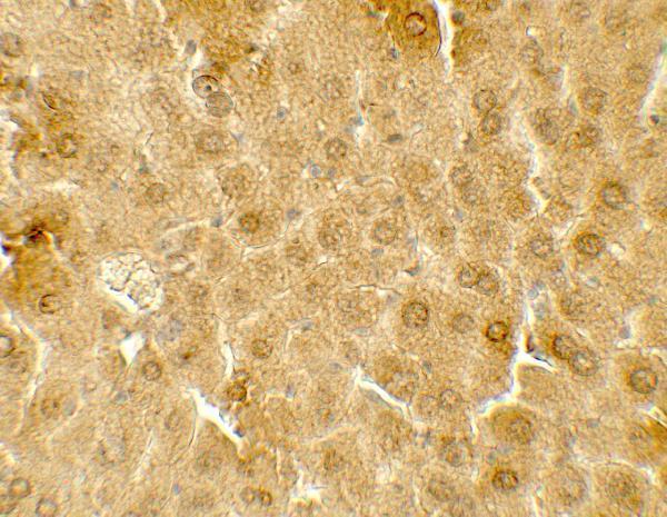
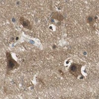


Comprehensive Human Protein Atlas IHC scoring per tissue (reliability: Enhanced — Medium consistency between antibody staining and RNA expression data. Pending external verification.). Rows are taken directly from the HPA tissue chart — click any row's HPA link to view the source.
| Tissue | Cell type | Level | Evidence | Source |
|---|---|---|---|---|
| Adrenal gland | Glandular cells | High | Protein (IHC) | HPA → |
| Testis | Elongated or late spermatids | High | Protein (IHC) | HPA → |
| Bone marrow | Hematopoietic cells | Medium | Protein (IHC) | HPA → |
| Cerebellum | Purkinje cells | Medium | Protein (IHC) | HPA → |
| Cerebral cortex | Neuronal cells | Medium | Protein (IHC) | HPA → |
| Tissue | Cell type | Level | Evidence | Source |
|---|---|---|---|---|
| Appendix | Glandular cells | Not detected | Protein (IHC) | HPA → |
| Breast | Adipocytes | Not detected | Protein (IHC) | HPA → |
| Bronchus | Respiratory epithelial cells | Not detected | Protein (IHC) | HPA → |
| Colon | Endothelial cells | Not detected | Protein (IHC) | HPA → |
| Endometrium | Cells in endometrial stroma | Not detected | Protein (IHC) | HPA → |
Troubleshoot ME1 staining in paraffin sections by checking retrieval, background and cellular localisation before comparing signal intensity across samples.
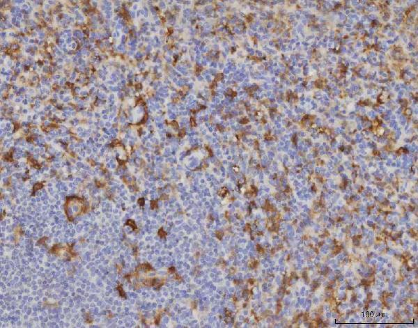
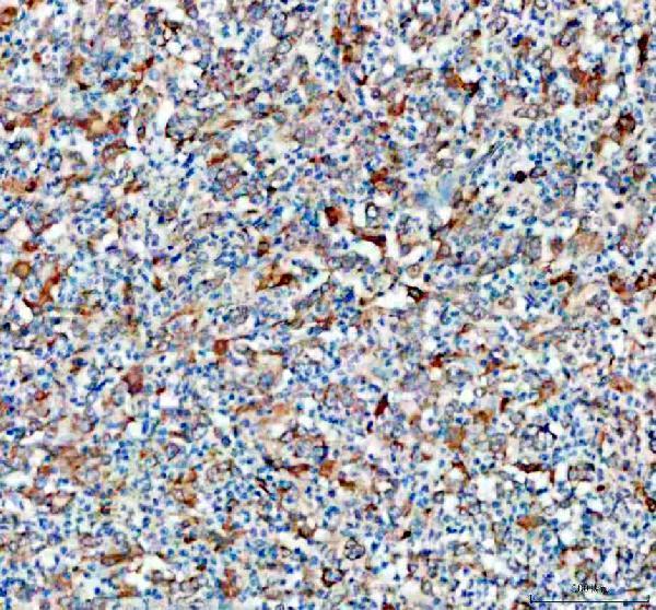
Anti-ME1 IHC images cover human liver cancer, spleen, and stomach cancer, rat testis, and mouse liver (catalog IHC captions); IF images cover A549 cells and human gastric cancer sections (A03449-3 IF captions).
M03449 shows paraffin-section IHC in human liver cancer, human spleen, and rat testis (M03449 IHC captions); A03449-3 shows paraffin-section IHC in human stomach cancer (A03449-3 IHC caption). A03449 shows IHC in mouse liver (A03449 IHC caption), while A03449-3 also shows IF in A549 cells and paraffin-embedded human gastric cancer tissue (A03449-3 IF captions).
Which to pick: For human or rat paraffin-section IHC, M03449 is a mouse monoclonal (clone 5E5F7; M03449 catalog) with images from both species (M03449 IHC captions); A03449-3 is a rabbit option with a human stomach cancer paraffin-section image (A03449-3 IHC caption). For IF/ICC, choose A03449-3, which lists both applications and has IF images from A549 cells and human gastric cancer sections (A03449-3 applications and IF captions). For cross-species planning, all three list human, mouse, and rat reactivity (catalog reactivity), while A03449 provides a mouse liver IHC image at 5 μg/mL (A03449 IHC caption); fixatives are unreported in the IHC captions (catalog IHC captions).