Click image to see more details
-

-
-
-
-
+2
Product Info Summary
| SKU: | A03449-3 |
|---|---|
| Size: | 100 μg/vial |
| Reactive Species: | Human, Mouse, Rat |
| Host: | Rabbit |
| Application: | ELISA, Flow Cytometry, IF, IHC, ICC, WB |
Customers Who Bought This Also Bought

Product info
Product Name
Anti-ME1 Antibody Picoband®
SKU/Catalog Number
A03449-3
Size
100 μg/vial
Form
Lyophilized
Description
Boster Bio Anti-ME1 Antibody Picoband® catalog # A03449-3. Tested in ELISA, Flow Cytometry, IF, ICC/IF, IHC, WB applications. This antibody reacts with Human, Mouse, Rat. The brand Picoband indicates this is a premium antibody that guarantees superior quality, high affinity, and strong signals with minimal background in Western blot applications. Only our best-performing antibodies are designated as Picoband, ensuring unmatched performance.
Storage & Handling
At -20°C for one year from date of receipt. After reconstitution, at 4°C for one month. It can also be aliquotted and stored frozen at -20°C for six months. Avoid repeated freezing and thawing.
Cite This Product
Anti-ME1 Antibody Picoband® (Boster Biological Technology, Pleasanton CA, USA, Catalog # A03449-3)
Host
Rabbit
Contents
Each vial contains 4 mg Trehalose, 0.9 mg NaCl, 0.2 mg Na2HPO4.
Clonality
Polyclonal
Isotype
Rabbit IgG
Immunogen
E.coli-derived human ME1 recombinant protein (Position: M1-Q572).
Cross-reactivity
No cross-reactivity with other proteins.
Reactive Species
A03449-3 is reactive to ME1 in Human, Mouse, Rat
Observed Molecular Weight
64 kDa
Calculated molecular weight
64.2 kDa
Background of ME1
NADP-dependent malic enzyme is a protein that in humans is encoded by the ME1 gene. This gene encodes a cytosolic, NADP-dependent enzyme that generates NADPH for fatty acid biosynthesis. The activity of this enzyme, the reversible oxidative decarboxylation of malate, links the glycolytic and citric acid cycles. The regulation of expression for this gene is complex. Increased expression can result from elevated levels of thyroid hormones or by higher proportions of carbohydrates in the diet.
Antibody Validation
Boster validates all antibodies on WB, IHC, ICC, Immunofluorescence, and ELISA with known positive control and negative samples to ensure specificity and high affinity, including thorough antibody incubations.
Application & Images
Applications
A03449-3 is guaranteed for ELISA, Flow Cytometry, IF, IHC, ICC, WB Boster Guarantee
Recommend Dilution
| Application | Dilution | Species |
|---|---|---|
| Western blot | 0.25-0.5 μg/ml | Human, Mouse, Rat |
| Immunohistochemistry(Paraffin-embedded Section) | 2-5 μg/ml | Human |
| Immunocytochemistry/Immunofluorescence | 5 μg/ml | Human |
| Immunofluorescence | 5 μg/ml | Human |
| Flow Cytometry(Fixed) | 1-3 μg/1x106 cells | Human |
| ELISA | 0.1-0.5 μg/ml | - |
Tested application
Suggested blocking solution with 5% non-fat milk or BSA; (*)Recommended protein loading: 20-40 µg per lane
Use TE buffer pH 9.0 for antigen retrieval; (*) citrate buffer pH 6.0 is an alternative.
Validation Images & Assay Conditions
Click image to see more details
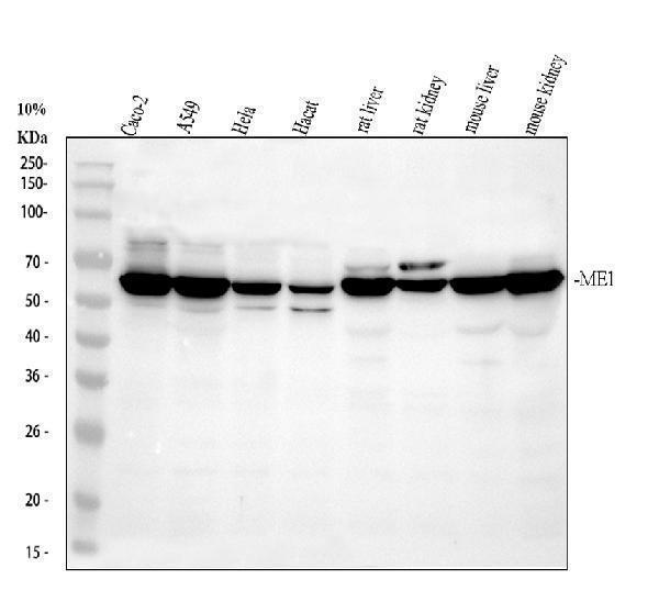
Western blot analysis of ME1 using anti-ME1 antibody (A03449-3).
Electrophoresis was performed on a 10% SDS-PAGE gel at 80V (Stacking gel) / 120V (Resolving gel) for 2 hours. The sample well of each lane was loaded with 30 ug of sample under reducing conditions.
Lane 1: human CACO-2 whole cell lysates,
Lane 2: human A549 whole cell lysates,
Lane 3: human Hela whole cell lysates,
Lane 4: human Hacat whole cell lysates,
Lane 5: rat liver tissue lysates,
Lane 6: rat kidney tissue lysates,
Lane 7: mouse liver tissue lysates,
Lane 8: mouse kidney tissue lysates.
After electrophoresis, proteins were transferred to a nitrocellulose membrane at 150 mA for 50-90 minutes. Blocked the membrane with 5% non-fat milk/TBS for 1.5 hour at RT. The membrane was incubated with rabbit anti-ME1 antigen affinity purified polyclonal antibody (A03449-3) at 0.5 μg/mL overnight at 4°C, then washed with TBS-0.1%Tween 3 times with 5 minutes each and probed with a goat anti-rabbit IgG-HRP secondary antibody (Catalog # BA1054) at a dilution of 1:5000 for 1.5 hour at RT. The signal is developed using an ECL Plus Western Blotting Substrate (Catalog # AR1196-200) with Tanon 5200 system. A specific band was detected for ME1 at approximately 64 kDa. The expected band size for ME1 is at 64 kDa.

Click image to see more details
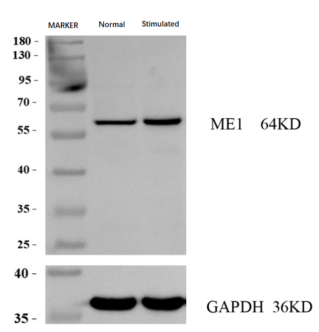
Western blot analysis of ME1 using anti-ME1 antibody (A03449-3).
Electrophoresis was performed on a 10% SDS-PAGE gel at 80V (Stacking gel) / 120V (Resolving gel) for 2 hours. The sample well of each lane was loaded with 30 ug of sample under reducing conditions.
Lane 1: human CACO-2 whole cell lysates,
Lane 2: LPS-Stimulated human A549 whole cell lysates.
After electrophoresis, proteins were transferred to a nitrocellulose membrane at 150 mA for 50-90 minutes. Blocked the membrane with 5% non-fat milk/TBS for 1.5 hour at RT. The membrane was incubated with rabbit anti-ME1 antigen affinity purified polyclonal antibody (A03449-3) at 1:2000 overnight at 4°C, then washed with TBS-0.1%Tween 3 times with 5 minutes each and probed with a goat anti-rabbit IgG-HRP secondary antibody (Catalog # BA1054) at a dilution of 1:10000 for 1 hour at RT. The signal is developed using an ECL Plus Western Blotting Substrate (Catalog # AR1196-200) with ChemiDoc MP system. A specific band was detected for ME1 at approximately 64 kDa. The expected band size for ME1 is at 64 kDa.

Click image to see more details
IHC analysis of ME1 using anti-ME1 antibody (A03449-3).
ME1 was detected in a paraffin-embedded section of human stomach cancer tissue. Heat mediated antigen retrieval was performed in EDTA buffer (pH 8.0, epitope retrieval solution). The tissue section was blocked with 10% goat serum. The tissue section was then incubated with 2 μg/ml rabbit anti-ME1 Antibody (A03449-3) overnight at 4°C. Peroxidase Conjugated Goat Anti-rabbit IgG was used as secondary antibody and incubated for 30 minutes at 37°C. The tissue section was developed using HRP Conjugated Rabbit IgG Super Vision Assay Kit (Catalog # SV0002) with DAB as the chromogen.

Click image to see more details
IF analysis of ME1 using anti-ME1 antibody (A03449-3) and anti-Tubulin Alpha antibody (M03989-3).
ME1 was detected in immunocytochemical section of A549 cell. Enzyme antigen retrieval was performed using IHC enzyme antigen retrieval reagent (AR0022) for 15 mins. The cells were blocked with 10% goat serum. And then incubated with 5 μg/mL rabbit anti-ME1 Antibody (A03449-3) and mouse anti-Tubulin Alpha antibody (M03989-3) overnight at 4°C. DyLight®488 Conjugated Goat Anti-Rabbit IgG (BA1127) and Cy3 Conjugated Goat Anti-Mouse IgG (BA1031) were used as secondary antibody at 1:500 dilution and incubated for 30 minutes at 37°C. Visualize using a fluorescence microscope and filter sets appropriate for the label used.

Click image to see more details
IF analysis of ME1 using anti-ME1 antibody (A03449-3).
ME1 was detected in a paraffin-embedded section of human gastric cancer tissue. Heat mediated antigen retrieval was performed in EDTA buffer (pH 8.0, epitope retrieval solution). The tissue section was blocked with 10% goat serum. The tissue section was then incubated with 5 μg/mL rabbit anti-ME1 Antibody (A03449-3) overnight at 4°C. Cy3 Conjugated Goat Anti-Rabbit IgG (BA1032) was used as secondary antibody at 1:500 dilution and incubated for 30 minutes at 37°C. The section was counterstained with DAPI. Visualize using a fluorescence microscope and filter sets appropriate for the label used.

Click image to see more details
Flow Cytometry analysis of A549 cells using anti-ME1 antibody (A03449-3).
Overlay histogram showing A549 cells stained with A03449-3 (Blue line). To facilitate intracellular staining, cells were fixed with 4% paraformaldehyde and permeabilized with permeabilization buffer. The cells were blocked with 10% normal goat serum. And then incubated with rabbit anti-ME1 Antibody (A03449-3, 1 μg/1x106 cells) for 30 min at 20°C. DyLight®488 conjugated goat anti-rabbit IgG (BA1127, 5-10 μg/1x106 cells) was used as secondary antibody for 30 minutes at 20°C. Isotype control antibody (Green line) was rabbit IgG (1 μg/1x106) used under the same conditions. Unlabelled sample without incubation with primary antibody and secondary antibody (Red line) was used as a blank control.
Specific Publications For Anti-ME1 Antibody Picoband® (A03449-3)
Loading publications
Recommended Resources
Here are featured tools and databases that you might find useful.
- Boster's Pathways Library
- Protein Databases
- Bioscience Research Protocol Resources
- Data Processing & Analysis Software
- Photo Editing Software
- Scientific Literature Resources
- Research Paper Management Tools
- Molecular Biology Software
- Primer Design Tools
- Bioinformatics Tools
- Phylogenetic Tree Analysis
Customer Reviews
Have you used Anti-ME1 Antibody Picoband®?
Share your experimental results or join a short interview to earn up to $1,000 in product credits or other rewards.
1 Reviews For Anti-ME1 Antibody Picoband®
The antibody shows a clear target band at the correct position, with minimal background, and the results are excellent.
Excellent
| SKU | A03449-3 |
|---|---|
| Application | Western Blot |
| Sample | Human keratinocytes isolated from skin |
| Sample Processing Description | Human Caco-2 cell line |
| Other Reagents | RIPA lysis buffer, Protease inhibitor, Electrophoresis buffer, Transfer buffer, Blocking buffer |
| Primary Antibody | ME1 Antibody Picoband® |
| Primary Incubation | 1:2000, overnight at 4 ℃ |
| Secondary Antibody | HRP Goat Anti-Rabbit IgG |
| Secondary Incubation | 1:10000, 1 hour in room temperature |
| Detection | Substrate: ECL, Imaging system:ChemiDoc MP |
| Results Summary | The protein level of ME1 in LPS-stimulated CACO-2 cells is higher than that in normal CACO-2 cells. |
Yuantao Mao, Shandong First Medical University
Verified customer
Submitted 2025-12-10
Customer Q&As
Have a question?
Find answers in Q&As, reviews.
Can't find your answer?
Submit your question
