This website uses cookies to ensure you get the best experience on our website.


- Table of Contents
Plan chromogenic MYC IHC on paraffin sections using nuclear and nucleolar tissue staining as the benchmark (HPA tissue IHC). Start with the IHC-validated total-MYC antibody M00026-1 at 1:50 (datasheet M00026-1), and assess glial and stromal cells separately (HPA tissue IHC).

Expected localisation, validated protocols, controls and antibodies — the at-a-glance facts below, then the full design guide.
| Expected localisation | Nuclear and nucleolar staining in tissue (HPA tissue IHC) | |
| Staining pattern | Nuclear and nucleolar staining in most cells; glia and stroma spared (HPA tissue IHC) | |
| Antigen retrieval | Tris-EDTA pH 9.0 HIER, 95–98 °C, 20 min (rule: nuclear antigen) | |
| Positive control | Caudate+4 more · see all | |
| Negative control | None in HPA (detected in all 45 tissues); use no-primary + isotype controls |
| Fixation | Keep fixation consistent; overfixation may mask epitopes (standard IHC practice) | |
| Caveat | Staining and RNA expression have medium consistency (HPA tissue IHC) | |
| Regulation | No expression inducer is annotated (UniProt) | |
| Isoform / epitope | 3 isoforms; epitope differences are unspecified (UniProt) |
The catalog antibody’s IHC-P protocol is followed by published MYC IHC methods for mantle cell lymphoma, breast cancer specimens, and large B-cell lymphoma (PMC3871984; PMC3276715; PMC6365198).
| Sample | FFPE tissue sections (standard) |
| Fixation | 10% neutral-buffered formalin, 24 h, standard processing (standard) |
| Sectioning | 4–5 µm sections on charged slides (standard) |
| Deparaffinisation | Xylene, graded ethanol series to water (standard) |
| Antigen retrieval | Heat-induced epitope retrieval in Tris-EDTA buffer, pH 9.0, 20 min at 95–98 °C (standard rule: nuclear antigen) |
| Peroxidase block | 3% H2O2, 10 min, room temperature (standard) |
| Blocking | 10% normal serum of the secondary host, 30 min, room temperature (standard) |
| Primary antibody | Rabbit anti-MYC, 1:200-1:1000 (datasheet M00026-6) |
| Primary incubation | Overnight at 4 °C (standard) |
| Detection | HRP-polymer secondary, DAB chromogen 5–10 min (standard) |
| Counterstain | Hematoxylin, blue, dehydrate and mount (standard) |
| Expected result | MYC-positive staining in neuronal cells of caudate (HPA tissue IHC: High). HPA tissue profile: Ubiquitous nuclear and nucleolar expression in essentially all cells except in glial- and stroma cells. No signal in the no-primary control. |
MYC is mainly nuclear, including the nucleoplasm and nucleolus (UniProt P01106). HPA reports nuclear and nucleolar staining in essentially all cells except glial and stromal cells; its tissue IHC rating is Approved, with medium consistency against RNA data (HPA tissue IHC). MYC has no transmembrane segment (UniProt P01106 topology).
| Nuclear/nucleolar stain in cervix squamous cells (HPA: High). | Fits the expected MYC pattern (HPA tissue IHC; UniProt P01106 localization). |
| Predominantly membranous staining. | Investigate artefact: MYC has no transmembrane segment (UniProt P01106 topology). |
| Strong staining confined to glial or stromal cells. | Check cross-reactivity or endogenous activity (HPA tissue IHC profile; IHC practice). |
| Diffuse colour across cells and tissue background. | Check blocking, washing and chromogen background (IHC practice). |
| No nuclear stain in cervix squamous cells (HPA: High). | Check retrieval, antibody dilution and detection controls (IHC practice). |
| Fixation and cross-linking (IHC practice) | Overfixation may mask nuclear epitopes; compare fixation conditions (IHC practice). |
| Antigen retrieval (IHC practice) | Insufficient retrieval can weaken nuclear staining in paraffin sections (IHC practice). |
| 3 isoforms (UniProt P01106) | Confirm which isoforms the antibody recognizes; epitope coverage is unspecified (UniProt P01106; IHC practice). |
| Endogenous peroxidase (IHC practice) | Unblocked activity can produce misleading chromogenic colour (IHC practice). |
| Species reactivity (IHC practice) | Confirm antibody reactivity for the specimen species (IHC practice). |
| IF/ICC: where should signal appear? | Primarily in the nucleoplasm (HPA ICC-IF: enhanced nucleoplasmic localization). |
| Situation | Likely cause | Next action |
|---|---|---|
| No signal in cervix squamous cells (HPA: High). | Masked epitope or weak detection (IHC practice). | Optimize retrieval and check antibody and detection controls (IHC practice). |
| Weak nuclear signal (HPA tissue IHC: nuclear pattern). | Fixation or retrieval may reduce epitope access (IHC practice). | Review fixation and optimize retrieval (IHC practice). |
| Membranous colour dominates. | Possible nonspecific signal; MYC lacks a membrane span (UniProt P01106 topology). | Check antibody specificity and detection controls (IHC practice). |
| Diffuse tissue background. | Incomplete blocking or excess antibody (IHC practice). | Improve blocking and washing; titrate antibody (IHC practice). |
| Colour appears without primary antibody. | Endogenous enzyme activity or detection background (IHC practice). | Block peroxidase and inspect the no-primary control (IHC practice). |
| Strong glial or stromal staining (HPA tissue IHC profile). | Possible cross-reactivity or endogenous activity (IHC practice). | Compare no-primary control and a known-positive tissue (IHC practice). |



Comprehensive Human Protein Atlas IHC scoring per tissue (reliability: Approved — Medium consistency between antibody staining and RNA expression data.). Rows are taken directly from the HPA tissue chart — click any row's HPA link to view the source.
| Tissue | Cell type | Level | Evidence | Source |
|---|---|---|---|---|
| Caudate | Neuronal cells | High | Protein (IHC) | HPA → |
| Cerebellum | Purkinje cells | High | Protein (IHC) | HPA → |
| Cervix | Squamous epithelial cells | High | Protein (IHC) | HPA → |
| Hippocampus | Neuronal cells | High | Protein (IHC) | HPA → |
| Lung | Alveolar cells | High | Protein (IHC) | HPA → |
| Tissue | Cell type | Level | Evidence | Source |
|---|---|---|---|---|
| None in HPA: MYC is detected in all 45 scored tissues. Use a no-primary (secondary-only) and an isotype control instead. | ||||
Troubleshoot MYC staining by checking nuclear localisation, specimen handling and controls before comparing chromogenic IHC scores (UniProt P01106; HPA tissue IHC).
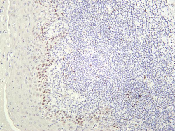
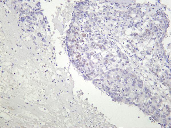
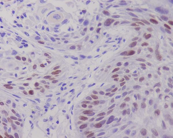
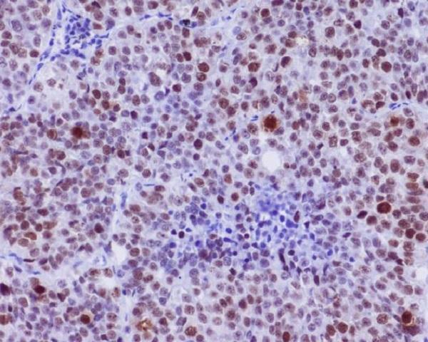
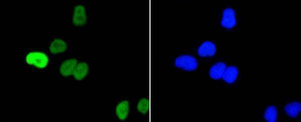
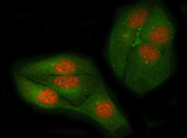
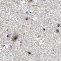
IHC figures show MYC staining in human tonsil and lung tumors; IF/ICC figures show HeLa cells (figure captions). Catalog-listed reactivity includes human, mouse, and rat (catalog: reactivity).
M00026-6 has an IHC figure from human tonsil, M00026-1 from paraffin-embedded human lung cancer, and P00026-2 from paraffin-embedded human lung carcinoma stained for phospho-S62 MYC (figure captions). A00026S62-1 has a phospho-S62 IF/ICC figure in HeLa cells, and PB9092 has a MYC IF/ICC figure in HeLa cells (figure captions).
Which to pick: For FFPE IHC, choose M00026-1 for total MYC or P00026-2 for phospho-S62 MYC; both are rabbit monoclonals with paraffin-embedded human lung tumor figures (catalog: titles; figure captions). For IF/ICC, choose A00026S62-1 for phospho-S62 or PB9092 for total MYC; both have HeLa cell figures and list IF/ICC applications (catalog: titles/applications; figure captions). For work across human, mouse, and rat, M00026-6 is a recombinant rabbit monoclonal listed for IHC and IF in all three species, with an IHC figure from human tonsil (catalog: description/applications/reactivity; figure caption).