Click image to see more details
Product Info Summary
| SKU: | PB9092 |
|---|---|
| Size: | 100 μg/vial |
| Reactive Species: | Human, Mouse, Rat |
| Host: | Rabbit |
| Application: | IF, ICC, WB |
Customers Who Bought This Also Bought

Product info
Product Name
Anti-c-Myc Antibody Picoband®
SKU/Catalog Number
PB9092
Size
100 μg/vial
Form
Lyophilized
Description
Boster Bio Anti-c-Myc Antibody Picoband® catalog # PB9092. Tested in IF, ICC, WB applications. This antibody reacts with Human, Mouse, Rat. The brand Picoband indicates this is a premium antibody that guarantees superior quality, high affinity, and strong signals with minimal background in Western blot applications. Only our best-performing antibodies are designated as Picoband, ensuring unmatched performance.
Storage & Handling
Store at -20˚C for one year from date of receipt. After reconstitution, at 4˚C for one month. It can also be aliquotted and stored frozen at -20˚C for six months. Avoid repeated freeze-thaw cycles.
Cite This Product
Anti-c-Myc Antibody Picoband® (Boster Biological Technology, Pleasanton CA, USA, Catalog # PB9092)
Host
Rabbit
Contents
Each vial contains 4 mg Trehalose, 0.9 mg NaCl and 0.2 mg Na2HPO4.
Clonality
Polyclonal
Isotype
Rabbit IgG
Immunogen
E.coli-derived human c-Myc recombinant protein (Position: E257-A439). Human c-Myc shares 91% amino acid (aa) sequences identity with both mouse and rat c-Myc.
Cross-reactivity
No cross-reactivity with other proteins
Reactive Species
PB9092 is reactive to MYC in Human, Mouse, Rat
Observed Molecular Weight
60-65 kDa
Calculated molecular weight
48.8 kDa
Background of MYC
C-Myc is an oncogene that functions both in the stimulation of cell proliferation and in apoptosis. C-Myc elicits its oncogenic activity by causing immortalization, and to a lesser extent the transformation of cells, in addition to several other mechanisms. The c-MYC proto-oncogene encodes a transcription factor that is critical for cell growth and proliferation. It is one of the genes frequently altered in cancer cells in which it exhibits constitutive activity. Downregulation of c-Myc is critical for 2-Methoxyestradiol (2ME2)-induced oxidative stress and apoptosis in AML cells. And its up-regulation is important for promoting lymphocyte cell division, and demonstrating that GFP-c-Myc expression is a marker of proliferating lymphocytes in vivo.
Antibody Validation
Boster validates all antibodies on WB, IHC, ICC, Immunofluorescence, and ELISA with known positive control and negative samples to ensure specificity and high affinity, including thorough antibody incubations.
Application & Images
Applications
PB9092 is guaranteed for IF, ICC, WB Boster Guarantee
Recommend Dilution
| Application | Dilution | Species |
|---|---|---|
| Western blot | 0.1-0.5μg/ml | Human, Mouse, Rat |
| Immunocytochemistry/Immunofluorescence | 5 μg/ml | Human |
Tested application
Suggested blocking solution with 5% non-fat milk or BSA; (*)Recommended protein loading: 20-40 µg per lane
Validation Images & Assay Conditions
Click image to see more details
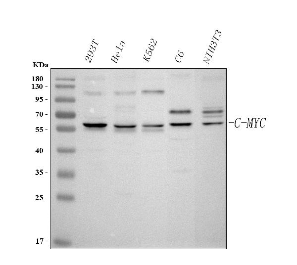
Western blot analysis of c-Myc using anti-c-Myc antibody (PB9092).
Electrophoresis was performed on a 5-20% SDS-PAGE gel at 70V (Stacking gel) / 90V (Resolving gel) for 2-3 hours. The sample well of each lane was loaded with 30 ug of sample under reducing conditions.
Lane 1: human 293T whole cell lysates,
Lane 2: human Hela whole cell lysates,
Lane 3: human K562 whole cell lysates,
Lane 4: rat C6 whole cell lysates,
Lane 5: mouse NIH/3T3 whole cell lysates.
After electrophoresis, proteins were transferred to a nitrocellulose membrane at 150 mA for 50-90 minutes. Blocked the membrane with 5% non-fat milk/TBS for 1.5 hour at RT. The membrane was incubated with rabbit anti-c-Myc antigen affinity purified polyclonal antibody (Catalog # PB9092) at 0.5 μg/mL overnight at 4°C, then washed with TBS-0.1%Tween 3 times with 5 minutes each and probed with a goat anti-rabbit IgG-HRP secondary antibody at a dilution of 1:5000 for 1.5 hour at RT. The signal is developed using an Enhanced Chemiluminescent detection (ECL) kit (Catalog # EK1002) with Tanon 5200 system. A specific band was detected for c-Myc at approximately 60-65 kDa. The expected band size for c-Myc is at 49 kDa.

Click image to see more details
IF analysis of c-Myc using anti-c-Myc antibody (PB9092) and anti-Beta Tubulin antibody (M01857-3).
c-Myc was detected in immunocytochemical section of Hela cell. Enzyme antigen retrieval was performed using IHC enzyme antigen retrieval reagent (AR0022) for 15 mins. The cells were blocked with 10% goat serum. And then incubated with 5 μg/mL rabbit anti-c-Myc Antibody (PB9092) and mouse anti-Beta Tubulin antibody (M01857-3) overnight at 4°C. Cy3 Conjugated Goat Anti-Rabbit IgG (BA1032) and DyLight®488 Conjugated Goat Anti-Mouse IgG (BA1126) were used as secondary antibody at 1:500 dilution and incubated for 30 minutes at 37°C. Visualize using a fluorescence microscope and filter sets appropriate for the label used.
Specific Publications For Anti-c-Myc Antibody Picoband® (PB9092)
Loading publications
Recommended Resources
Here are featured tools and databases that you might find useful.
- Boster's Pathways Library
- Protein Databases
- Bioscience Research Protocol Resources
- Data Processing & Analysis Software
- Photo Editing Software
- Scientific Literature Resources
- Research Paper Management Tools
- Molecular Biology Software
- Primer Design Tools
- Bioinformatics Tools
- Phylogenetic Tree Analysis
Customer Reviews
Have you used Anti-c-Myc Antibody Picoband®?
Share your experimental results or join a short interview to earn up to $1,000 in product credits or other rewards.
0 Reviews For Anti-c-Myc Antibody Picoband®
Customer Q&As
Have a question?
Find answers in Q&As, reviews.
Can't find your answer?
Submit your question
5 Customer Q&As for Anti-c-Myc Antibody Picoband®
Question
We bought anti-c-Myc antibody for Flow Cytometry on cervix last year. I am using human, and We intend to use the antibody for IHC-P next. I would like examining cervix as well as uterus in our next experiment. Could you please give me some suggestion on which antibody would work the best for IHC-P?
Verified Customer
Verified customer
Asked: 2019-11-05
Answer
I have checked the website and datasheets of our anti-c-Myc antibody and it appears that PB9092 has been validated on human in both Flow Cytometry and IHC-P. Thus PB9092 should work for your application. Our Boster satisfaction guarantee will cover this product for IHC-P in human even if the specific tissue type has not been validated. We do have a comprehensive range of products for IHC-P detection and you can check out our website bosterbio.com to find out more information about them.
Boster Scientific Support
Answered: 2019-11-05
Question
My colleagues were happy with the WB result of your anti-c-Myc antibody. However we have been able to see positive staining in promyelocytic leukemia nucleus using this antibody. Is that expected? Could you tell me where is MYC supposed to be expressed?
L. Johnson
Verified customer
Asked: 2018-11-22
Answer
According to literature, promyelocytic leukemia does express MYC. Generally MYC expresses in nucleus, nucleoplasm. Regarding which tissues have MYC expression, here are a few articles citing expression in various tissues:
Cervix carcinoma, Pubmed ID: 17081983, 18669648, 20068231
Cervix carcinoma, and Erythroleukemia, Pubmed ID: 23186163
Cervix, Placenta, and Testis, Pubmed ID: 15489334
Promyelocytic leukemia, Pubmed ID: 3540591
Uterus, Pubmed ID: 14702039
Boster Scientific Support
Answered: 2018-11-22
Question
We have seen staining in rat promyelocytic leukemia. Do you have any suggestions? Is anti-c-Myc antibody supposed to stain promyelocytic leukemia positively?
L. Brown
Verified customer
Asked: 2017-12-08
Answer
From literature promyelocytic leukemia does express MYC. From Uniprot.org, MYC is expressed in thoracic mammary gland, uterus, cervix, placenta testis, promyelocytic leukemia, cervix carcinoma, cervix carcinoma erythroleukemia, among other tissues. Regarding which tissues have MYC expression, here are a few articles citing expression in various tissues:
Cervix carcinoma, Pubmed ID: 17081983, 18669648, 20068231
Cervix carcinoma, and Erythroleukemia, Pubmed ID: 23186163
Cervix, Placenta, and Testis, Pubmed ID: 15489334
Promyelocytic leukemia, Pubmed ID: 3540591
Uterus, Pubmed ID: 14702039
Boster Scientific Support
Answered: 2017-12-08
Question
We are currently using anti-c-Myc antibody PB9092 for rat tissue, and we are content with the IHC-P results. The species of reactivity given in the datasheet says human, rat. Is it likely that the antibody can work on horse tissues as well?
Verified Customer
Verified customer
Asked: 2017-08-21
Answer
The anti-c-Myc antibody (PB9092) has not been tested for cross reactivity specifically with horse tissues, though there is a good chance of cross reactivity. We have an innovator award program that if you test this antibody and show it works in horse you can get your next antibody for free. Please contact me if I can help you with anything.
Boster Scientific Support
Answered: 2017-08-21
Question
We are currently using anti-c-Myc antibody PB9092 for rat tissue, and we are content with the IHC-P results. The species of reactivity given in the datasheet says human, rat. Is it likely that the antibody can work on horse tissues as well?
M. Edwards
Verified customer
Asked: 2016-05-03
Answer
The anti-c-Myc antibody (PB9092) has not been tested for cross reactivity specifically with horse tissues, though there is a good chance of cross reactivity. We have an innovator award program that if you test this antibody and show it works in horse you can get your next antibody for free. Please contact me if I can help you with anything.
Boster Scientific Support
Answered: 2016-05-03
