NMI / N-myc-interactor · IHC design guide

Design Immunohistochemistry for NMI

Plan chromogenic NMI IHC in paraffin sections using the catalog antibody at 0.5–1 μg/mL (datasheet PB9340). Assess cytoplasmic staining in glandular cells (HPA tissue IHC), while considering nuclear relocalization and macrophage secretion when interpreting signal (UniProt).

Evidence assembled Oct 2026 · For research use; verify linked source records and product datasheet before use
Immunohistochemistry protocol sheet for NMI (IHC for NMI): expected localisation Cytoplasmic staining in most tissues (HPA tissue IHC), antibody PB9340, validated IHC image, and IHC protocol steps
Printable NMI IHC protocol sheet — expected localisation Cytoplasmic staining in most tissues (HPA tissue IHC), antibody PB9340, controls and protocol steps. Open the full NMI IHC guide →

NMI Immunohistochemistry Experimental Design Guide

Expected localisation, validated protocols, controls and antibodies — the at-a-glance facts below, then the full design guide.

Must know before staining
Expected localisation Cytoplasmic staining in most tissues (HPA tissue IHC)
Staining pattern Cytoplasmic staining, high in gut glandular cells (HPA tissue IHC)
Antigen retrieval EDTA pH 8.0 HIER, heat-mediated (datasheet PB9340)
Positive control ⓘ Appendix+4 more · see all
Negative control ⓘ Adipose tissue+4 more · see all
Important caveats
Reasons your staining may differ from the expected pattern.
Fixation Keep fixation consistent across sections (standard IHC practice; not target-specific). Selected-image fixative and duration unreported (datasheet PB9340); verify before use.
Caveat Secretion can decouple tissue staining from RNA (HPA tissue IHC)
Regulation IFN-α increases nuclear NMI (UniProt)
Isoform / epitope No isoforms; the 1–307 chain is intracellular or secreted (UniProt)
Section 1

Recommended NMI IHC & IF Protocols

The catalog antibody protocol uses EDTA pH 8.0 retrieval (datasheet PB9340). The four published IHC protocols below provide tissue specific staining conditions (PMC7043944; PMC5639741; PMC5921859; PMC11079014).

Recommended immunohistochemistry (IHC-P) protocol parameters
SampleParaffin-embedded human colon cancer tissue; fixative not specified (datasheet PB9340)
FixationImage fixative and duration unreported (datasheet PB9340); verify before use.
Sectioning4–5 µm sections on charged slides (standard)
DeparaffinisationXylene, graded ethanol series to water (standard)
Antigen retrievalHeat retrieval: EDTA pH 8.0 (datasheet PB9340); 20 min, 95–100 °C (standard)
Peroxidase block3% H2O2, 10 min, room temperature (standard)
Blocking10% goat serum (datasheet PB9340)
Primary antibodyRabbit anti-NMI, 0.5-1μg/ml (datasheet PB9340)
Primary incubationOvernight at 4 °C (datasheet PB9340)
DetectionStreptavidin-biotin complex (SABC), DAB chromogen (datasheet PB9340)
CounterstainHematoxylin, blue, dehydrate and mount (standard)
Expected resultNMI-positive staining in glandular cells of appendix (HPA tissue IHC: High). HPA tissue profile: Cytoplasmic expression in most tissues. No signal in the no-primary control.
💡Decision noteStart with EDTA pH 8.0 heat retrieval for the catalog antibody (datasheet PB9340). For the published lung protocol, use its reported pH 6 microwave retrieval (PMC5639741).
Section 2

What Is the Expected NMI Staining Pattern?

NMI staining is predominantly cytoplasmic across tissues, with strong signal reported in gastrointestinal glandular cells and cardiomyocytes (HPA: tissue IHC). Nucleoplasmic signal is also plausible (HPA: ICC-IF; UniProt Q13287: nucleus). NMI has no transmembrane segment, so a membrane-only pattern is unexpected (UniProt Q13287: topology). Interpret tissue results cautiously: HPA rates its IHC data Approved but reports low consistency between antibody staining and RNA expression.

What am I looking at on my slide?
Strong cytoplasmic staining in glandular cells of colon, stomach, or small intestine.This matches the reported high staining in those cells (HPA: tissue IHC). Assess the identified glandular cells and their cytoplasm; a strong color deposit elsewhere in the section does not establish that the expected cell population is positive (general IHC practice).
Cytoplasmic staining with some nuclear signal in identifiable positive cells.Both compartments are biologically plausible (UniProt Q13287: cytoplasm and nucleus; HPA: ICC-IF nucleoplasm and cytosol). UniProt reports increased nuclear localization after IFN-alpha treatment; nuclear staining alone cannot establish that treatment occurred. Record compartments separately when scoring (general IHC practice).
Staining appears confined to cell borders, with little cytoplasmic signal.A membrane-only pattern conflicts with the reported cytoplasmic tissue profile and lack of a transmembrane segment (HPA: tissue IHC; UniProt Q13287: topology). Consider nonspecific edge staining or a detection artifact, then reassess against an expected positive area (general IHC practice).
Strong staining occurs mainly in cells reported as undetected, or color covers the tissue diffusely.Adipocytes and esophageal squamous cells are reported as not detected (HPA: tissue IHC). Signal in these cells raises cross-reactivity or endogenous detection activity as possibilities; diffuse color without cellular boundaries raises background. Neither finding alone proves antibody specificity (general IHC practice).
No discernible signal in a section expected to contain high-staining glandular cells.Colon, duodenum, and rectum glandular cells are reported as high (HPA: tissue IHC). Check that the relevant cells are present, then review the staining run and controls (general IHC practice). A negative result needs caution because HPA reports low antibody-staining/RNA consistency and possible secretion.
💡Expected NMI appearanceCall the result positive when identifiable gastrointestinal glandular cells show clear, predominantly cytoplasmic staining at the reported high level (HPA: tissue IHC); nucleoplasmic signal may coexist (HPA: ICC-IF), whereas uniform haze, membrane-only color, or signal restricted to reported undetected cells should prompt review (general IHC practice).
How each factor affects the staining
Tissue and cell selectionHPA reports high staining in appendix, colon, duodenum, rectum, small intestine, and stomach glandular cells, and in cardiomyocytes (HPA: tissue IHC). Breast glandular cells are medium; adipocytes and esophageal squamous cells are not detected. Score the specified cell type, not the whole tissue.
Compartment and cell stateCytoplasmic NMI can form punctate granular structures, and nuclear localization increases after IFN-alpha treatment (UniProt Q13287: subcellular location). HPA supports cytosol and approves nucleoplasm in ICC-IF. A mixed pattern can be credible, but these records do not predict its proportion in every paraffin section.
Secretion and tissue-level concordanceMacrophages express NMI and can release it extracellularly during injury or pathogen invasion (UniProt Q13287: tissue specificity and function). HPA notes that secretion can separate protein location from RNA location and rates tissue IHC Approved with low staining/RNA consistency. Extracellular color still needs suitable controls.
Topology and processingNMI has no transmembrane segment or annotated signal peptide, and its listed chain spans residues 1–307 (UniProt Q13287: topology and processing). These annotations support interpreting intracellular staining; they do not identify this antibody’s epitope or establish an antigen retrieval condition.
Antibody validation scopeThe listed antibody HPA008588 is Approved for IHC and Supported for ICC (HPA: antibodies). These statuses support the reported applications but do not make every cell-specific signal definitive. HPA’s tissue reliability note remains relevant when a section differs from its reference images.
Why is my staining missing, weak or wrong?
SituationLikely causeNext action
Expected glandular cells are present but show no staining.A run or detection problem is possible; HPA reports high glandular staining at several gastrointestinal sites (HPA: tissue IHC).Review section morphology, run controls, primary-antibody use, and chromogen development (general IHC practice). Compare the same cell type in a reported high-staining tissue before interpreting an isolated negative.
The section shows broad, weak brown haze.Diffuse background can obscure cell boundaries and mimic cytoplasmic signal (general IHC practice).Inspect the negative control and review blocking, wash steps, and detection conditions (general IHC practice). Score only signal assignable to intact cells; HPA’s expected profile is cytoplasmic expression, not indiscriminate tissue-wide color (HPA: tissue IHC).
Color is strongest in cells listed as not detected.Cross-reactivity or endogenous detection activity is possible when signal predominates in adipocytes or esophageal squamous cells (HPA: tissue IHC; general IHC practice).Compare a negative control and a reported high-staining cell population on the same run (general IHC practice). Treat the unexpected population as unresolved until the cellular pattern and controls support it.
Signal is mainly at cell borders or extracellular spaces.A membrane-only pattern lacks support from NMI topology (UniProt Q13287); extracellular NMI is possible after macrophage secretion (UniProt Q13287: function).Assess whether intact cells also show the expected cytoplasmic pattern and review controls (HPA: tissue IHC; general IHC practice). Do not assign extracellular color to NMI from location alone.
Nuclear staining seems stronger than expected.Nucleoplasmic localization is supported in ICC-IF, and IFN-alpha can increase nuclear NMI (HPA: ICC-IF; UniProt Q13287: subcellular location).Score nuclear and cytoplasmic compartments separately and check the relevant treatment history (general IHC practice). Avoid inferring IFN-alpha exposure or a technical failure solely from nuclear color.
What should IF/ICC show?HPA reports nucleoplasm and cytosol, with nucleoplasm Approved and cytosol Supported; listed ICC-IF images include A-431, U-251MG, and U2OS (HPA: ICC-IF).Use those locations as the IF/ICC interpretation reference (HPA: ICC-IF). Consult the separate IF/ICC guide for its workflow; the IHC tissue staining profile alone does not establish an IF/ICC protocol.

Sample controls for NMI IHC & IF

🧪Run colon first and score its glandular cells for NMI staining (HPA: High in colon glandular cells). Use esophagus squamous epithelial cells as the negative tissue (HPA: Not detected in esophagus squamous epithelial cells); on the colon slide, non-glandular cells should provide an internal background reference only where they show no specific staining (standard IHC practice).
Positive control tissue: Appendix (Glandular cells, HPA High)
Negative control tissue: Adipose tissue (HPA Not detected)
ICC-IF cell lines (HPA subcellular resource): HPA ICC-IF images show NMI in A-431, U-251MG, U2OS, with annotated localisation: Nucleoplasm (approved), Cytosol (supported) (HPA subcellular).
Technical controls: Include a no-primary, secondary-only control and a host-matched rabbit IgG isotype control (caption: rabbit anti-NMI primary; standard IHC practice); confirm specificity with NMI-knockout material if available (standard IHC practice). Check endogenous peroxidase and biotin background in colon because the shown chromogenic method uses a streptavidin-biotin complex and DAB (caption: SABC/DAB detection; standard IHC practice).
⚠️Feasibility: No target-specific fixation window or fixation effect is reported, and the selected PB9340 paraffin-section caption does not state the fixative (caption: fixative unreported). That caption uses heat retrieval in EDTA at pH 8.0, so retrieval is documented for the illustrated IHC result, but its necessity has not been established (caption: EDTA heat retrieval). The supplied evidence does not show that frozen sections or IF are easier; assess glandular and adjacent non-glandular regions separately to avoid treating background as NMI staining (HPA: High in colon glandular cells; standard IHC practice).

HPA tissue IHC evidence for NMI

Comprehensive Human Protein Atlas IHC scoring per tissue (reliability: Approved — Low consistency between antibody staining and RNA expression data. At least one protein variant secreted, tissue location of RNA and protein might differ and correlation is complex.). Rows are taken directly from the HPA tissue chart — click any row's HPA link to view the source.

Positive expression · recommended positive controls

TissueCell typeLevelEvidenceSource
Appendix Glandular cells High Protein (IHC) HPA →
Colon Glandular cells High Protein (IHC) HPA →
Duodenum Glandular cells High Protein (IHC) HPA →
Heart muscle Cardiomyocytes High Protein (IHC) HPA →
Rectum Glandular cells High Protein (IHC) HPA →

Undetected expression · recommended negative controls

TissueCell typeLevelEvidenceSource
Adipose tissue Adipocytes Not detected Protein (IHC) HPA →
Bone marrow Hematopoietic cells Not detected Protein (IHC) HPA →
Esophagus Squamous epithelial cells Not detected Protein (IHC) HPA →
Ovary Ovarian stroma cells Not detected Protein (IHC) HPA →
Soft tissue Fibroblasts Not detected Protein (IHC) HPA →
Section 3

Advanced NMI IHC Tips

Troubleshoot NMI staining in paraffin sections by checking retrieval, cell compartment and controls before comparing signal across tissues (datasheet PB9340; UniProt Q13287; HPA tissue IHC).

What retrieval should I try first when NMI staining is weak?
Start with heat-mediated retrieval in EDTA at pH 8.0 for paraffin sections (datasheet PB9340). Keep the retrieval method, heating conditions and section handling consistent across the test and control slides, then compare staining in glandular cells of colon tissue, where NMI staining is reported as high (HPA: colon glandular cells). The selected image used 1 μg/ml primary antibody overnight at 4°C, so check those conditions before attributing weak signal to retrieval (datasheet PB9340). If staining remains weak, test an alternative retrieval condition on paired sections while watching for tissue damage and increased background (standard IHC practice).
Could fixation explain weak or uneven NMI staining?
The selected NMI image documents a paraffin section but does not state its fixative or fixation duration, so target-specific fixation sensitivity is unknown (datasheet PB9340). Record the fixative and processing history for each block, and compare similarly processed sections before changing the antibody concentration or retrieval conditions (standard IHC practice). In paired sections, check whether staining varies with section depth, preservation or tissue edges while keeping EDTA at pH 8.0 retrieval and the 1 μg/ml primary condition constant (datasheet PB9340; standard IHC practice). Do not use an apparent staining difference alone to assign a fixation mechanism; verify morphology and controls first (standard IHC practice).
How should I assess cytoplasmic versus nuclear NMI staining?
Score cytoplasmic and nuclear staining separately, because NMI is reported in both compartments and its cytoplasmic signal can be punctate (UniProt Q13287). Nuclear localisation can increase after IFN-alpha treatment, so record treatment status before comparing nuclear fractions between samples (UniProt Q13287). HPA reports predominantly cytoplasmic tissue staining and identifies the nucleoplasm and cytosol in its subcellular data; those observations provide context, not a required pattern for every section (HPA tissue IHC; HPA subcellular). At 1 μg/ml primary antibody, compare compartment patterns with a matched negative control and intact cell morphology before calling a faint nuclear signal positive (datasheet PB9340; standard IHC practice).
Could an NMI isoform or hidden epitope explain discordant staining?
The supplied record lists 0 isoforms, one chain spanning residues 1–307, and no transmembrane segment (UniProt Q13287). It also lists a phosphoserine at residue 16 and NID regions at 103–192 and 201–292, but the antibody epitope is not supplied (UniProt Q13287; datasheet PB9340). Consequently, do not assign a lost stain to a particular domain or modification without epitope information or a direct validation experiment (standard IHC practice). First compare matched sections under the documented EDTA pH 8.0 retrieval condition, then evaluate whether any revised retrieval preserves morphology and the expected compartment pattern (datasheet PB9340; UniProt Q13287).
How can IF help resolve an ambiguous chromogenic NMI pattern?
Use IF as a separate localisation check: NMI is reported in cytosol and nucleoplasm, with possible punctate cytoplasmic signal (HPA subcellular; UniProt Q13287). In colon sections, multiplex an epithelial marker to identify glandular cells, which show high NMI staining in the tissue atlas (HPA: colon glandular cells; standard IF practice). Choose a fluorophore channel with low measured tissue autofluorescence, and compare single-label, no-primary and multiplex controls before interpreting overlap (standard IF practice). Because NMI lacks a transmembrane segment and the reported compartments are intracellular, permeabilise sufficiently to access cytosolic and nuclear epitopes; optimise this for the IF preparation rather than transferring the IHC retrieval condition of EDTA pH 8.0 (UniProt Q13287; datasheet PB9340; standard IF practice).
What should I check when DAB staining is diffuse?
The selected tissue image used 10% goat serum block, 1 μg/ml primary antibody overnight at 4°C, a biotinylated secondary for 30 minutes at 37°C, and SABC with DAB (datasheet PB9340). If diffuse colour obscures cells, inspect a no-primary control and review primary concentration, blocking, washing and development time on paired sections (standard IHC practice). Include controls for endogenous peroxidase and endogenous biotin when using this detection chemistry, since either can contribute signal unrelated to the primary antibody (standard IHC practice). Judge any adjustment against preserved morphology and the predominantly cytoplasmic tissue pattern, rather than lower background alone (HPA tissue IHC; standard IHC practice).
How should I quantify NMI across paraffin sections? ⚠ ANSWER MARKED FOR VERIFICATION
Define the cell population and tissue area before scoring; HPA reports high NMI staining in colon glandular cells but no detection in adipocytes, so pooled tissue intensity can obscure cell-specific results (HPA tissue IHC). For cellular staining, report the percentage of positive cells and an H-score from consistent intensity categories, recording cytoplasmic and nuclear scores separately (standard IHC practice; UniProt Q13287 localisation). For scattered positive cells, report density per mm² of viable tissue and normalise counts to the annotated tissue area (standard IHC practice). Keep EDTA pH 8.0 retrieval and imaging or scoring thresholds constant across the compared slides, and exclude damaged or necrotic regions (datasheet PB9340; standard IHC practice).
When is an apparent NMI-positive area likely to be artefactual?
Look for staining within intact cells in a plausible compartment: NMI can be cytoplasmic, punctate or nuclear, while HPA describes cytoplasmic expression in most tissues (UniProt Q13287; HPA tissue IHC). Compare the cell type with tissue context; HPA reports high signal in colon glandular cells but no detection in several other listed populations, including adipocytes (HPA tissue IHC). Edge-restricted colour, necrotic deposits or signal in a no-primary control warrants review of section quality, endogenous peroxidase and the biotin-based DAB workflow (datasheet PB9340; standard IHC practice). Treat a discordant result cautiously because HPA rates tissue staining Approved while noting low consistency with RNA expression and possible secretion-related differences in location (HPA tissue IHC).
Boster reagents

Best NMI / N-myc-interactor IHC Antibodies

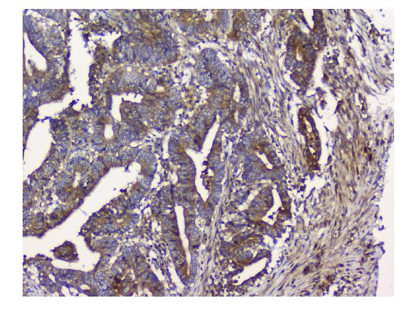
Anti-NMI antibodies have IHC images from human paraffin sections (PB9340 and M02768 image captions) and IF images from human cells (M02768-1 image caption).

Real IHC data IHC analysis of NMI using anti-NMI antibody (PB9340). NMI was detected in paraffin-embedded section of human colon cancer tissue. Heat mediated antigen retrieval was performed in EDTA buffer (pH8.0, epitope retrieval solution). The tissue section was blocked with 10% goat serum. The tissue section was then incubated with 1μg/ml rabbit anti-NMI Antibody (PB9340) overnight at 4°C. Biotinylated goat anti-rabbit IgG was used as secondary antibody and incubated for 30 minutes at 37°C. The tissue section was developed using Strepavidin-Biotin-Complex (SABC) (Catalog # SA1022) with DAB as the chromogen.
Anti-N myc interactor/NMI Antibody ®
Cat # PB9340
Real IHC data IHC analysis of NMI using anti-NMI antibody (M02768). NMI was detected in paraffin-embedded section of human lung cancer tissue. Heat mediated antigen retrieval was performed in EDTA buffer (pH8.0, epitope retrieval solution). The tissue section was blocked with 10% goat serum. The tissue section was then incubated with 1μg/ml mouse anti-NMI Antibody (M02768) overnight at 4°C. Biotinylated goat anti-mouse IgG was used as secondary antibody and incubated for 30 minutes at 37°C. The tissue section was developed using Strepavidin-Biotin-Complex (SABC) (Catalog # SA1021) with DAB as the chromogen.
Anti-NMI Antibody ® (monoclonal, 2F3)
Cat # M02768
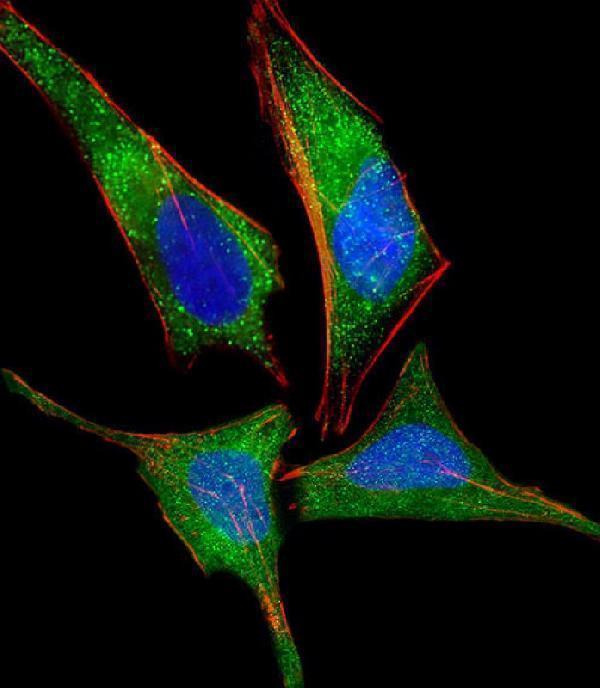
Real IF data Immunofluorescent analysis of 4% paraformaldehyde-fixed, 0.1% Triton X-100 permeabilized HeLa (human cervical epithelial adenocarcinoma cell line) cells labeling NMI with M02768-1 at 1/25 dilution, followed by Dylight® 488-conjugated goat anti-mouse IgG secondary antibody at 1/200 dilution (green). Immunofluorescence image showing cytoplasm and nucleus staining on HeLa cell line. Cytoplasmic actin is detected with Dylight® 554 Phalloidin at 1/100 dilution (red).The nuclear counter stain is DAPI (blue).
Anti-NMI Antibody
Cat # M02768-1

PB9340 shows IHC staining in a human colon cancer paraffin section, and M02768 shows IHC staining in a human lung cancer paraffin section (respective image captions). M02768-1 shows IF staining in paraformaldehyde-fixed HeLa cells (M02768-1 image caption).

Which to pick: For human paraffin-section IHC, choose rabbit PB9340 or mouse monoclonal M02768; each has an IHC figure from its own antibody (respective catalog entries and image captions). For IF/ICC, PB9340 and M02768 list both applications, while M02768-1 lists IF and shows cytoplasmic and nuclear staining in HeLa cells (catalog applications; M02768-1 image caption). M02768 lists mouse and rat reactivity, but its IHC figure shows human tissue; the fixative for both IHC figures is unreported (M02768 catalog reactivity; PB9340 and M02768 image captions).

Each figure is that product's own IHC / IF validation image from its datasheet.

References

  1. UniProt Consortium. UniProt entry Q13287 (NMI_HUMAN, N-myc-interactor).
  2. Human Protein Atlas. NMI tissue IHC expression (reliability: Approved).
  3. Human Protein Atlas. NMI subcellular location (ICC-IF): Localized to the nucleoplasm and cytosol..
  4. Human Protein Atlas. NMI antibody validation summary (1 antibodies).
  5. Downregulation of N-myc and STAT Interactor Protein Predicts Aggressive Tumor Behavior and Poor Prognosis in Invasive Ductal Carcinoma. Journal of breast cancer 2020 — PMC7043944.
  6. Downregulation of NMI promotes tumor growth and predicts poor prognosis in human lung adenocarcinomas. Molecular cancer 2017 — PMC5639741.
  7. Conditional knockout of N-Myc and STAT interactor disrupts normal mammary development and enhances metastatic ability of mammary tumors. Oncogene 2018 — PMC5921859.
  8. Targeting EMT using low-dose Teniposide by downregulating ZEB2-driven activation of RNA polymerase I in breast cancer. Cell death & disease 2024 — PMC11079014.
  9. PubMed PMID:8668343 — UniProt-cited evidence.
  10. PubMed PMID:19054851 — UniProt-cited evidence.
  11. PubMed PMID:15815621 — UniProt-cited evidence.