Click image to see more details
Product Info Summary
| SKU: | M02768-1 |
|---|---|
| Size: | 50 µl |
| Reactive Species: | Human |
| Host: | Mouse |
| Application: | Flow Cytometry, IF, WB |
Customers Who Bought This Also Bought

Product info
Product Name
Anti-NMI Antibody
SKU/Catalog Number
M02768-1
Size
50 µl
Description
Boster Bio Anti-NMI Antibody (Catalog # M02768-1). Tested in WB, Flow Cytometry, IF application(s). This antibody reacts with Human.
Storage & Handling
Maintain refrigerated at 2-8°C for up to 2 weeks. For long-term storage, store at -20°C in small aliquots to prevent freeze-thaw cycles.
Cite This Product
Anti-NMI Antibody (Boster Biological Technology, Pleasanton CA, USA, Catalog # M02768-1)
Host
Mouse
Contents
Purified monoclonal antibody supplied in PBS with 0.09% (W/V) sodium azide.
Clonality
Monoclonal
Clone Number
1580CT730.43.59
Isotype
IgG1,k
Immunogen
This NMI antibody is generated from a mouse immunized with a recombinant protein of human NMI.
Reactive Species
M02768-1 is reactive to NMI in Human
Calculated molecular weight
35.1 kDa
Background of NMI
May be involved in augmenting coactivator protein recruitment to a group of sequence-specific transcription factors. Augments cytokine-mediated STAT transcription. Enhances CBP/p300 coactivator protein recruitment to STAT1 and STAT5.
Antibody Validation
Boster validates all antibodies on WB, IHC, ICC, Immunofluorescence, and ELISA with known positive control and negative samples to ensure specificity and high affinity, including thorough antibody incubations.
Application & Images
Applications
M02768-1 is guaranteed for Flow Cytometry, IF, WB Boster Guarantee
Assay Dilutions Recommendation
The recommendations below provide a starting point for assay optimization. The actual working concentration varies and should be decided by the user.
IF: 1:25
WB: 1:2000
FC: 1:25
Validation Images & Assay Conditions
Click image to see more details
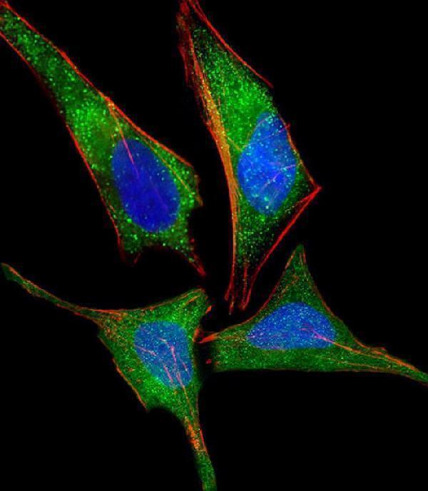
Immunofluorescent analysis of 4% paraformaldehyde-fixed, 0.1% Triton X-100 permeabilized HeLa (human cervical epithelial adenocarcinoma cell line) cells labeling NMI with M02768-1 at 1/25 dilution, followed by Dylight® 488-conjugated goat anti-mouse IgG secondary antibody at 1/200 dilution (green). Immunofluorescence image showing cytoplasm and nucleus staining on HeLa cell line. Cytoplasmic actin is detected with Dylight® 554 Phalloidin at 1/100 dilution (red).The nuclear counter stain is DAPI (blue).

Click image to see more details
Anti-NMI Antibody at 1:2000 dilution + A431 whole cell lysate
Lysates/proteins at 20 µg per lane.
Secondary
Goat Anti-mouse IgG, (H+L), Peroxidase conjugated at 1/10000 dilution.
Predicted band size : 35 kDa
Blocking/Dilution buffer: 5% NFDM/TBST.

Click image to see more details
Overlay histogram showing K562 cells stained with M02768-1 (green line). The cells were fixed with 2% paraformaldehyde (10 min) and then permeabilized with 90% methanol for 10 min. The cells were then icubated in 2% bovine serum albumin to block non-specific protein-protein interactions followed by the antibody (M02768-1, 1:25 dilution) for 60 min at 37ºC. The secondary antibody used was Goat-Anti-Mouse IgG, DyLight® 488 Conjugated Highly Cross-Adsorbed at 1/200 dilution for 40 min at 37ºC. Isotype control antibody (blue line) was mouse IgG1 (1g/1x10^6 cells) used under the same conditions. Acquisition of >10, 000 events was performed.
Specific Publications For Anti-NMI Antibody (M02768-1)
Loading publications
Recommended Resources
Here are featured tools and databases that you might find useful.
- Boster's Pathways Library
- Protein Databases
- Bioscience Research Protocol Resources
- Data Processing & Analysis Software
- Photo Editing Software
- Scientific Literature Resources
- Research Paper Management Tools
- Molecular Biology Software
- Primer Design Tools
- Bioinformatics Tools
- Phylogenetic Tree Analysis
Customer Reviews
Have you used Anti-NMI Antibody?
Share your experimental results or join a short interview to earn up to $1,000 in product credits or other rewards.
0 Reviews For Anti-NMI Antibody
Customer Q&As
Have a question?
Find answers in Q&As, reviews.
Can't find your answer?
Submit your question
