NOX4 · Western blot design guide

Design a Western Blot for NOX4

Real validated NOX4 Western blot protocols, expected-band and isoform facts, troubleshooting for weak or shifted signal, and recommended anti-NOX4 WB antibodies. Everything you need to plan the experiment before you commit precious samples.
Last reviewed: May 2026 · Scientific review: Boster Bio technical team
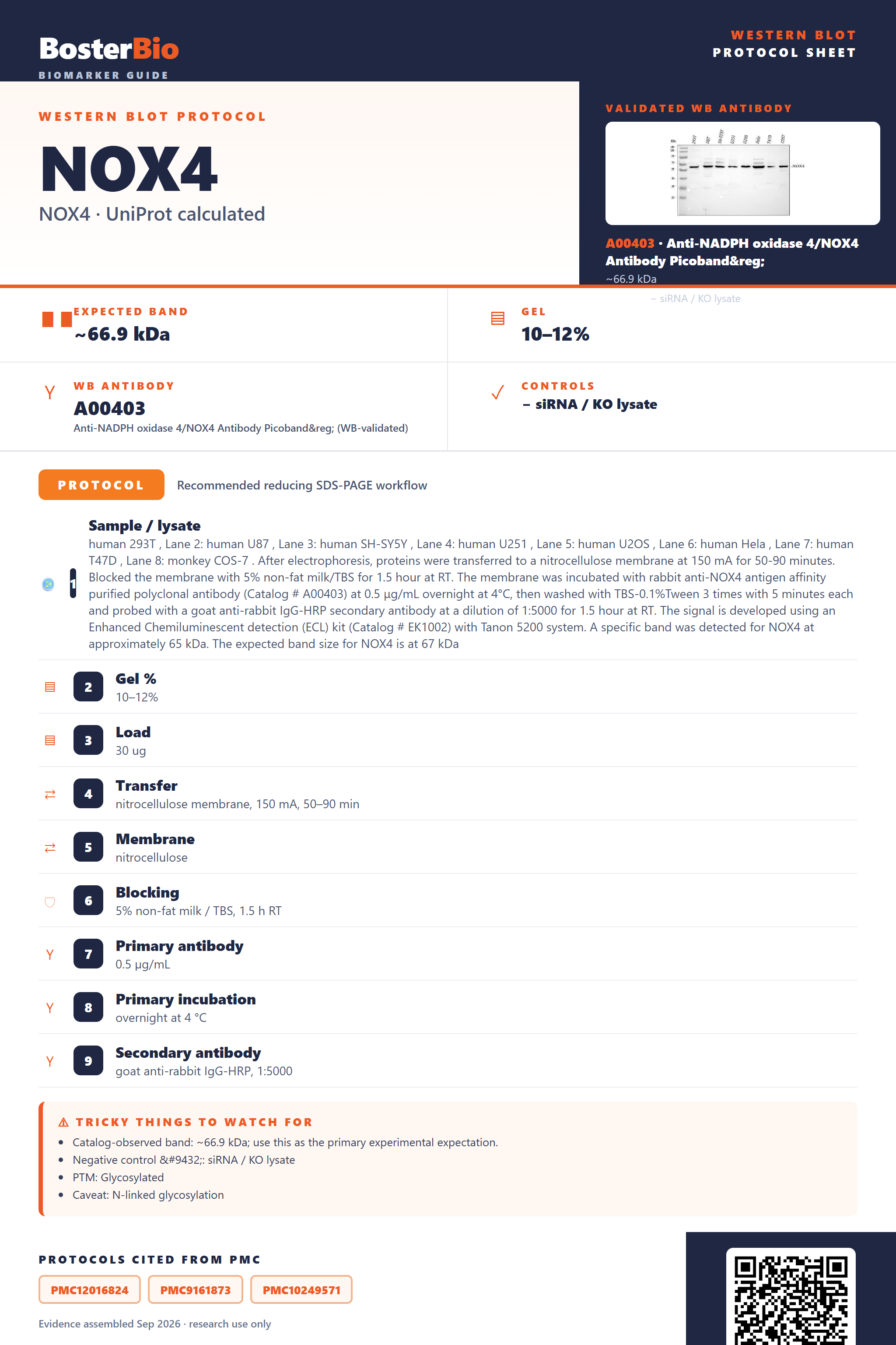
Western blot protocol sheet for NOX4: expected band ~66.9 kDa, antibody A00403, and PMC-cited SDS-PAGE protocol steps
NOX4 Western blot protocol sheet — expected band ~66.9 kDa, antibody A00403, controls and PMC citations. Open the full NOX4 WB guide →

NOX4 Western Blot Experimental Design Guide

Expected bands, validated protocols, controls and antibodies — the at-a-glance facts below, then the full design guide.

Must know before running
Expected band ~66.9 kDa
Observed band 67-80 kDa
Gel 10–12%
Negative control ⓘ siRNA / KO lysate
Important caveats
Reasons your observed band may differ from the expected size.
PTM Glycosylated
Caveat N-linked glycosylation
Regulation LPS-induced
Isoform 9 isoform(s)
Section 1

Real Curated NOX4 Western Blot Protocols

Literature-validated Western blot parameters for NOX4 — gel percentage, transfer, blocking, antibody incubation and detection, extracted from published methods.

Recommended Western blot protocol parameters
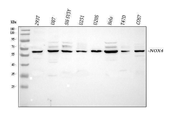
Sample / lysatehuman 293T , Lane 2: human U87 , Lane 3: human SH-SY5Y , Lane 4: human U251 , Lane 5: human U2OS , Lane 6: human Hela , Lane 7: human T47D , Lane 8: monkey COS-7 . After electrophoresis, proteins were transferred to a nitrocellulose membrane at 150 mA for 50-90 minutes. Blocked the membrane with 5% non-fat milk/TBS for 1.5 hour at RT. The membrane was incubated with rabbit anti-NOX4 antigen affinity purified polyclonal antibody (Catalog # A00403) at 0.5 μg/mL overnight at 4°C, then washed with TBS-0.1%Tween 3 times with 5 minutes each and probed with a goat anti-rabbit IgG-HRP secondary antibody at a dilution of 1:5000 for 1.5 hour at RT. The signal is developed using an Enhanced Chemiluminescent detection (ECL) kit (Catalog # EK1002) with Tanon 5200 system. A specific band was detected for NOX4 at approximately 65 kDa. The expected band size for NOX4 is at 67 kDa
Gel %10–12%
Load30 ug
Transfernitrocellulose membrane, 150 mA, 50–90 min
Membranenitrocellulose
Blocking5% non-fat milk / TBS, 1.5 h RT
Primary antibody0.5 µg/mL
Primary incubationovernight at 4 °C
Secondary antibodygoat anti-rabbit IgG-HRP, 1:5000
WashTBS-0.1% Tween, 3 × 5 min
DetectionECL
Exposure / imagingTanon 5200
Observed band65 kDa
Section 2

What Is the Expected NOX4 Western Blot Band Size?

NOX4 has a 66.9 kDa core mass but runs at 67-80 kDa on blots due to N-glycosylation at Asn133 and Asn230, with its 9 splice isoforms adding potential band complexity.

What am I looking at on my blot?
single band around 67-80 kDathe native, N-glycosylated form of NOX4 running above its 66.9 kDa core mass
diffuse or smeared band spanning roughly 67-80 kDa rather than a sharp lineheterogeneous carbohydrate loading on the two N-glycosylation sites (Asn133, Asn230)
closely spaced doublet within the 67-80 kDa rangepartial versus full occupancy of the two annotated N-glycosylation sites
extra band(s) at a molecular weight distinct from the main 67-80 kDa banddetection of a different NOX4 splice isoform among the 9 annotated isoforms
sharp band sitting right at 66.9 kDa with no upward shiftan unmodified, non-glycosylated NOX4 backbone, e.g. bacterially expressed recombinant protein
💡Expected NOX4 appearanceExpect NOX4 to run as a single band at approximately 67-80 kDa in most lysates, reflecting the 66.9 kDa core protein plus added mass from N-glycosylation at Asn133 and Asn230.
How each factor affects band size
predicted core mass (66.9 kDa)sets the baseline unmodified backbone size before any post-translational modification is added
N-glycosylation at Asn133 and Asn230adds carbohydrate mass that shifts the apparent band up into the observed 67-80 kDa range
nine annotated splice isoforms (1-9)alternative splicing can generate additional bands at sizes relatively smaller or larger than the canonical form
non-glycosylated bacterial recombinant standardlacking glycosylation machinery, it runs closer to the unmodified 66.9 kDa mass, lower than native glycosylated NOX4
Why is my band missing or off?
SituationLikely causeNext action
Band higher than expectedheavy N-glycosylation at Asn133 and Asn230 pushes the band toward the upper end of the 67-80 kDa rangereference the empirical 67-80 kDa range rather than the raw 66.9 kDa predicted mass, and confirm with glycosidase treatment if needed
Broad smear instead of sharp bandheterogeneous glycan occupancy and branching at the two N-glycosylation sitestreat lysate with PNGase F or run a longer, lower-voltage separation to resolve the glycoforms into a tighter band
Multiple bandscross-reactivity with different NOX4 splice isoforms among the 9 annotated forms, or partial glycosylation statesverify the dominant 67-80 kDa band matches the expected pattern and check the antibody epitope against isoform sequences
Band lower than expectedusing a non-glycosylated bacterial-expressed recombinant NOX4 as the size referencecompare native lysate bands only to glycosylated, mammalian-expressed NOX4 controls, not bacterial recombinant standards
Weak or no signalNOX4 is a multi-pass membrane protein residing in the ER and cell membrane, so incomplete solubilization can leave it in the insoluble pelletuse a lysis buffer with sufficient detergent for membrane protein extraction and avoid over-clearing the lysate before loading

Sample controls for NOX4 Western blot

🧪For positive controls for NOX4 in Western blot, you can use lysate from NOX4-overexpressing HEK293 cells, since no endogenous high-expressing tissue is available from the supplied evidence.
Positive control: NOX4-overexpressing HEK293 cells
Negative control: ubiquitously expressed; use siRNA knockdown or KO line
Loading controls: Run GAPDH and β-actin blots alongside, plus a total-protein stain (e.g., stain-free gel, Ponceau S, or REVERT) for normalization.
⚠️Feasibility: No HPA expression profile is available for NOX4, so a confirmed endogenous positive/negative tissue pair can't be identified; combine an overexpression lysate for the positive control with siRNA/CRISPR knockout for specificity, and use membrane-protein-compatible lysis (e.g., stronger detergent) given its ER/cell-membrane, multi-pass topology.

Comprehensive Human Protein Atlas IHC scoring per tissue. Rows are taken directly from the HPA tissue chart — click any row's HPA link to view the source.

Positive expression · recommended positive controls

TissueCell typeLevelEvidenceSource

Undetected expression · recommended negative controls

TissueCell typeLevelEvidenceSource
Section 3

Advanced NOX4 Western Blot Tips

Deeper troubleshooting and optimisation questions for NOX4, answered from its protein features.

Why does NOX4 run higher than its predicted 66.9 kDa mass?
NOX4 carries 2 N-glycosylation sites and is a multi-pass membrane protein embedded in the ER and plasma membrane. Glycosylation plus incomplete SDS denaturation of transmembrane domains typically shifts apparent mobility upward, explaining the 67-80 kDa observed band versus the 66.9 kDa calculated mass. Confirm identity with a NOX4-specific antibody and, if needed, PNGase F treatment to collapse the band toward predicted size.
Could isoform diversity affect the NOX4 band pattern?
NOX4 has 9 annotated isoforms from alternative splicing, so lysates from different tissues or cell lines may show additional bands above or below the primary species. Select an antibody epitope mapped to a region common to the isoforms of interest, and note that isoform-specific bands are a normal biological outcome, not necessarily a technical artifact.
What causes NOX4 induction relevant to Western blot signal?
NOX4 expression is transcriptionally induced by TGF-beta and hypoxia-responsive signaling in many cell types, so basal levels can be low. Include a known inducing treatment as a positive control to confirm antibody sensitivity, and always pair with an unstimulated control to correctly interpret changes in band intensity.
How should blocking be optimized for glycosylated NOX4?
Because NOX4 carries N-linked glycans, milk-based blockers containing lectin-binding glycoproteins can cause nonspecific background on the blot. Use a BSA-based blocking buffer instead of milk to avoid competing glycan interactions, and extend blocking time slightly given the membrane-embedded, glycoprotein nature of the target.
What transfer method to use for NOX4 Western blot?
NOX4 is a multi-pass transmembrane protein localized to the ER and plasma membrane, which can transfer poorly with standard rapid semi-dry systems. Use wet (tank) transfer with extended time and include SDS in the transfer buffer to improve elution of this hydrophobic membrane protein from the gel onto the membrane.
How should NOX4 signal be quantitated across samples?
Because NOX4 is a low-abundance, membrane-embedded oxidoreductase, use a membrane-protein loading control rather than a cytoplasmic housekeeping protein, since NOX4 partitions with detergent-solubilized membrane fractions. Quantify band intensity within the linear range of detection and normalize to the membrane marker for accurate comparison across conditions.
What explains unexpected extra bands on a NOX4 blot?
NOX4 interacts with and stabilizes CYBA/p22phox and is annotated with disulfide bonds, both of which can generate oligomeric or complex-associated species under non-reducing conditions. Extra higher-molecular-weight bands may reflect NOX4-p22phox complexes or incompletely reduced oligomers, while lower bands may reflect isoform variants; a reducing, denaturing SDS-PAGE protocol should resolve most of these to the monomeric species.
Boster reagents

Best NOX4 Western Blot Antibodies

BosterBio's NOX4 antibodies are among the best-performing WB antibodies on the market — well cited, thoroughly validated, and orthogonally cross-validated against negative tissues and complementary methods.

Real WB data Western blot analysis of NOX4 using anti-NOX4 antibody (A00403). Electrophoresis was performed on a 5-20% SDS-PAGE gel at 70V (Stacking gel) / 90V (Resolving gel) for 2-3 hours. The sample well of each lane was loaded with 30 ug of sample under reducing conditions. Lane 1: human 293T whole cell lysates, Lane 2: human U87 whole cell lysates, Lane 3: human SH-SY5Y whole cell lysates, Lane 4: human U251 whole cell lysates, Lane 5: human U2OS whole cell lysates, Lane 6: human Hela whole cell lysates, Lane 7: human T47D whole cell lysates, Lane 8: monkey COS-7 whole cell lysates. After electrophoresis, proteins were transferred to a nitrocellulose membrane at 150 mA for 50-90 minutes. Blocked the membrane with 5% non-fat milk/TBS for 1.5 hour at RT. The membrane was incubated with rabbit anti-NOX4 antigen affinity purified polyclonal antibody (Catalog # A00403) at 0.5 μg/mL overnight at 4°C, then washed with TBS-0.1%Tween 3 times with 5 minutes each and probed with a goat anti-rabbit IgG-HRP secondary antibody at a dilution of 1:5000 for 1.5 hour at RT. The signal is developed using an Enhanced Chemiluminescent detection (ECL) kit (Catalog # EK1002) with Tanon 5200 system. A specific band was detected for NOX4 at approximately 65 kDa. The expected band size for NOX4 is at 67 kDa.
Anti-NADPH oxidase 4/NOX4 Antibody Picoband®
Cat # A00403
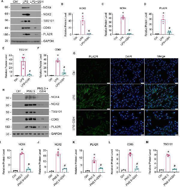
Real WB data PM2.5 induces oxidative stress and PLA2R overexpression in bronchial epithelium. (A–F) Levels of oxidative stress, PLA2R expression, and EVs production in Beas-2B cells stimulated by LPS, with or without GSH intervention. Western blot (A) and quantitative data (B–F) are presented. (B, C) NOX2 and NOX4 are oxidative stress markers from the NADPH oxidase family. (D) PLA2R expression levels. (E, F) TSG101 and CD63 are markers for EVs. All are significantly upregulated by LPS, while GSH effectively reverses this upregulation. (* p < 0.05 versus control; #P < 0.05 versus LPS). (G) Representative micrographs show immunofluorescence staining of PLA2R in Beas-2B cells treated with LPS, with or without GSH. Scale bar = 100 μm. (H–M) Levels of oxidative stress, PLA2R expression, and EVs production in Beas-2B cells stimulated by RM8785, with or without GSH intervention. Representative Western blot (H) and quantitative data (I–M) are provided. The expression levels of NOX4 (I) , NOX2 (J) , PLA2R (K) , CD63 (L) , and TSG101 (M) are all significantly upregulated by RM8785, while GSH effectively reverses this upregulation. (* p < 0.05 versus control; #P < 0.05 versus PM2.5). <b>Index in PubMed under a CC BY license. PMID: <a href='https://www.frontiersin.org/journals/pharmacology/articles/10.3389/fphar.2024.1516111/full'>39744137</a></b>
Anti-NADPH oxidase 4/NOX4 Antibody Picoband®
Cat # PA1929

Our recommended anti-NOX4 antibodies are top-performing, extensively cited reagents, thoroughly validated by Western blot and orthogonally cross-checked against negative tissue and complementary detection methods, delivering confident, reproducible detection of NOX4 expression across sample types.

Which to pick: For NOX4 WB, favor A00403 — its cue is an authentic NOX4 Western blot validation image. PA1929's listed image actually depicts PLA2R/oxidative-stress work, not a NOX4 blot, so treat it as secondary until NOX4-specific data is confirmed.

Source: BosterBio NOX4 gene-info card — filtered to Western-blot-capable antibodies; each card shows that product's actual WB validation figure.

References

  1. UniProt Consortium. UniProt entry Q9NPH5.
  2. Human Protein Atlas. NOX4 tissue expression.